Methods and Algorithms For Cell Enumeration in a Low-Cost Cytometer

a low-cost, cytometer technology, applied in the direction of instruments, computing, measurement devices, etc., can solve the problems of inaccessibility to routine clinical use of flow cytometry and similar complex analytical systems, lack of robustness, and exhibited rather complex results, so as to achieve low cost, low cost, and simple design

Inactive Publication Date: 2011-03-03
VERIDEX LCC
View PDF42 Cites 35 Cited by
  • Summary
  • Abstract
  • Description
  • Claims
  • Application Information

AI Technical Summary

Benefits of technology

[0018]Further advantages provided by this invention are the functional simplicity in design, ruggedness, compactness, AC or DC power options, and substantially lower purchase and operating costs relative to conventional commercial devices with comparable performance characteristics. The features and improvements of the devices of this invention, exemplified as compact clinical cell cytometer, make them particularly useful for operation in primitive laboratories or under field conditions prevalent in resource-poor countries.
[0019]A further improvement is in the individual illumination / light capture components of the cytometer. The LEDs are positioned to illuminate along the long axis of the cartridge at a mean angle of incidence of 45 degrees. The turret provides up to four wavelengths, depending on the intensity required to illuminate the specimen. The present configuration is for two different wavelengths. This ensures maximum illumination and light capture. The use of solid state illumination devices ensures that the light source will outlive the life of the instrument, providing a distinct advantage in field use. The filter changer operates through a slider crank having an eccentric bearing to align the individual filters. The slider crank is optimized for a small space and minimal expense. The preferred number of emission filters is two, but multiple filters are contemplated with the present application.
[0020]A further improvement is the elimination of an active Z-stage adjustment when imaging the target cells. Target cells are maintained in focus along the Z-plain with the incorporation of a spring loaded mechanism on the holding device that references the slide or cartridge against the position tabs (monuments) that maintains a fixed distance between the cells and objective lens. The spring support acts to apply a upward force to the cartridge / sample holder when positioned onto the holder. Accordingly, monuments come down onto the sample holder that push against the springs and force the sample to be in a pre-set plane. This eliminates variations in tolerances in the sample holder and cartridge. Thus, any need for changes in focus are eliminated along the Z-direction. A similar principle is used with the slide configuration.

Problems solved by technology

Flow cytometry and similar complex analytical systems remain largely inaccessible for routine clinical use in resource-poor countries due to high instrument and reagents costs, lack of technical support, lack of robustness requiring frequent service, and the need for AC power.
In addition to the above-cited full sized flow cytometry systems available from Becton Dickinson and Beckman-Coulter, these vendors also sell scaled down less expensive versions, which still suffer from the other cited limitations.
Similar limitations apply to the compact flow cytometer device CYFLOW® from Partec GmbH, (Munster, Germany) and to the Guava Personal Cytometer (Burlingame, Calif.).
U.S. Pat. No. 6,097,485 (assigned to Integrated Wave Guides, Brookings, S. Dak.) discloses an ultra-miniature personal flow cytometer (pFCM) claimed to be of lower cost, but still exhibiting rather complex, electronic circuitry, optical designs, data reduction, all of which contribute to unacceptable complexity for a third world setting.
All these systems use the flow concept, which obviously complicates the instrumental design.
These scaled down versions of flow cytometry systems do not meet the clear need for a truly simple, compact, rugged, battery-operable and affordable cell analyzer.
They generally require sophisticated techniques, which involve the use of instruments that are expensive both in terms of initial cost and maintenance as well as requiring highly trained personnel.
This makes the conventional systems unsuitable for use in laboratories of resource-poor countries.
Obviously, this does not have a high enough throughput to be an effective analyzer, especially in remote settings.
While very effective for classifying objects in images, it requires a large amount of computational resources.
Furthermore, human input and subsequent analysis is still necessary.
None are capable of detecting and enumerating a target population within a biological specimen as defined herein.
Furthermore, none appear to be portable or high throughput devices.

Method used


Image

  • Methods and Algorithms For Cell Enumeration in a Low-Cost Cytometer
  • Methods and Algorithms For Cell Enumeration in a Low-Cost Cytometer
  • Methods and Algorithms For Cell Enumeration in a Low-Cost Cytometer

Examples


example 1

[0048]Cell enumeration is commonly performed using expensive flow cytometry (FC) or less accurate manual count methods. Here, we present an automated microscope device to obtain absolute cell counts based on fluorescence with cell identification not prone to operator to operator variability of manual cell counts.

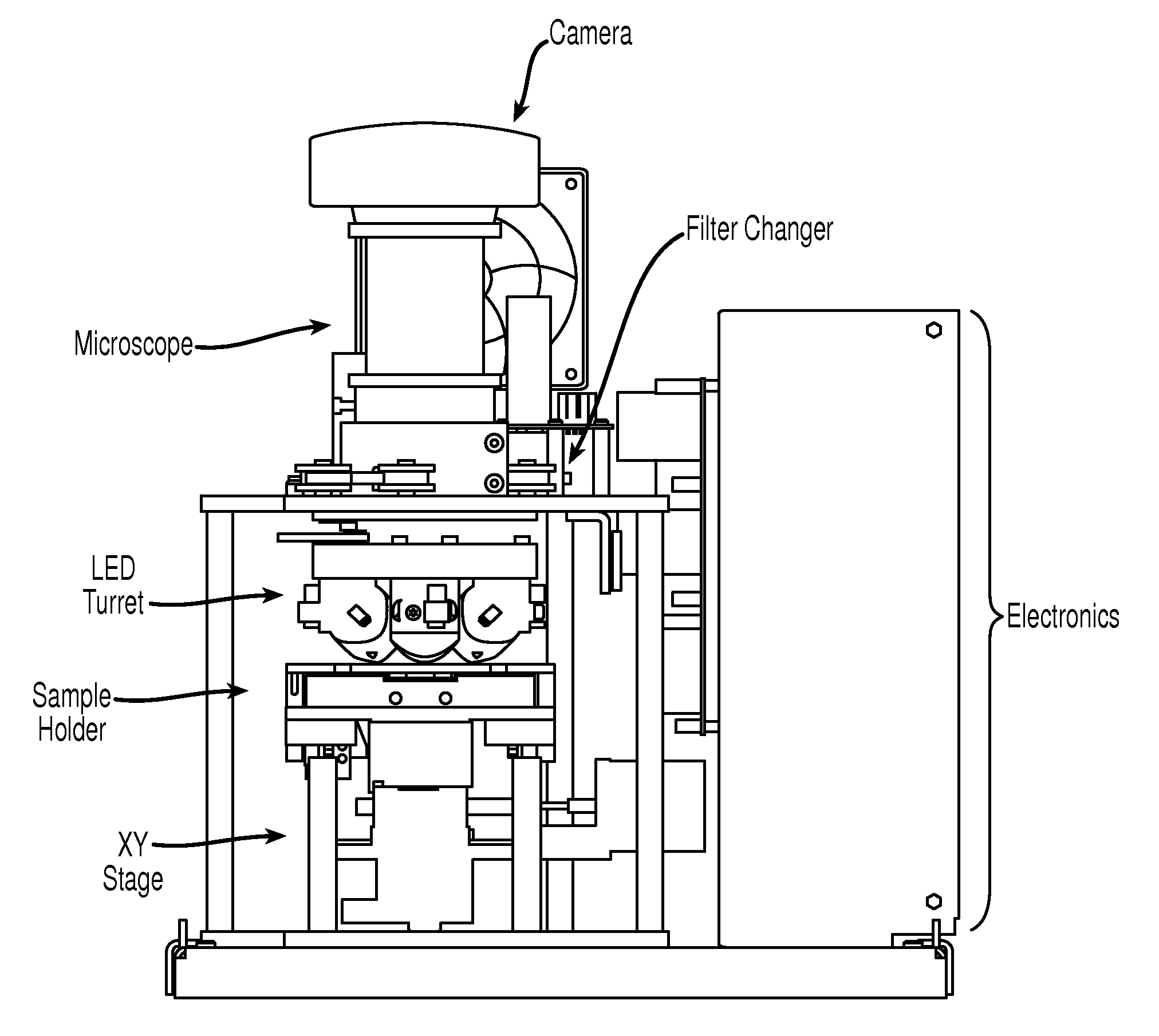
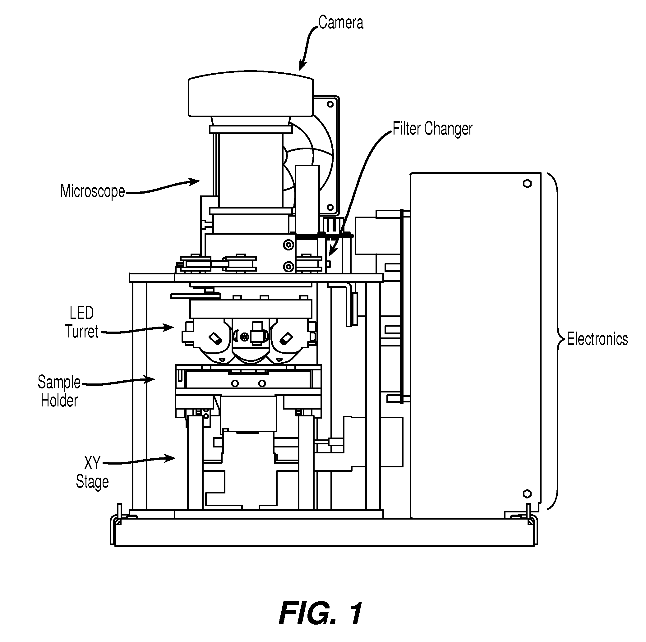
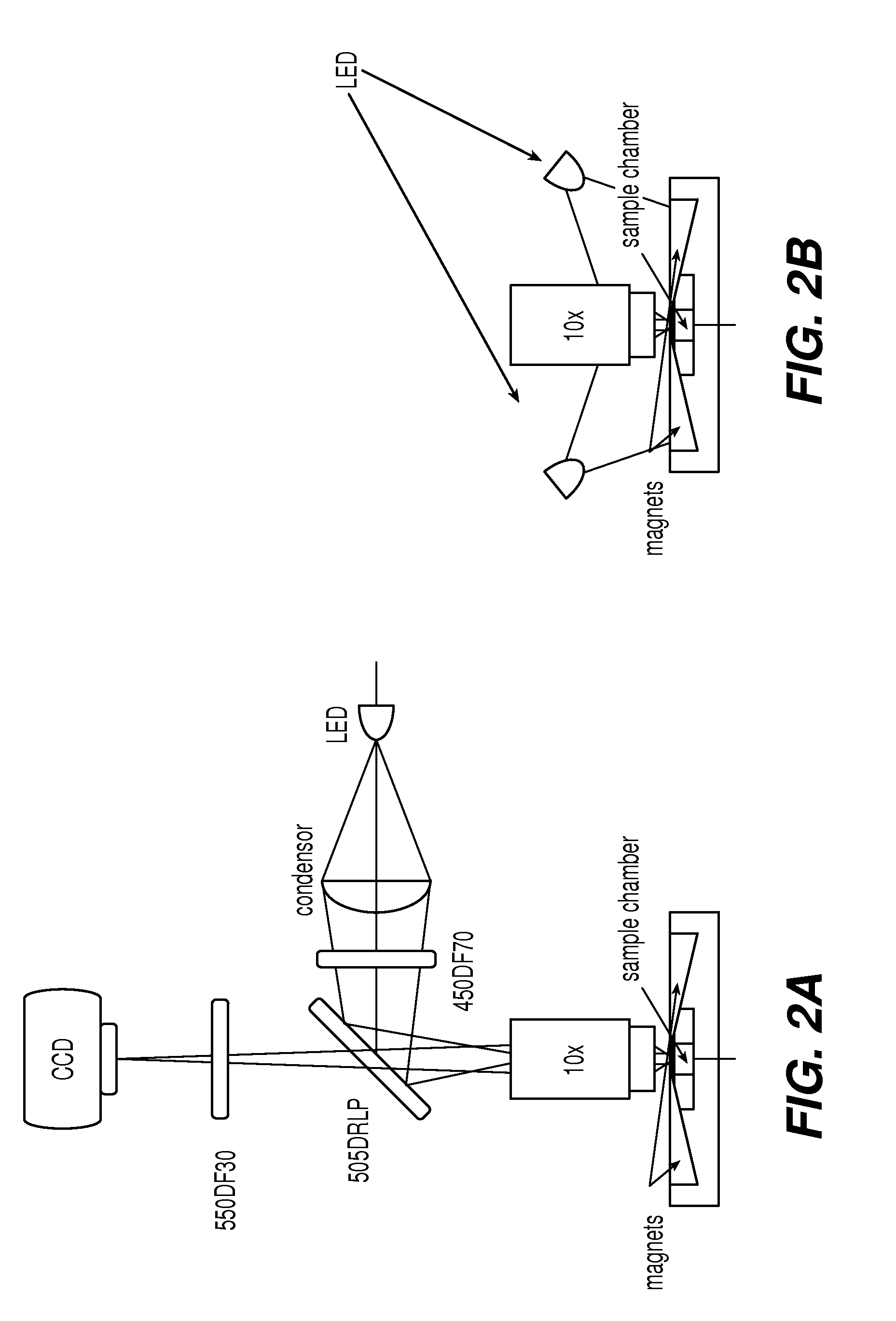
[0049]The device of the present invention, EASYCOUNT®, is a simple fluorescence microscope coupled to an on board microcomputer. It has a 5× objective (NA 0.1) and 4 high power LEDs for excitation at two different wavelengths. Each LED is equipped with an excitation filter and a collection lens for the stray light. The LEDs are placed next to the objective lens and illuminates the sample directly. Available illumination wavelengths are 490, 530 and 590 nm. Detection is performed by capturing a fluorescent image from a field of view of 0.95×1.3 mm on a 16 bit camera. The light is collected by the objective and selected by emission filters. The illumination and detection wavel...

example 2

[0053]Cells from the prostate cell line PC3-9 were harvested and resuspended in RPMI culture media. Cell count and viability determination by hemacytometer involved mixing cells 1:1 with trypan blue. Cells to be counted were mixed 1:1 with a cocktail containing acridine orange and ethidium bromide. Samples were then loaded onto a slide, placed on the EASYCOUNT® platform, and viable and non-viable counts were obtained. Cells at concentrations ranging from 2×10⁴ / ml through 5×10⁶ / ml were analyzed for total cell counts using both the hemacytometer and EASYCOUNT®. Regression analysis of the data showed an R2 of 0.999 with a slope of 1.2. At the upper end of the titration curve, hemacytometer counts required an additional dilution step which was not necessary with EASYCOUNT®. Regression analysis showed an R2 of 0.97 with a slope of 0.99. Correlation of sample viability involved making mixtures of cells with viabilities of approximately 0, 25, 50, 75, and 100%. Non-viable cells were prepar...

example 3

[0054]Counting residual leukocytes in leukodepleted blood products is done using either a specialized hemacytometer (NAGEOTTE™) or by flow cytometry on nucleic acid-stained samples. NAGEOTTE™ counting is tedious and labor-intensive. Flow cytometry requires expensive instrumentation, a skilled operator, and has low sample throughput. The fluorescent imaging system of the present invention is useful for counting low numbers of residual leukocytes in leukodepleted blood products.

[0055]One hundred microliters of blood were mixed with anti-CD45-ferrofluid and a fluorescent stain to visualize the cells. A variety of fluorescents stains were evaluated among which nucleated acid dyes and anti-CD45-PE. Leukocytes were magnetically mounted in a cartridge, used in the fluorescent imaging device CELLTRACKS®, and then the cartridge was placed in a device for magnetic orientation of magnetized targets, MAGNEST®, and imaged using either an EASYCOUNT® cell counting device or a fluorescent microscop...


PUM


Abstract

The enumeration of cells in fluids by flow cytometry is widely used across many disciplines such as assessment of leukocyte subsets in different bodily fluids or of bacterial contamination in environmental samples, food products and bodily fluids. For many applications the cost, size and complexity of the instruments prevents wider use, for example, CD4 analysis in HIV monitoring in resource-poor countries. The novel device, methods and algorithms disclosed herein largely overcome these limitations. Briefly, all cells in a biological sample are fluorescently labeled, but only the target cells are also magnetically labeled. In addition, non-magnetically labeled cells are imaged for viability in a modified slide configuration. The labeled sample, in a chamber or cuvet, is placed between two wedge-shaped magnets to selectively move the magnetically labeled cells to the observation surface of the cuvet. An LED illuminates the cells and a CCD camera captures the images of the fluorescent light emitted by the target cells. Image analysis performed with a novel algorithm provides a count of the cells on the surface that can be related to the target cell concentration of the original sample. The compact cytometer system provides a rugged, affordable and easy-to-use technique, which can be used in remote locations.

Description

CROSS REFERENCE TO RELATED APPLICATIONS[0001]This application is a divisional application from non-provisional application U.S. Ser. No. 11 / 434,321, filed 12 May 2006, which is continuation-in-part of U.S. Ser. No. 10 / 903,798, filed Jul. 30, 2004, incorporated by reference herein and claims priority, in part, of International Application PCT / US03 / 04468, filed 14 Feb. 2003, which is based upon U.S. Provisional Application No. 60 / 357,170, filed 14 Feb. 2002, now abandoned.FIELD OF THE INVENTION[0002]This invention relates generally to simple and low cost electronic optical devices, methods and algorithms for enumeration of microscopic particles distributed in a two-dimensional plane. The novel counting techniques are particularly applicable to enumerating magnetically selected fluorescent cells in complex biological specimens such as blood by means of an inexpensive cell cytometer. Further, the present invention is applicable in the analysis of specific sample characteristics such as,...

Claims


Application Information

Patent Timeline
Patent Type & Authority Applications(United States)
IPC IPC(8): G06K9/00
CPCG01N15/1475G01N33/54333G01N33/5094
Inventor COUMANS, FRANK A.W.GREVE, JANMODICA, FRANK P.TERSTAPPEN, LEON W.M.M.TIBBE, ARJAN G.J.VERRANT, JOHN A.
Owner VERIDEX LCC
Why Eureka
Try Eureka