Plasmonic System for Detecting Binding of Biological Molecules

a plasmonic system and molecular technology, applied in the field of surface plasmonic sensing compositions, methods and devices for the detection of molecular binding on membrane surfaces, can solve the problems of no commercial platform that provides a way to screen these kinds of interactions in a phospholipid membrane environment, and the difficulty of working with membrane proteins outside of the membrane environment, etc., to achieve the effect of simple fabrication and readout, and easy parallelization

Inactive Publication Date: 2012-08-16
RGT UNIV OF CALIFORNIA
View PDF3 Cites 38 Cited by
  • Summary
  • Abstract
  • Description
  • Claims
  • Application Information

AI Technical Summary

Benefits of technology

[0015]One object of the invention is to provide a nano-plasmonic sensing device having simplicity of fabrication and of readout. In one embodiment, the manufacture of the basic sensor surface is based on a series of solution-based deposition and wash steps, and the

Problems solved by technology

Furthermore, membrane proteins are notoriously difficult to work with outside of the membrane environment; supported membranes offer a strategy to handle these.
There are no commercial platforms that provide a way to screen these kinds of interactions in a phospholipid membrane environment.
The most common, comparable, way of probing molecular interactions for drug discovery in vitro is surface plasmon resonance (SPR), which involves very expensive instrumentation a

Method used


Image

  • Plasmonic System for Detecting Binding of Biological Molecules
  • Plasmonic System for Detecting Binding of Biological Molecules
  • Plasmonic System for Detecting Binding of Biological Molecules

Examples


example 1

A Nanocube-Plasmonic Sensor

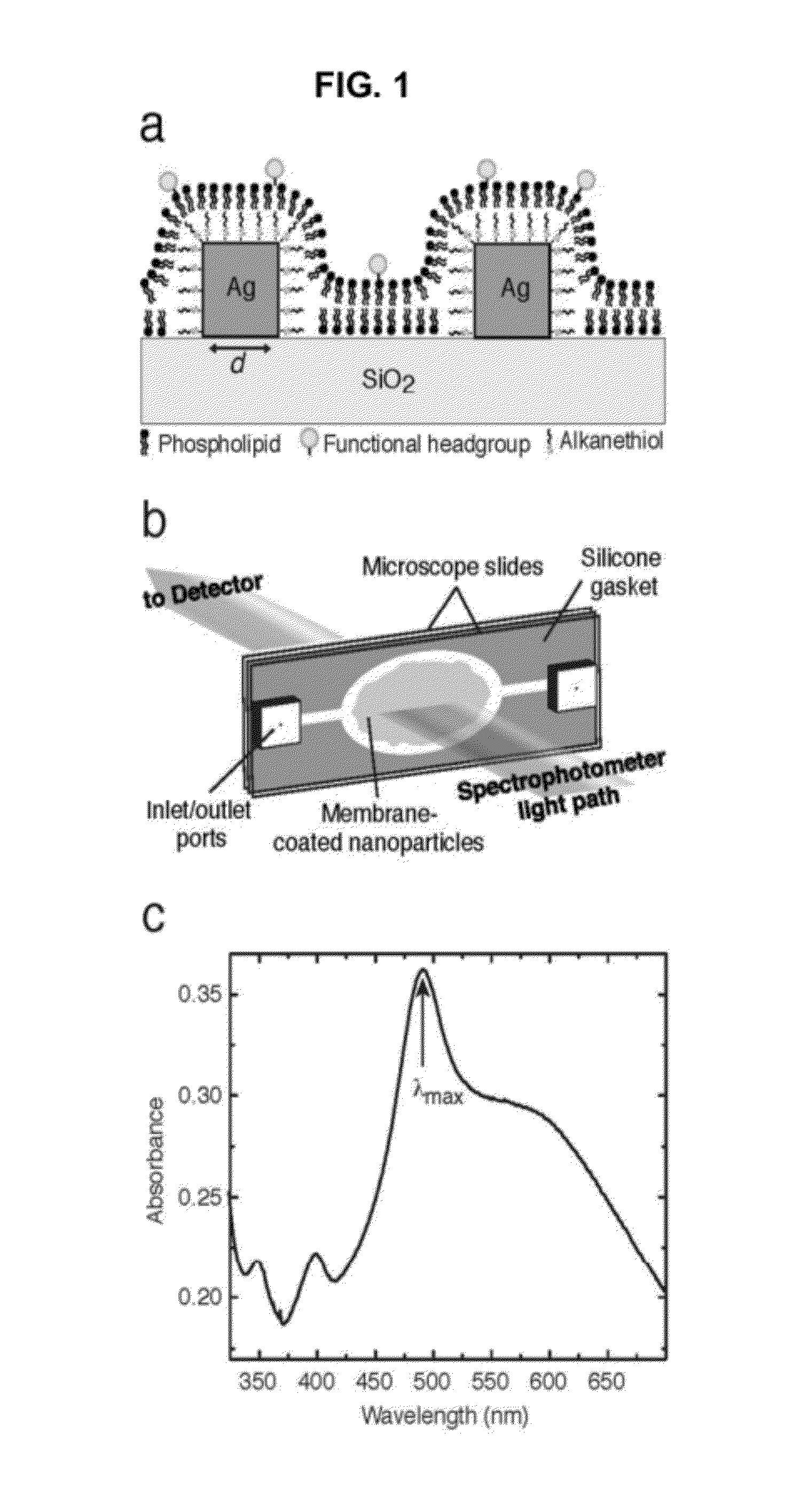
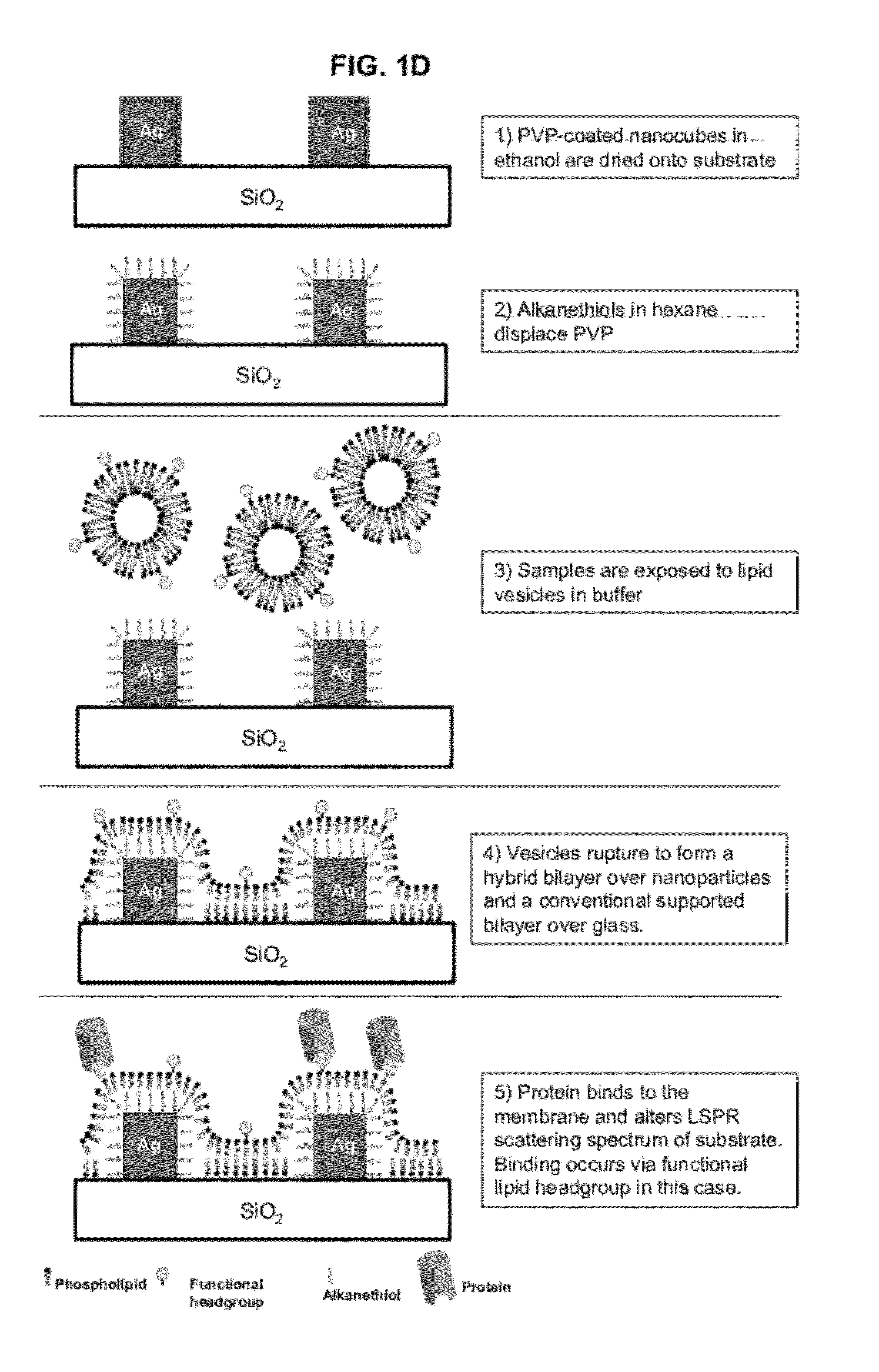
[0090]A multiplexable, label-free sensor device to measure interfacial binding of an analyte at a phospholipid membrane surface was made comprising a glass slide with a randomly ordered array of ˜100 nm wide silver nanocubes{Tao:2006,Tao:2007} (at ˜10-100 cubes / μm2 density), coated by a hybrid lipid bilayer and surrounded by a normal lipid bilayer surface (see FIG. 1). The silver nanocubes are made according to the methods described in A. Tao, P. Sinsermsuksakul, and P. Yang. Tunable plasmonic lattices of silver nanocrystals. Nature Nanotechnology, 2(7):435-440, July 2007, and] A. Tao, P. Sinsermsuksakul, and P. D. Yang. Polyhedral silver nanocrystals with distinct scattering signatures. Angewandte Chemie-International Edition, 45(28):4597-4601, 2006, both of which are hereby incorporated by reference for all purposes. The lipids themselves, or biomolecules embedded into the bilayers, determine the analyte specificity of the device. Binding occurs either t...

example 2

Detecting Molecular Binding with a Nanocube-Plasmonic Sensor

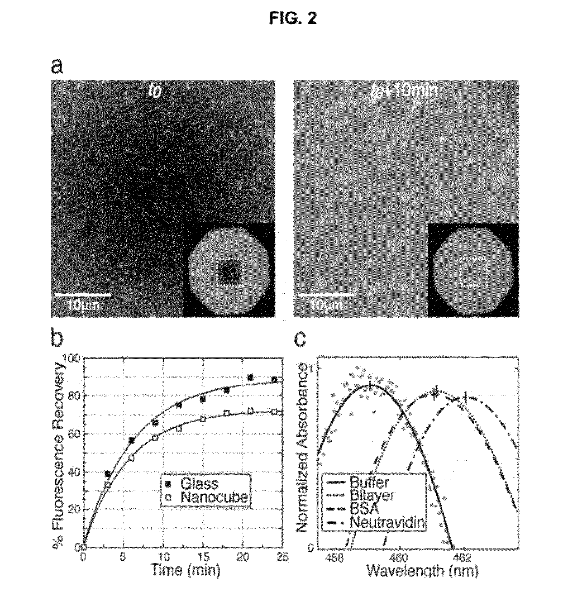
[0098]The nanocubes are seen clearly in fluorescence microscopy images as objects that appear brighter than the surrounding fluorescent supported bilayer (FIG. 2a). There are several potential causes for the high relative fluorescence intensity. Nanocubes provide an excess of local surface area compared to the flat substrate. However, the nanocubes are approximately 4-fold brighter than would be expected based purely on the geometry of a monolayer-coated 100 nm cube (see FIG. 9). One explanation for this is that it is possible for fluorophores to energetically couple to nearby plasmonic fields, resulting in a localized enhancement of fluorescence intensity, even for fluorophores without good spectral overlap between their excitation spectrum and the plasmonic scattering profile[Haes, A.; Zou, S.; Zhao, J.; Schatz, G.; VanDuyne, R. Journal of the American Chemical Society 2006, 128, 10905-10914; Zhang, J.; Fu, Y.; Chowdhury,...

example 3

Sensitivity of Nanocube-Plasmonic Sensor

[0107]An estimate of sensor noise is found by considering data from the negative control bilayer (without DOGS-NTA-Ni), shown in FIG. 3 (open squares), where protein binding to the membrane does not occur. The first sixty measurements have a standard deviation of 0.02 nm, which corresponds to a mass density of 1.5 ng cm−2 by applying the sensitivity of 170 nm cm2 ng−1. This also results in a calculated limit of detection (3× noise)[Homola, J. Chemical Reviews 2008, 108, 462-493] of 4.5 ng cm−2. The 0.02 nm value also provides an upper limit to the noise of the polynomial peak fitting method described above—the true resolution is likely much finer. While the limit of detection of supported bilayers formed in microfabricated nanoscale holes in metal films on glass is reported to be 0.1 ng cm−2 [Dahlin, A. B.; Tegenfeldt, J. O.; Hook, F. Analytical Chemistry 2006, 78, 4416-4423], the numbers quoted for the nanocube membrane sensor here represent ...


PUM


Abstract

Detection and characterization of molecular interactions on membrane surfaces is important to biological and pharmacological research. In one embodiment, silver nanocubes interfaced with glass-supported model membranes form a label-free sensor that measures protein binding to the membrane. The present device and technique utilizes plasmon resonance scattering of nanoparticles, which are chemically coupled to the membrane. In contrast to other plasmonic sensing techniques, this method features simple, solution-based device fabrication and readout. Static and dynamic protein/membrane binding are monitored and quantified.

Description

CROSS-REFERENCE TO RELATED APPLICATIONS[0001]This application claims priority to PCT International Application No. PCT / US2010 / 023375, filed on Feb. 5, 2010, hereby incorporated by reference in its entirety, which claims priority to U.S. Provisional Patent Application, 61 / 150,680, filed on Feb. 6, 2009, and U.S. Provisional Patent Application, 61 / 174,855, filed on May 1, 2009, each of which is incorporated by reference in its entirety.STATEMENT OF GOVERNMENTAL SUPPORT[0002]This invention was made with government support under Contract No. DE-AC02-05CH11231 awarded by the U.S. Department of Energy. The government has certain rights in the invention.REFERENCE TO SEQUENCE LISTING, TABLE, OR COMPUTER PROGRAM APPENDIX[0003]Not applicable.BACKGROUND OF THE INVENTION[0004]1. Field of the Invention[0005]The present invention relates to the fields of surface plasmonic sensing compositions, methods and devices for the detection of molecular binding on membrane surfaces.[0006]2. Related Art[000...

Claims


Application Information

Patent Timeline
IPC IPC(8): G01N21/75G01N21/47G01N21/59G01N21/55B82Y15/00
CPCG01N33/54346G01N21/554
Inventor GALUSH, WILLIAM J.SHELBY, SARAH A.MULVIHILL, MARTIN J.TAO, ANDREA R.YANG, PEIDONGGROVES, JOHN T.
Owner RGT UNIV OF CALIFORNIA
Why Eureka
Try Eureka