Methods And Compositions For Cellular Imaging And Cancer Cell Detection Using Light Harvesting Conjugated Polymer-Biomolecular Conjugates

a technology of biomolecular conjugates and cellular imaging, which is applied in the field of compositions for cellular imaging and cancer cell detection using light harvesting conjugated polymerbiomolecular conjugates, can solve the problems of severe cytotoxicity, low photobleaching threshold, and difficulty in implementing such a strategy using cpes

Inactive Publication Date: 2013-05-02
NAT UNIV OF SINGAPORE
View PDF2 Cites 8 Cited by
  • Summary
  • Abstract
  • Description
  • Claims
  • Application Information

AI Technical Summary

Problems solved by technology

However, each of these materials has disadvantages (e.g., low photobleaching thresholds for organic and genetic fluorophores, severe cytotoxicity for QDs under oxidative conditions, and, for live cell imaging, microinjection or electroporation techniques are often necessary...

Method used


Image

  • Methods And Compositions For Cellular Imaging And Cancer Cell Detection Using Light Harvesting Conjugated Polymer-Biomolecular Conjugates
  • Methods And Compositions For Cellular Imaging And Cancer Cell Detection Using Light Harvesting Conjugated Polymer-Biomolecular Conjugates
  • Methods And Compositions For Cellular Imaging And Cancer Cell Detection Using Light Harvesting Conjugated Polymer-Biomolecular Conjugates

Examples


example 1

Synthesis of Biomolecule-Functionalized HCPEs

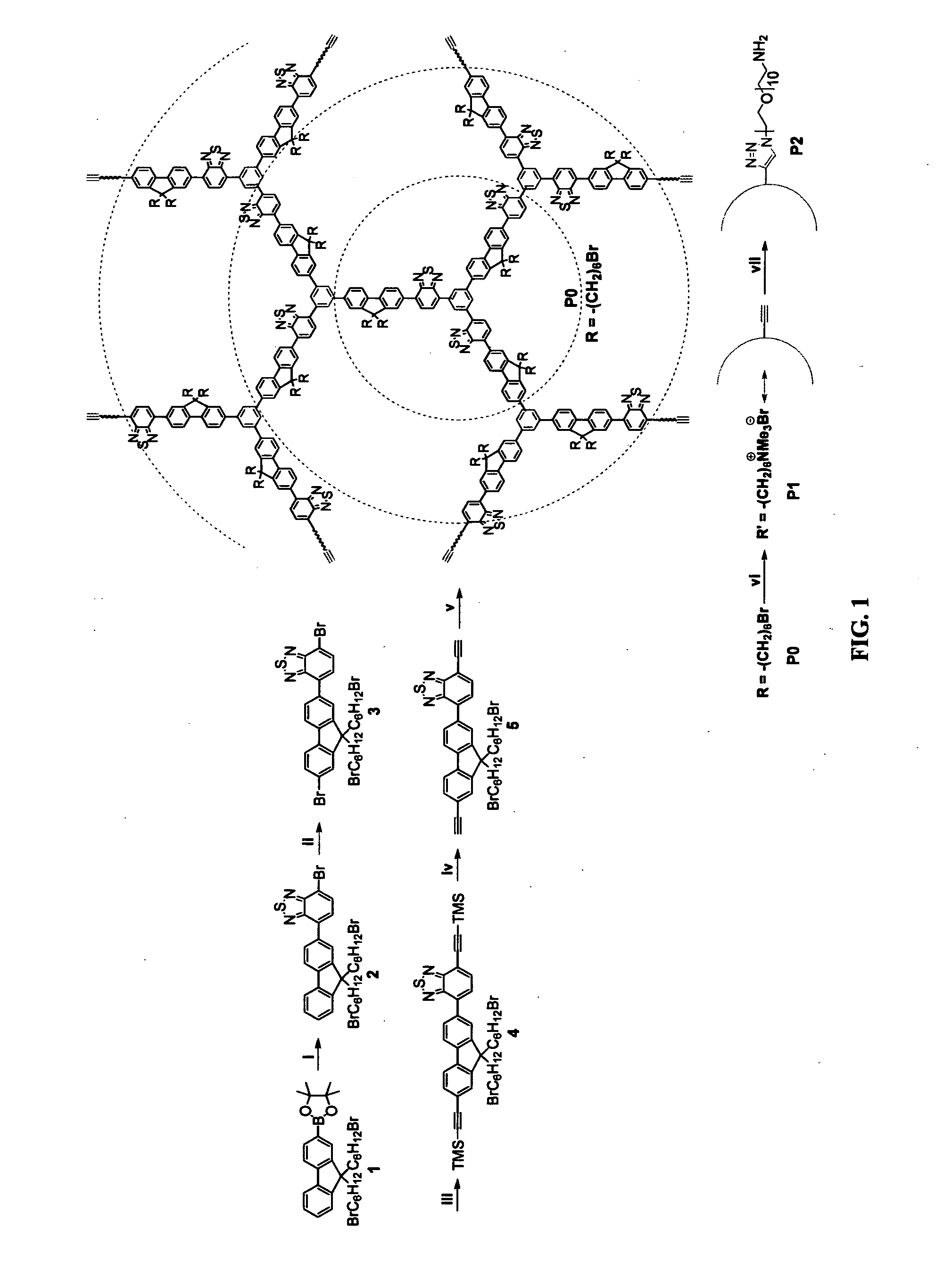
[0176]An affibody-attached hyperbranched conjugated polyelectrolyte (HCPE) was used for targeted fluorescence imaging of human epidermal growth factor receptor 2 (HER2) positive cancer cells. Early-stage detection of HER2 is of clinical significance in personalizing cancer treatment, because HER2 expression levels are closely related to tumor behavior and clinical outcome. Anti-HER2 affibody instead of commonly-used HER2-specific antibody (herceptin) was chosen as the recognition element, in view of its higher affinity for HER2 and smaller size (approximately 7 kDa) compared to herceptin (approximately 150 KDa). The HCPE (P2) used for bioconjugation was endowed with a unique core-shell molecular architecture to minimize nonspecific interactions with biomolecules and to facilitate bioconjugation and targeted cellular imaging.

[0177]The core-shell HCPE (P2) had a hyperbranched conjugated polymer as the fluorescent core and linear poly(ethyle...

example 2

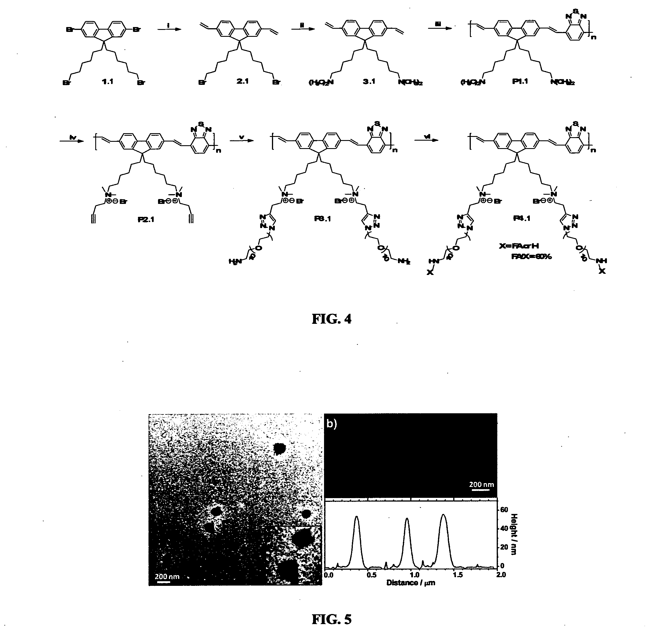
Synthesis of a Folid Acid-Functionalized Molecular Brush

[0199]The molecular brush (P4.1) was synthesized via a stepwise “grafting onto” method involving click chemistry. P4.1 formed core-shell spherical nanoparticles in aqueous solution, wherein the PEG grafting chains constituted the shell layer encapsulating the charged, conjugated backbones. Such a self-assembled nanostructure not only resulted in a high PL quantum yield in aqueous solution (11%), but also led to minimal nonspecific interactions with biomolecules and suppressed nonspecific cellular uptake. These desirable biochemical and optical properties make P4.1 an effective FR / NIR cellular probe for discrimination and visualization of MCF-7 cancer cells from NIH-3T3 normal cells in a high contrast and selective manner. In view of its high photostability and low cytotoxicity, such a molecular brush based cellular nanoprobe holds great promises as an alternative to current stains such as QDs and silica nanoparticles for clinic...

example 3

Self-Assembly Properties of P2

[0212]High-resolution transmission electron microscopy (HR-TEM) shows that P2 self-assembles into spherical nanoparticles with an average diameter of 30 nm in aqueous solution. Moreover, these nanospheres possess a core-shell nanostructure, wherein the dark interior and the gray exterior correspond to the domains enriched with electron-rich conjugated segments and saturated PEG chains, respectively. Such a core-shell nanostructure is beneficial to both bioconjugation and cell imaging, as PEG shells could serve as a protective layer.


PUM

PropertyMeasurementUnit
Quantum yieldaaaaaaaaaa
Stabilityaaaaaaaaaa
Fluorescenceaaaaaaaaaa

Abstract

The present invention relates to conjugated polyelectrolyte (CPE) or oligoelectrolyte (COE) compounds represented by general structural formulae (I)-(IV), or a salt thereof and methods of using these compounds to detect targets in samples. In particular, the methods include: (1) exposing a sample to a compound of structural formula (I), (II) or (IV) or a salt thereof, allowing the compound to bind to a target and detecting a signal produced by the compound; (2) functionalizing a solid support with a ligand, incubating the sample with a charged CPE or COE and detecting the fluorescence of the solid support and thereby detecting the target or (3) functionalizing a surface of a solid support with a charged ligand, thereby creating a charge on the surface of the solid support; incubating the ligand-functionalized solid support with a sample, whereupon binding of the target, the charge on the surface of the solid support switches; incubating the sample with CPE or COE that has a complementary charge to the charge of the target-bound surface; and detecting the fluorescence of the solid support and thereby detecting the target. The compounds of the present invention possess high photoluminescence quantum yields in biological media, low cytotoxicity, and excellent environmental stability and photostability and can be used in biosensor and bioimaging applications.

Description

RELATED APPLICATIONS[0001]This application claims the benefit of U.S. Provisional Application No. 61 / 359,737, filed on Jun. 29, 2010 and U.S. Provisional Application No. 61 / 487,880, filed on May 19, 2011. The entire teachings of the above applications are incorporated herein by reference.BACKGROUND OF THE INVENTION[0002]Fluorescent cellular probes with high selectivity and sensitivity are of central importance not only for fundamental biology and pathophysiology, but also for clinical diagnosis and therapy. Various materials including organic fluorophores, fluorescent proteins and semiconductor quantum dots (QDs) have been extensively applied for cellular imaging. However, each of these materials has disadvantages (e.g., low photobleaching thresholds for organic and genetic fluorophores, severe cytotoxicity for QDs under oxidative conditions, and, for live cell imaging, microinjection or electroporation techniques are often necessary to deliver the fluorescent probes). Recently, con...

Claims


Application Information

Patent Timeline
IPC IPC(8): C07K17/02
CPCB82Y15/00C07D285/10G01N33/582C09K2211/1416C07K17/02C09K11/06G01N33/588
Inventor LIU, BINPU, KANYILI, KAICAI, LIPINGWANG, YANYANDING, DAN
Owner NAT UNIV OF SINGAPORE
Why Eureka
Try Eureka