

Optical control of cardiac function
a technology of optical control and cardiac function, applied in the direction of light therapy, peptide/protein ingredients, therapy, etc., can solve the problems of ventricular arrest and/or fibrillation, death, compromising cardiac output,
- Summary
- Abstract
- Description
- Claims
- Application Information
AI Technical Summary
Benefits of technology
Problems solved by technology
Method used
Image

Examples
example 1
Development and Characterization of a Cell Delivery System for Non-Viral Optogenetics
[0080]Plasmid Preparation:
[0081]The plasmid, pcDNA3.1 / hChR2(H134R)-EYFP, was obtained from Addgene. It was expanded in bacteria (DH5a) on a LB+ampicillin agar plate overnight at 37° C. Selected colonies were further grown in LB-ampicillin medium with agitation, and the plasmid DNA was extracted into TE buffer using the Qiagen High Speed kit (Qiagen, Valencia, Calif.). Plasmid DNA (dsDNA) was quantified using the absorption ratio at 260 nm vs. 280 nm. After confirming the identity of the plasmid (by gel and spectrophotometry), it was stored at −20° C. in TE buffer at the obtained concentration (typically 2-4 μg / ml), and later diluted to 1 μg / ml for transfection.
[0082]Transfection:
[0083]HEK293 cells (ATCC, Manassas, Va.) were used as a model non-excitable cell and were transfected with the plasmid using Lipofectamine 2000 (Invitrogen) as directed: 4 μg of DNA and 10 μg of Lipofectamine in 250 μl mediu...
example 2
Optically-Excitable Cardiac Syncytium: Primary Cardiomyocyte Cell Culture and Co-Culture with HEK-ChR2 cells
[0091]Primary Cardiomyocyte Cell Culture:
[0092]Neonatal Sprague-Dawley rats were sacrificed and cardiomyocytes were isolated by an approved Stony Brook University IACUC protocol as previously described [37]. Briefly, the ventricular portion of the hearts was excised and washed free of blood. The tissue was cut into small pieces and enzymatically digested with trypsin at 4° C. (1 mg / ml, USB, Cleveland, Ohio), then with collagenase at 37° C. (1 mg / ml, Worthington, Lakewood, N.J.) the next morning. Cardiac fibroblasts were removed by a two-stage pre-plating process. In some transfection experiments, these cardiac fibroblasts were used in conjunction with electroporation.
[0093]Co-Culture of Cardiomyocytes with HEK-ChR2 Cells:
[0094]Cardiomyocytes were plated onto fibronectin-coated glass coverslips at high density: 4×10⁵ cells / cm2 for the control myocyte group and 3.5×10⁵ cells / cm2...
example 3
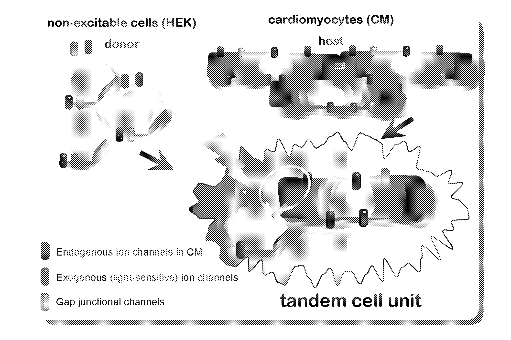
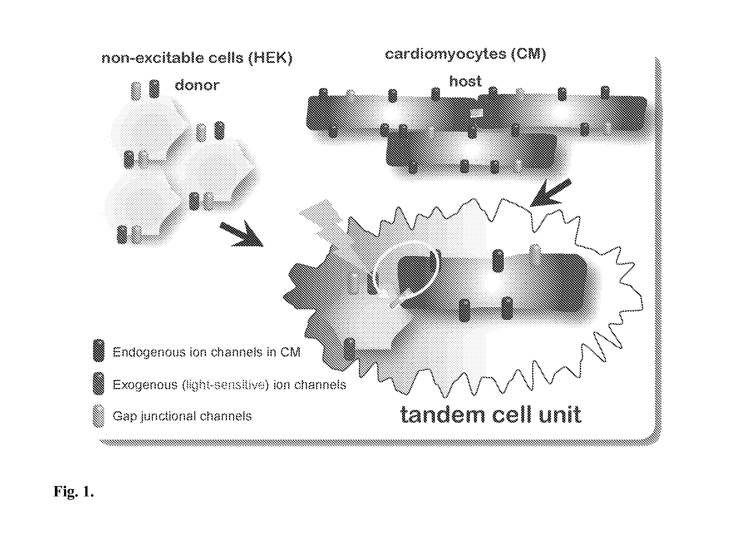
Demonstration of Tandem Cell Unit (TCU) Functionality in Cell Pairs of Adult Canine Ventricular Myocytes and HEK-ChR2
[0105]Adult mongrel dogs were euthanized as per IACUC protocol at Stony Brook University by intravenous injection of sodium pentobarbitone (80 mg / kg body weight) and the heart was removed. Canine ventricular cells were isolated using a modified Langendorff procedure [38] perfusing a wedge of the left ventricle through a coronary artery with 0.5 mg ml-1 collagenase (Worthington) and 0.08 mg ml−1 protease (Sigma) for 10 min before tissue digestion. Prior to plating, isolated cardiomyocytes were stored in Kraft-Brühe (KB) solution (in mM: KCl, 83; K2HPO4, 30; MgSO4, 5; Na-Pyruvic Acid, 5; β-OH-Butyric Acid, 5; Creatine, 5; Taurine, 20; Glucose, 10; EGTA, 0.5; KOH, 2; and Nae-ATP, 5; pH was adjusted to 7.2 with KOH) at room temperature. The canine ventricular myocytes were plated onto laminin-coated glass coverslips (10 μg / ml, Invitrogen) and incubated at 37° C. to ensure...
PUM
| Property | Measurement | Unit |
|---|---|---|
| Ratio | aaaaa | aaaaa |
| Light | aaaaa | aaaaa |
Abstract
Description
Claims
Application Information