Method for imaging a site of arthritis in an animal

imaging technology, applied in the field of imaging a site of arthritis in an animal, can solve the problems of reducing quality of life, severe pain, loss of joint function, etc., and achieve the effect of high fusogenic affinity, detection and evaluation

Active Publication Date: 2014-03-27
QI XIAOYANG
View PDF1 Cites 2 Cited by
  • Summary
  • Abstract
  • Description
  • Claims
  • Application Information

AI Technical Summary

Benefits of technology

[0008]The present disclosure demonstrates that a nanovesicle comprising a membrane-associated lysosomal protein (saposin C; “SapC”) incorporated into a phospholipid has a high fusogenic affinity for phosphatidylserine-rich domains on the surfaces of target cell membranes. It is believed that the nanovesicles target surface-exposed phosphatidylserine on the membranes of cells associated with arthritis, allowing for detection of local tissue damage associated with arthritis. In plasma membranes, phosphatidylserine is normally present only on the inner leaflet but is “flipped” to the outer leaflet upon the presence of cell damage. Incorporation of the fluorophore in the nanovesicles allows for the in vivo visualization of the fluorophore in targeted tissue and provides a technique to detect and evaluate the onset and progression of arthritic disease in an animal. Furthermore, the use of the nanovesicle in optical imaging methods provides great promise for analyzing events occurring early in the pathogenesis of arthritis and arthritic disease.
[0010]The present disclosure describes several advantages compared to current methods utilized for the imaging, detection, and assessment of arthritis in an animal. For instance, embodiments of the present disclosure can be applied via optical imaging, which uses probes that, when excited, emit light that can penetrate the skin more readily than probes with lower emission wavelengths. Thus, the use of optical imaging can facilitate advantageous signal detection in vivo. Some advantages of optical imaging include a decrease in radiation exposure compared to X-ray imaging, a clear targeting and localization of molecular probes associated with disease pathogenesis, and a minimal acquisition time compared to other radiographic methods.

Problems solved by technology

Rheumatoid arthritis is a chronic inflammatory disease in animals characterized by the destruction of cartilage and bone, frequently resulting in severe pain.
Ultimately, untreated rheumatoid arthritis in patients can result in loss of joint function, reduction in quality of life, and, in extreme cases, morbidity.
However, reliable and manageable tools for evaluation of early joint inflammation and the onset and progression of arthritic disease are lacking.
Furthermore, although radiographic imaging progression may be able to assess arthritic joint damage, soft tissue inflammation and destruction are not readily detected by radiography.

Method used


Image

  • Method for imaging a site of arthritis in an animal
  • Method for imaging a site of arthritis in an animal
  • Method for imaging a site of arthritis in an animal

Examples


example 1

K / BxN Arthritis Model

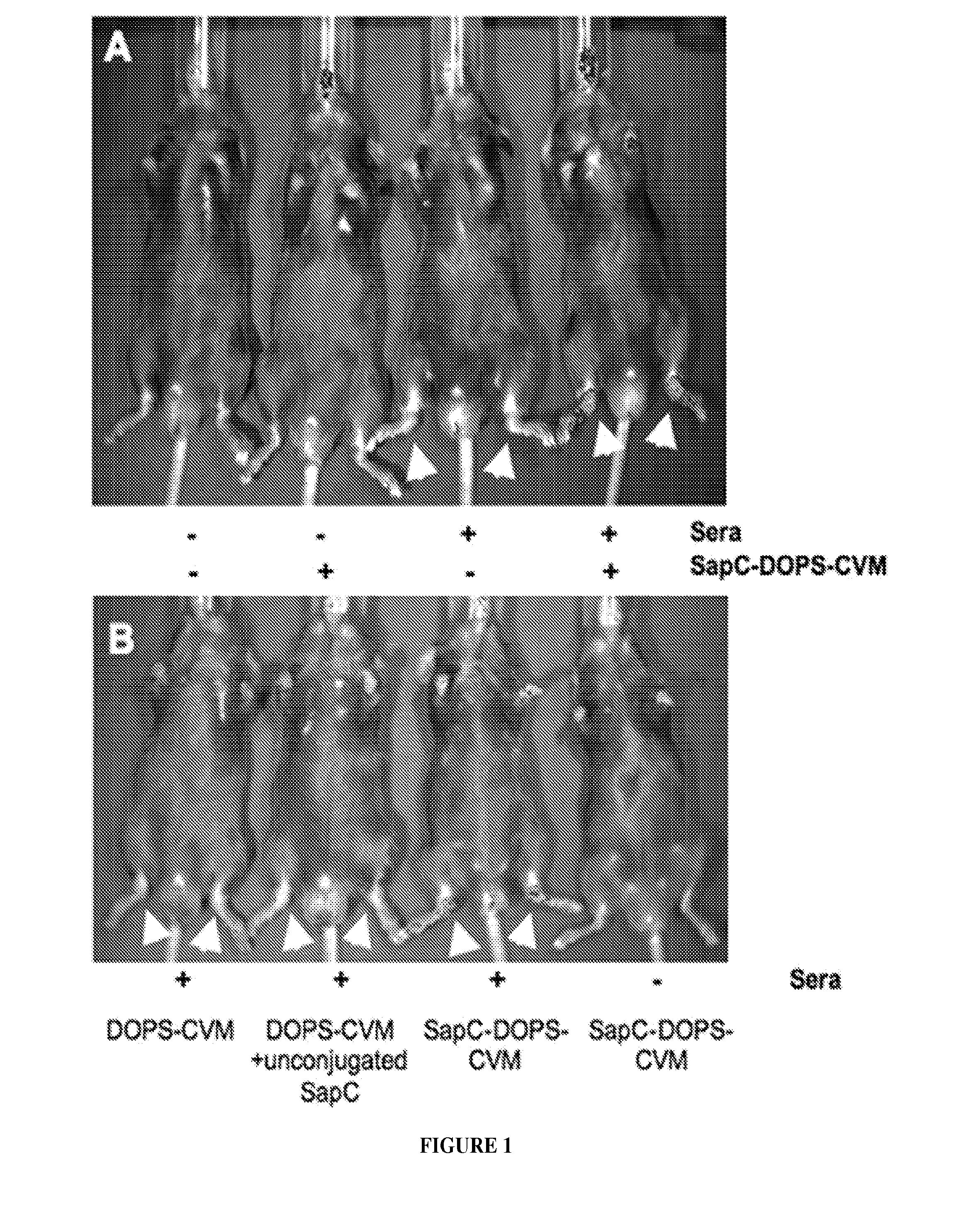
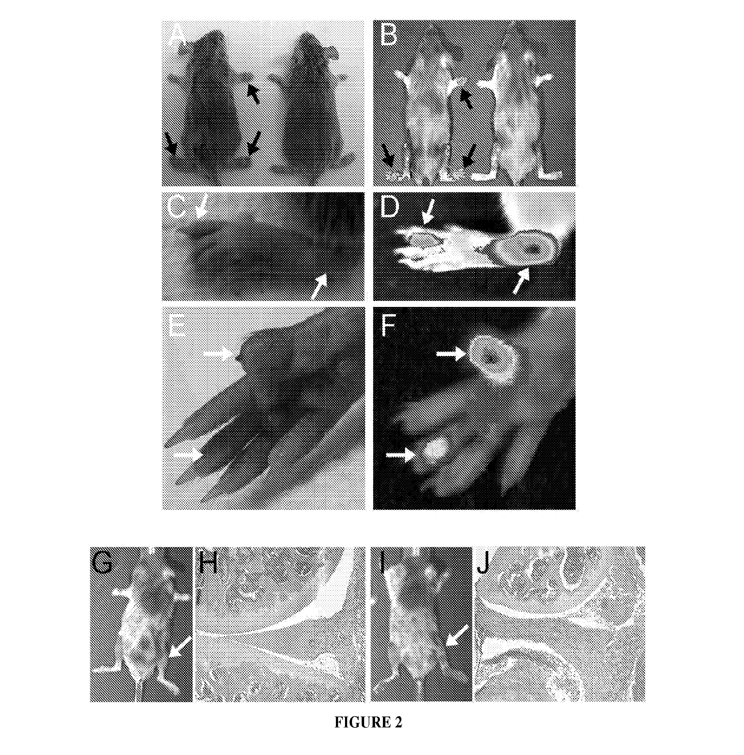
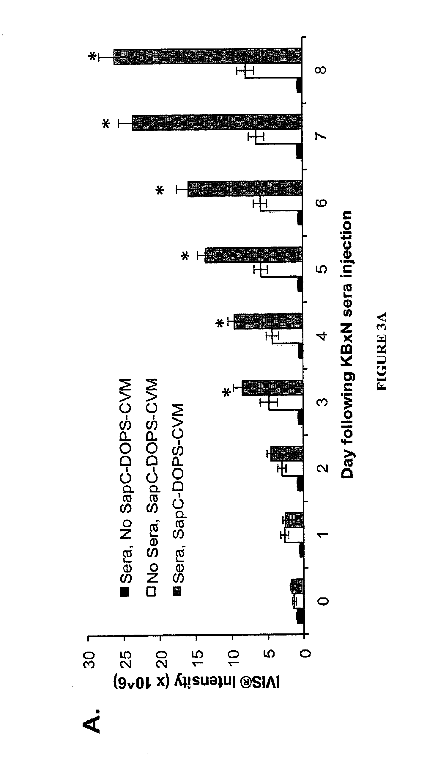
[0195]Sera can be collected from KRN x NOD F1 mice, and 150 μl sera can be given by intraperitoneal injection to C57Bl / 6J mice at day 0 of disease. Calipers can be used to measure paw thickness and ankle thickness in the mice. For ankle thickness, the elliptical area of mouse ankles can be calculated using internalleolar measurements and measurement of the area between the dorsal talus and calcaneus. In this arthritis model, 100% penetrance is normally observed, with animal hind paws being the most highly affected.

example 2

Collagen-Induced Arthritis (CIA) Model

[0196]DBA / 1J male mice (eight weeks of age) can be injected intradermally at the base of the tail with 100 μg of bovine type II collagen (CII) (Elastin Products Co., Inc.) in complete Freund's adjuvant (CFA) on day 1. The mice can be given a similar challenge on day 21 following the initial CII injection. Mice can be evaluated for macroscopic arthritis using an arthritic index macroscopic scoring system ranging from 0 to 4 (0=no detectable arthritis, 1=swelling and / or redness of paw or one digit, 2=two joints involved, 3=three joints involved and 4=severe arthritis of the entire paw and digit). Compared to the K / BxN arthritis model, between 70-100% of mice in the CIA model develop overt arthritic disease and various joints will display arthritic disease.

example 3

Imaging of Nanoparticles Labeled with CVM Using IVIS®

[0197]An aliquot of CellVue® Maroon (CVM, Molecular Targeting Technologies Inc., West Chester, Pa.) in ethanol can be mixed with phospholipid solvent for bath sonication preparation by the procedure as previously described (see Kaimal V et al., “Saposin C Coupled Lipid Nanovesicles Enable Cancer-Selective Optical and Magnetic Resonance Imaging,” Mol Imaging Biol (2010) and Qi X, “Cancer-selective targeting and cytotoxicity by liposomal-coupled lysosomal saposin C protein,” Clin Cancer Res, 15: 5840-5851 (2009). Nanovesicles labeled with CVM can be separated from free CVM dye using a Sephadex™ G25 column (PD-10, Amersham Pharmacia Biotech, Piscataway, N.J.). Nanovesicles labeled with CVM (SapC=4.2 mg / kg, DOPS=2 mg / kg, CVM=6 μmol) and control agents can be administered to mice i.p or via tail vein injection in a volume of 200 μL. Real-time live images can be taken using an IVIS 200 Series imaging system with an XFO-6 fluorescent kit...


PUM

PropertyMeasurementUnit
wavelength rangeaaaaaaaaaa
wavelengthaaaaaaaaaa
diameteraaaaaaaaaa

Abstract

The present disclosure demonstrates that a nanovesicle comprising a membrane-associated lysosomal protein (saposin C; “SapC”) incorporated into a phospholipid has a high fusogenic affinity for phosphatidylserine-rich domains on the surfaces of target cell membranes. It is believed that the nanovesicles target surface exposed phosphatidylserine on the membranes of cells associated with arthritis, allowing for detection of local tissue damage associated with arthritis. In plasma membranes, phosphatidylserine is normally present only on the inner leaflet but is “flipped” to the outer leaflet upon the presence of cell damage. Incorporation of the fluorophore in the nanovesicles allows for the in vivo visualization of the fluorophore in targeted tissue and provides a technique to detect and evaluate the onset and progression of arthritic disease in an animal. Furthermore, the use of the nanovesicle in optical imaging methods provides great promise for analyzing events occurring early in the pathogenesis of arthritis and arthritic disease.

Description

CROSS REFERENCE TO RELATED APPLICATIONS[0001]This application claims the benefit under 35 U.S.C. §119(e) of U.S. Provisional Application Ser. No. 61 / 483,813 filed on May 9, 2011, the entire disclosure of which is incorporated herein by reference.GOVERNMENT RIGHTS[0002]This invention was made with government support under grant numbers RR026314 and AR047363 awarded by the National Institutes of Health. The U.S. Government has certain rights in the invention.TECHNICAL FIELD[0003]This invention pertains to the field of imaging a site of arthritis in an animal using a nanovesicle comprising a fluorophore, and methods for detection and assessment using the same.BACKGROUND AND SUMMARY OF THE INVENTION[0004]Arthritis is a joint disorder that involves inflammation of one or more joints. There are over 100 different forms of arthritis. The most common symptom of individuals affected with arthritis is joint pain. Arthritic pain is often constant and may be localized to the affected joint or j...

Claims


Application Information

Patent Timeline
Patent Type & Authority Applications(United States)
IPC IPC(8): A61K49/00
CPCA61K49/0056A61K9/5146A61B5/0071A61B5/4528A61B2503/40A61B5/004A61K49/0017
Inventor THORNTON, SHERRY L.QI, XIAOYANGFLICK, MATTHEW J.
Owner QI XIAOYANG
Why Eureka
Try Eureka