Magnetic separation using nanoparticles

a technology of nanoparticles and magnetic separation, applied in the field of magnetic separation, can solve the problems of multiple organ dysfunction and death, and achieve the effects of short incubation time, high selective separation, and rapid binding between nanoparticles and target species

Active Publication Date: 2014-07-31
MASSACHUSETTS INST OF TECH +1
View PDF5 Cites 14 Cited by
  • Summary
  • Abstract
  • Description
  • Claims
  • Application Information

AI Technical Summary

Benefits of technology

[0005]When bis-M-DPA modified nanoparticles are mixed with the liquid, e.g., blood, rapid binding occurs between the bis-M-DPA ligands on one or more nanoparticles and bacterial contaminants in the liquid. A motive force, e.g., a magnetic force, or other means of separation, e.g., size-based separation, centrifugal force separation, or flow cytometry-based separation, can then be applied to the nanoparticles to separate them from the liquid. Some of the nanoparticles have bacteria or endotoxin bound thereto, and when the nanoparticles are removed, the contaminants are also removed with them from the liquid. Microfluidic systems can be used as platforms for nanoparticle-based separations, providing a mechanism for efficient and selective separation of contaminants from liquids of any kind.
[0018]The approaches described herein for bacterial contaminant removal, e.g., magnetic separation, from fluids have several advantages. For instance, magnetic separation using nanoparticles is generic and can be used on many target bacterial contaminant species, including Gram positive bacteria, Gram negative bacteria, and / or bacterial endotoxins. Separation using nanoparticles is also highly selective; the nanoparticles have negligible affinity toward normal mammalian cells and other materials that may be present in the fluids. Binding between nanoparticles and a target species is rapid and very short incubation times can be used. In addition, separation can be achieved at rapid flow rates through microfluidic systems, enabling efficient separation of bacterial contaminants from fluids. The synthetic ligands used are inexpensive and straightforward to synthesize, can be easily conjugated to nanoparticles, and are stable against denaturation at room temperature and even at body temperature.

Problems solved by technology

Bacterial sepsis is a serious clinical condition that can lead to multiple organ dysfunction and death despite timely treatment with antibiotics and fluid resuscitation.

Method used


Image

  • Magnetic separation using nanoparticles
  • Magnetic separation using nanoparticles
  • Magnetic separation using nanoparticles

Examples


example 1

Nanoparticle Synthesis and Characterization

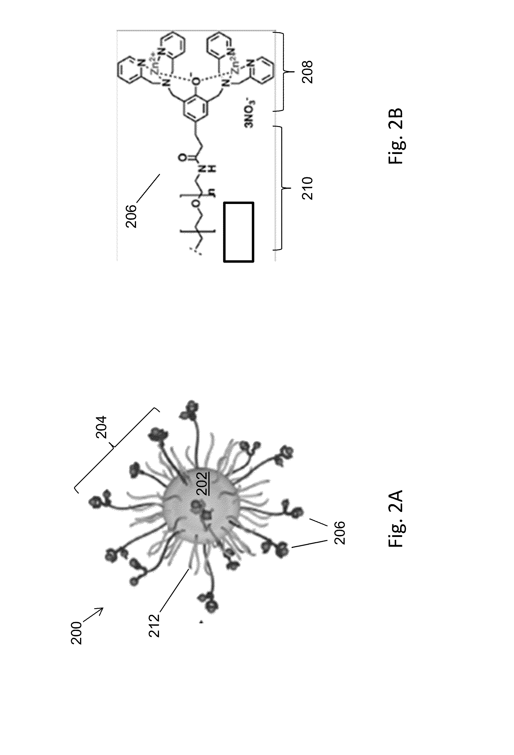
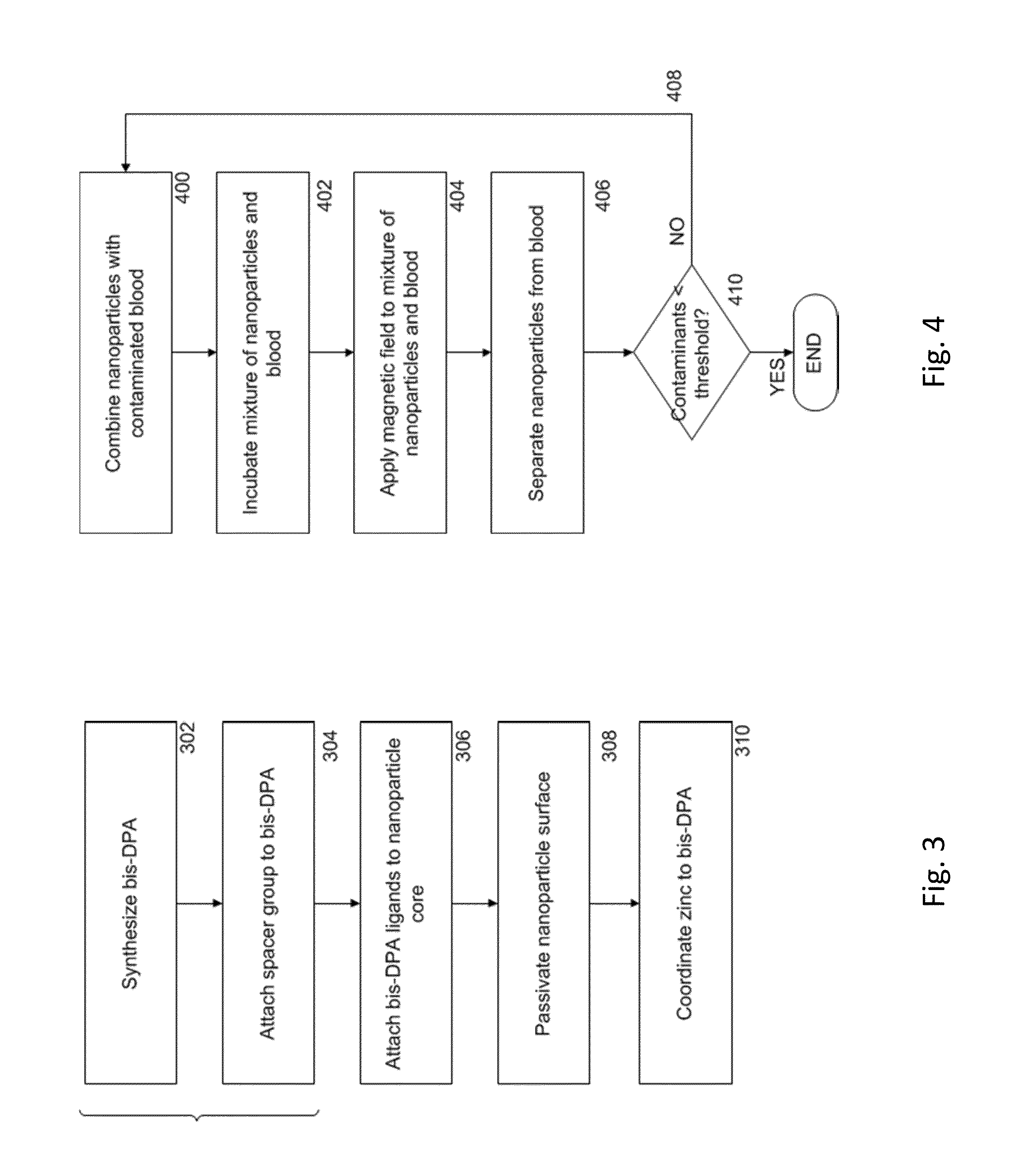
[0076]FIG. 8A shows a scheme 800 for the preparation of magnetic nanoparticles modified with bis-DPA-PEG-Zn (referred to herein as NPPEG-DPA-Zn). Bis-DPA with a PEG (MW=10 kDa) spacer (bis-DPA-PEG-COOH) was immobilized on the surface of 100 nm diameter amine-terminated Fe3O4 magnetic nanoparticles (approximately 300 amines per nanoparticle, SiMAG-Amine particles, chemicell GmbH, Berlin, Germany) through carbodiimide chemistry using EDC and (sulfo-NHS). Excess bis-DPA-PEG-COOH was activated by N-hydroxysulfosuccinimide (sulfo-NHS) with EDC (10-fold molar excess each) and added to a solution of nanoparticles. The nanoparticle solution was washed twice with carbonate buffer (50 mM sodium carbonate, pH 9.5) using a magnetic separator (MagnetoPURE, chemicell GmbH). After 2 hours of reaction at room temperature, the modified nanoparticles were washed twice with carbonated buffer.

[0077]Unreacted free amines on the surface of the nanoparticles were...

example 2

Fluorescence Visualization of E. Coli Binding to Nanoparticles

[0083]Fluorescence-labeled E. coli were used to demonstrate the binding capability of NPPEG-DPA-Zn. A bacterial cell culture was prepared by inoculating 25 mL of standard lysogeny broth (LB) with E. coli (E. coli strain Stbl3, Life Technologies, Grand Island, N.Y.) using standard sterile technique. This culture was incubated for 12-18 hours at 37° C. and 5.0% CO2 and agitated at 200 RPM in an orbital shaker incubator overnight. Before using the bacteria for experiments, cells were pelleted by centrifugation at 2,000 g for 5 minutes. The supernatant was discarded and the cell pellet was resuspended in phosphate buffered saline (PBS). The concentration of E. coli was re-adjusted to be 1.0×10⁹ CFU / mL (OD600=1.0).

[0084]To label the E. coli with the green fluorescent label SYTO 9™ (Life Technologies, Grand Island, N.Y.), which selectively labels live bacteria, the E. coli solution was incubated with 300× diluted SYTO 9 for 30 ...

example 3

Separation of E. Coli from a Buffer Solution

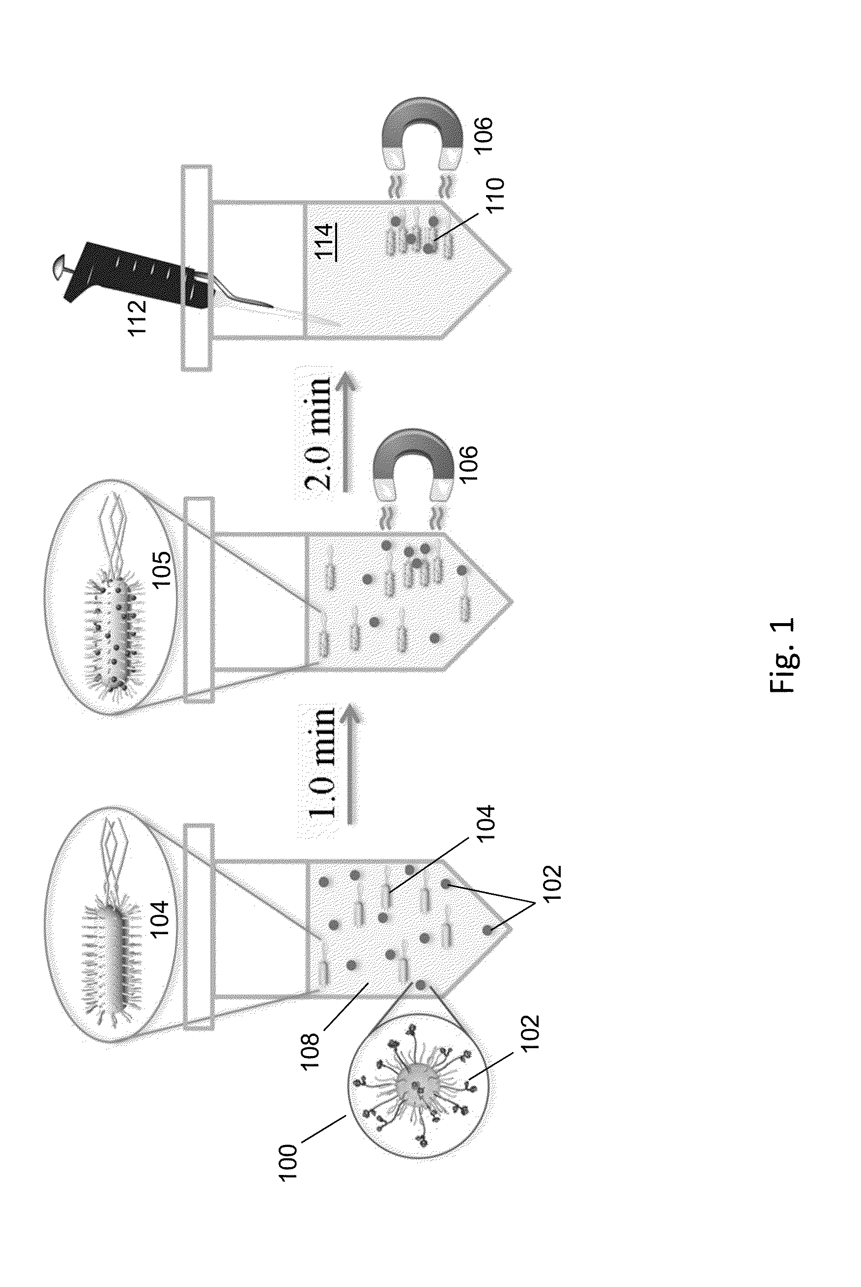
[0090]The efficiency of modified nanoparticles to separate a fixed concentration of E. coli (1.0×10⁷ CFU / mL) by magnetic separation was measured in phosphate buffered saline (PBS) with varying concentrations of NPPEG-DPA-Zn DPA-Zn and NPPEG (from 0 to 1.0×10¹¹ / mL). Each mixture of E. coli and nanoparticles was incubated in a 1.5 mL test tube for one minute at room temperature and a permanent magnet was placed on the side of the test tube for 2 minutes. 10 μL of the supernatant was collected and added to a cell counting chamber for analysis.

[0091]The number of bacteria in a 100× green fluorescence image of the supernatant was counted (using the NIH-provided freeware ImageJ) and converted to E. coli concentration in the solution. For this, a standard curve was generated that correlated the number of E. coli in the 100× green fluorescence image with the E. coli concentration in the solution (see below).

[0092]Referring to FIG. 11, a concentrat...


PUM

PropertyMeasurementUnit
molecular weightaaaaaaaaaa
flow rateaaaaaaaaaa
molecular weightaaaaaaaaaa

Abstract

Nanoparticles as described herein are configured to bind to bacterial contaminants, such as Gram positive bacteria, Gram negative bacteria, and endotoxins. The nanoparticles include a core comprising a magnetic material; and a plurality of ligands attached to the core. The ligands include, for example, bis(dipicolylamine) (“DPA”) coordinated with a metal ion, e.g., Zn2+ or Cu2+, to form, e.g., bis-Zn-DPA or bis-Cu-DPA, which can bind to the bacterial contaminants. The nanoparticles can be included in compositions for use in methods and systems to separate bacterial contaminants from liquids, such as liquids, such as blood, e.g., whole or diluted blood, buffer solutions, albumin solutions, beverages for human and / or animal consumption, e.g., drinking water, liquid medications for humans and / or animals, or other liquids.

Description

CLAIM OF PRIORITY[0001]This application claims priority to U.S. Provisional Application Ser. No. 61 / 758,046, filed on Jan. 29, 2013, the entire contents of which are hereby incorporated by reference.FIELD OF THE INVENTION[0002]The invention relates to magnetic separation, e.g., of bacteria and endotoxins, using nanoparticles.BACKGROUND OF THE INVENTION[0003]Bacterial sepsis is a serious clinical condition that can lead to multiple organ dysfunction and death despite timely treatment with antibiotics and fluid resuscitation. Gram-negative bacteria are common causative pathogens in bacterial sepsis. Common treatments for sepsis and septic shock include antibiotics, fluid resuscitation, and vasoactive medications. Other approaches, such as extracorporeal removal of inflammatory mediators, corticosteroids, and recombinant protein drug therapies have been introduced as experimental adjunct treatments. Antibody-based methods of separating bacteria from blood have also been developed.SUMMA...

Claims


Application Information

Patent Timeline
Patent Type & Authority Applications(United States)
IPC IPC(8): A61L2/18C07F15/02
CPCA61L2/18C07F15/02A61L2/00A61L9/00
Inventor LEE, JUNG-JAEJEONG, KYUNG JAEKOHANE, DANIEL S.
Owner MASSACHUSETTS INST OF TECH
Why Eureka
Try Eureka