Detection of membrane protein-therapeutic agent complexes by mass spectrometry

a membrane protein and complex technology, applied in the field of detection of complexes comprising membrane proteins bound to therapeutic agents, can solve the problems of affecting the delivery of targeted drugs, affecting the detection efficiency of targeted drugs, so as to achieve the effect of less sampl

Active Publication Date: 2014-08-28
OXFORD UNIV INNOVATION LTD
View PDF0 Cites 3 Cited by
  • Summary
  • Abstract
  • Description
  • Claims
  • Application Information

AI Technical Summary

Benefits of technology

[0015]A method of the present invention may allow therapeutic agents to be screened. In contrast to indirect methods such as fluorescence or calorimetry, the present method may allow therapeutic agents to be screened directly. In particular, a method may be used to screen for the binding of activators and transporter substrates which are difficult to screen using conventional in vivo methodologies. Moreover, unlike X-ray crystallography, the present methods are not complicated by the inherent structural flexibility of membrane protein-therapeutic agent complexes and may allow the dynamical behaviour of membrane proteins and their interaction with therapeutic agents to be studied. In addition, a method of the present invention may use significantly less sample than other analytical methods.

Problems solved by technology

On the one hand, its overexpression in tumour cells, impairing targeted drug delivery, is a major pitfall for actual chemotherapies.
Despite their importance, membrane proteins and ligand-bound complexes thereof are notoriously difficult to study.
However, the conformational flexibility of a membrane protein or a membrane protein-ligand complex is restricted within a crystal lattice and may distort the structural and / or ligand-binding properties of a protein.
Furthermore, difficulty is often encountered with the crystallisation of membrane proteins and membrane protein-ligand complexes.
However, these methods are low-resolution methods and usually require large quantities of protein.
In addition, data from these methods do not normally reveal ligand binding to membrane proteins, or allow observation on post-translational modifications.
In particular, the methods do not provide detailed information regarding drug binding to membrane proteins.
However, these methods do not provide structural or conformational information regarding the bound protein complex.
Until recently, mass spectrometry had not been considered suitable for detecting intact membrane proteins.
Despite these advances, the use of mass spectrometry to characterise the binding of membrane proteins to therapeutic agents such as drugs has not yet been demonstrated.

Method used


Image

  • Detection of membrane protein-therapeutic agent complexes by mass spectrometry
  • Detection of membrane protein-therapeutic agent complexes by mass spectrometry
  • Detection of membrane protein-therapeutic agent complexes by mass spectrometry

Examples


example 1

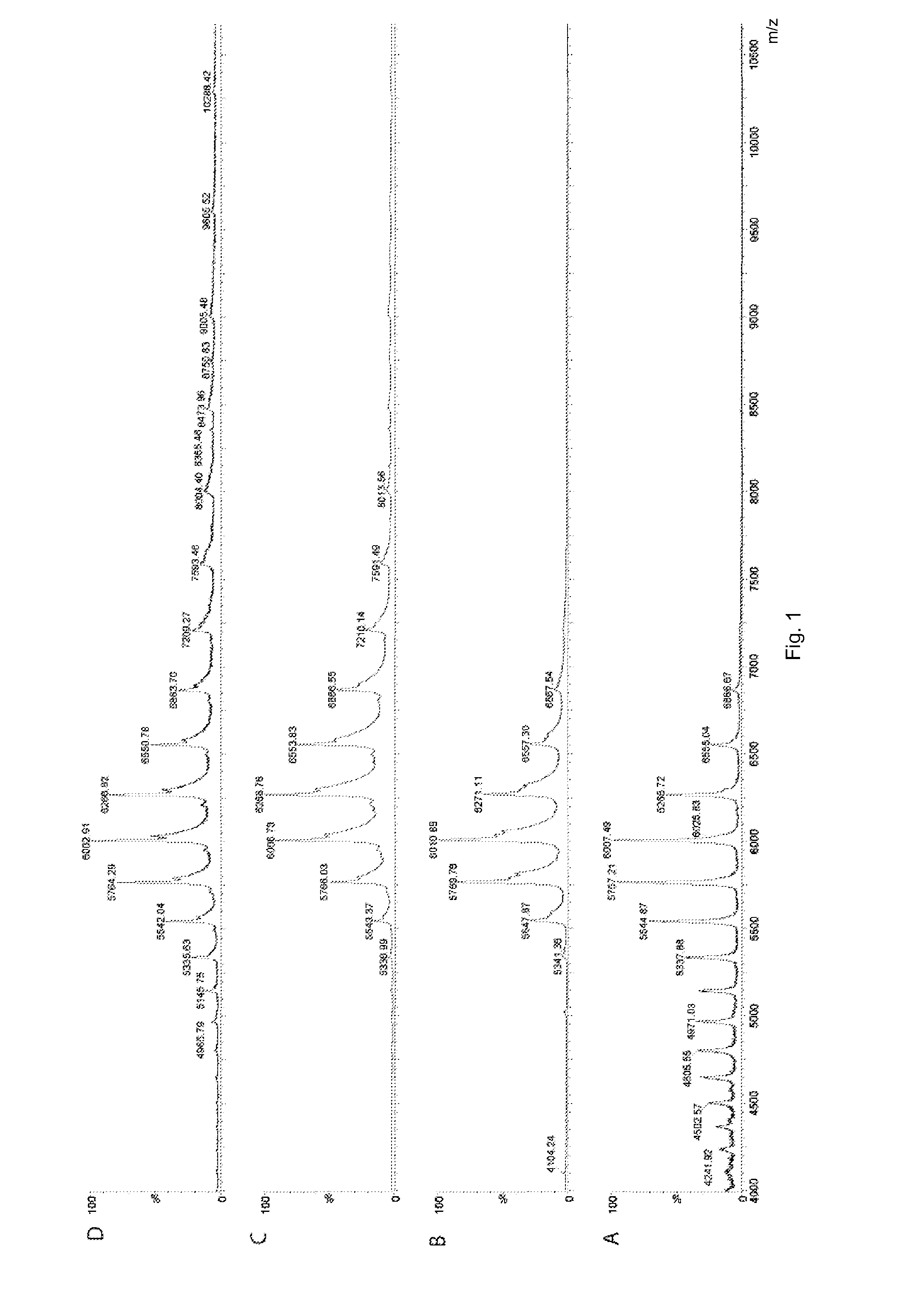
[0048]MS and IM-MS data of a membrane transporter, P-gp, binding to different drugs were acquired. The drugs included: two stereoisomers of a cyclic peptide inhibitor (QZ59Se, SSS; and QZ59Se, RRR); a further inhibitor (HT-55); and two activators (HT-35 and HT-122).

[0049]P-glycoprotein (P-gp) was expressed and purified as reported previously (Aller et al, Science, 2009, 323, 1718-1722). In this method, P-gp was expressed in P. pastoris strain and purified as a his-tagged protein from the membranes solubilised with Triton X-100. Buffer was exchanged by washing with buffer containing 20 mM imidazole, 0.04% sodium cholate and 0.0675% β-DDM. The protein was concentrated, subjected to gel filtration chromatography and assayed for purity by SDS-PAGE.

[0050]An aliquot (30 μl) of mouse P-gp solution (9 mg / ml) in 0.07% β-DDM, 0.04% sodium cholate, 100 mM NaCl, 0.2 mM TCEP, 20 mM Hepes, pH 7.5 was buffer-exchanged using a membrane filter and diluted 3-fold in 300 mM ammonium acetate, pH 7.5. S...

example 2

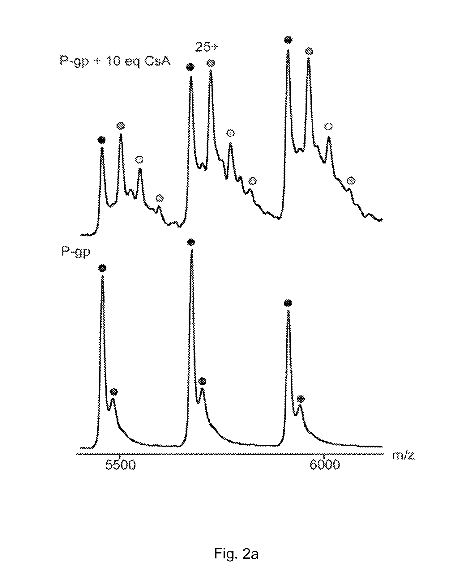
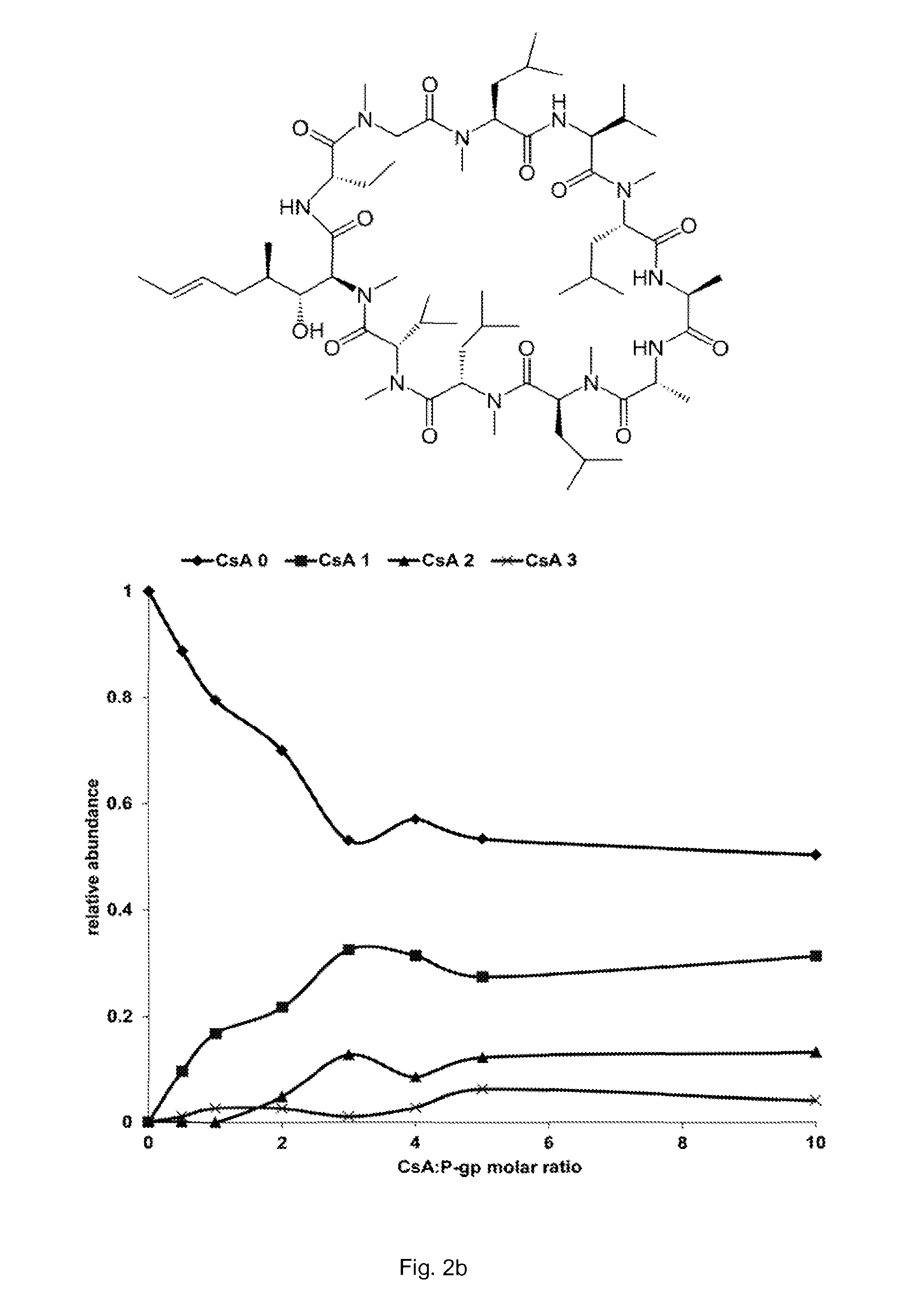
[0053]A similar experiment was performed to detect binding between cyclosporin A and P-gp. Cyclosporin A (CsA; 1202.6 Da) is an immunosuppressant and antifungal antibiotic known to inhibit P-gp ATPase activity with an IC50 of 3.4 μM. In addition, the concomitant binding of nucleotides, lipid and CsA to P-gp was also detected.

[0054]Materials and Methods

[0055]His-tagged P-glycoprotein was expressed in P.pastoris and purified according to a protocol described previously (see Aller et al, supra). Lipids were purchased from Avanti Polar Lipids. Cyclosporin A, ammonium acetate, ATP and ATPγS were purchased from Sigma-Aldrich.

[0056]Before analysis, P-gp was buffer exchanged into 200 mM ammonium acetate pH 7 supplemented with 0.02% n-dodecyl-β-D-maltoside (DDM), using micro BioSpin-6 devices (Bio-Rad). Lipids were first prepared at 2 mM in chloroform and then diluted to 20 μM in 200 mM ammonium acetate pH 7 supplemented with 0.02% DDM. Cyclosporin was first dissolved at 8 mM in methanol and...


PUM


Abstract

According to the present invention, there is provided a method of detecting a complex comprising a membrane protein bound to a therapeutic agent by mass spectrometry. The method comprises: (a) providing a solution comprising a detergent micelle in which said complex is contained; (b) providing a mass spectrometer comprising a nanoelectrospray ionisation source, a mass analyser and a detector; (c) vaporising the solution using the nanoelectrospray ionisation source under conditions such that the complex is released from the micelle; (d) ionising the complex; (e) resolving the ionised complex using the mass analyser; and (f) detecting the resolved complex using the detector. Also provided is a solution comprising a detergent micelle in which a complex is contained, wherein the complex comprises a membrane protein bound to a therapeutic agent.

Description

FIELD OF THE INVENTION[0001]The present invention relates to the detection of complexes comprising a membrane protein bound to a therapeutic agent. More particularly, the present invention relates to the detection of complexes comprising a membrane protein bound to a therapeutic agent by mass spectrometry.BACKGROUND OF THE INVENTION[0002]Membrane proteins are crucial in a wide range of biological functions including respiration, signal transduction and mediation of molecular traffic in and out of cells and organelles. In particular, membrane proteins play an important role in drug uptake and distribution and can therefore profoundly affect drug therapy and resistance to treatment by multiple drugs. By way of illustration, and without limitation, MDR1 P-glycoprotein (P-gp) is an ATP-driven low-specificity efflux pump playing a paramount role in the clearance of xenotoxins. This member of the ATPase Binding Cassette (ABC) transporters is a natural barrier against hydrophobic cytotoxic...

Claims


Application Information

Patent Timeline
Patent Type & Authority Applications(United States)
IPC IPC(8): H01J49/00H01J49/02
CPCH01J49/0027H01J49/02B01D15/3842G01N33/6848G01N33/94G01N2333/705
Inventor ROBINSON, CAROL V.WANG, SHEILABARRERA, NELSON P.
Owner OXFORD UNIV INNOVATION LTD
Why Eureka
Try Eureka