Method and apparatus for magnetic resonance imaging

a magnetic resonance imaging and apparatus technology, applied in wave based measurement systems, instruments, reradiation, etc., can solve the problems of long acquisition time, large scan time cost, and large scan time cost, and achieve the effect of reducing diffusion-weighting sensitivity and decreasing scan tim

Active Publication Date: 2014-09-18
VAPOSUN
View PDF2 Cites 47 Cited by
  • Summary
  • Abstract
  • Description
  • Claims
  • Application Information

AI Technical Summary

Benefits of technology

[0068]In contradistinction to the prior art methods, the present invention may use a phase shift of the non-phase encoded reference echo-signal accumulated due to applying the diffusion-weighting gradients in order to characterize bulk motion and tissue deformation and to compensate their effect for adjusting the magnetic resonance image. Based on comparison of symmetrical sequences (balanced with respect to first motion derivative) with different diffusion-weighting sensitivity (diffusion-weighting gradient magnitude), MR signal attenuation due to diffusion may be calculated in a case of sufficient diffusion-weighting sensitivity of at least one sequence. In a case of diffusion-weighted sequences with sufficiently reduced diffusion-weighting sensitivity, the asymmetrical sequences (unbalanced with respect to first motion derivative) may be used for distinguishing the perfusion component and thin sample structure. Particularly, the phase shift compensation may allow using the steady-state sequences with cyclical drift creating the drift-diffusion effect for decreasing scan time and / or for characterizing the fluid fraction. In a case of diffusion-weighting sensitivity being sufficient for slice-selection but may be insufficient for diffusion-weighting measurement, the symmetrical and asymmetrical sequences may be used for high-productivity MR imaging, such as using modified SE-EPI sequences with multi-slice acquisition (in this case, “diffusion-weighting” is mere a common name for the gradients applied in the specific manner than an indication of the purpose of the applying).

Problems solved by technology

However, the long repetition time results in long acquisition time.
However, for modern high-resolution diffusion sequences with standard SE-EPI protocol characterized by long readouts, total acquisition time approaches to 14-20 min and more, which is clinically unacceptable; the higher image resolution comes at a cost of sufficiently increased scan time and the blur associated with T2* spin-spin decay.
From the other hand, the gradient values are limited by the gradient coil performance limits and the safety issues; at that achieving sufficient b-value requires increasing Δ and TE.
For decreasing boundary blur and motion artifact effect, the diffusion time interval A may be limited, then obtaining quality images may be complicated.
However, DW-SSFP is practically very difficult to apply to in vivo imaging due to strong sensitivity to motion at repeated excitation technique.
However, the mutually-perpendicular multi-slice imaging is related to the noise amplification; it's extremely sensitive to motion artifacts, especially at diffusion-weighting; its applicability for DW-SSFP is strictly limited.
However, this method based on a phase shift by a fraction of FOV is very sensitive to a phase shift due to the second derivative of patient bulk motion; the motion compensation is required for avoiding motion artifacts.
However, applying the balanced pairs of phase-alternating spin-locking impulses and additional crusher gradient pulse is associated with higher noise and significant signal loss incurred when the balance is not fully restored; the motion compensation is desirable.
However, the blood flow rates vary in a broad diapason and can't be estimated directly based on this sequences; it's time-consuming to select the sensitivity values in a working range eliminating image flow artifacts while providing resulting signal attenuation sufficient for diffusion-weighting analysis.
While useful signal not containing the flow velocity derivatives is analyzed, practically it's difficult to distinguish perfusion flow from higher-order bulk motion derivatives and to eliminate diffusion signal component.
However, the motion artifacts are very difficult to avoid at the phase shifts more than π(180°), which is determined by diffusion sensitivity accordingly to Eq.
However, sufficient patient motion and tissue deformation may lead to the misregistration of subsequent image nodes (phase-encode lines in 2D images or slices in 3D images).
However, it's difficult to correctly assign phase shift to the correspondent location because the motion-disturbed phase characteristics of the navigator echoes are incompatible by phase in k-space.
The data acquisition by followed FLASH sequence is more time-consuming than widely used SE-EPI method; at that the image spatial resolution is impaired.
Summarizing, using the known high-productivity MRI techniques, such as SE-EPI, may lead to motion-related phase misregistration and artifacts.
The known methods of motion compensation don't sufficiently achieve the goals of artifact elimination at sufficient bulk motion, of distinguishing bulk motion from perfusion, etc.

Method used


Image

  • Method and apparatus for magnetic resonance imaging
  • Method and apparatus for magnetic resonance imaging
  • Method and apparatus for magnetic resonance imaging

Examples


Embodiment Construction

[0081]The advantages of the present invention may be more readily understood with reference to the following detailed description of the embodiments taken in conjunction with the accompanying drawings.

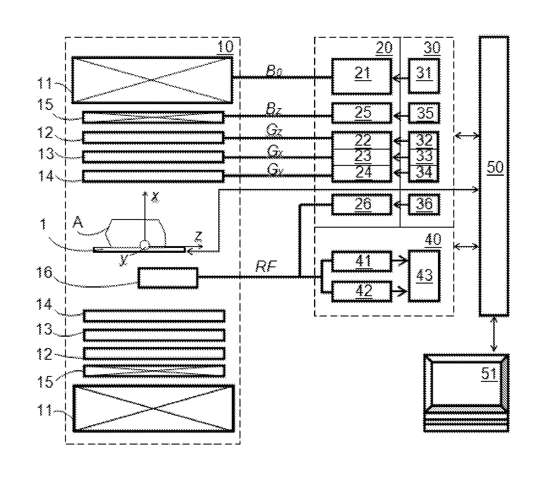
[0082]The DW-MRI method in accordance with the invention can be performed using a DW-MRI device as shown in FIG. 1. The DW-MRI device comprises a positioning system 1, a magnetic assembly unit 10, a power supply unit 20, system controls 30, a receiver unit (receiver) 40, a processor unit (computer) 50 having a periphery device 51. A subject (particularly patient) A is placed on a moving table of the patient positioning system 1 of the DW-MRI device, which is controlled by the computer 50.

[0083]The magnetic assembly unit 10 comprises a uniform magnetic coil means (a main magnetic coil set) 11 and controlled magnetic coil sets 12-15. The main magnetic coil set 11 is for generating a homogeneous longitudinal polarizing magnetic field B0 within an imaging volume within the subject A. Gradi...


PUM


Abstract

The method and system for correcting motion-induced phase errors in Magnetic Resonance Imaging (MRI) use a phase shift of the non-phase encoded reference echo-signal accumulated during the diffusion-weighting in order to characterize bulk motion and tissue deformation and to compensate their effect for correcting the diffusion / perfusion-weighted image. The sequences unbalanced with respect to the first motion derivative are used for distinguishing the perfusion component. The MRI apparatus provides additional excitation resonance-frequency ranges for forming the reference echo signals.

Description

CROSS-REFERENCE TO RELATED APPLICATIONS[0001]This patent application claims the benefit of priority, under 35 U.S.C. Section 119(e), U.S. Provisional patent application No. 61 / 777,174 “Method and apparatus for diffusion-weighted magnetic resonance imaging” to Pavel Levin, filed on 2013 Mar. 12.STATEMENT REGARDING FEDERALLY SPONSORED RESEARCH OR DEVELOPMENT[0002]Not applicable.REFERENCE TO SEQUENCE LISTING, A TABLE, OR A COMPUTER PROGRAM LISTING, COMPACT DISC APPENDIX[0003]Not applicable.TECHNICAL FIELD OF THE INVENTION[0004]The field of the invention is methods and systems for magnetic resonance imaging (MRI) applied particularly to whole-body, brain and other body part magnetic resonance (MR) imaging as well as to sample MR characterization. More specifically, the invention may be related to methods and systems for correcting motion-induced phase errors, particularly in high-productivity MRI, in Diffusion- and / or Perfusion-Weighted Magnetic Resonance Imaging (further generalized as...

Claims


Application Information

Patent Timeline
Patent Type & Authority Applications(United States)
IPC IPC(8): G01R33/565G01R33/563
CPCG01R33/56509G01R33/56341G01R33/5602G01N24/081G01N24/084G01R33/56366
Inventor LEVIN, PAVEL
Owner VAPOSUN
Why Eureka
Try Eureka