Detection kit for influenza a virus

a technology of immunochromatography and kit, which is applied in the field of detection kit for influenza a virus, can solve the problems of inability to obtain accurate determination, inability to obtain accurate diagnosis, and long time-consuming diagnosis, and achieve the effects of reducing the determination of false negative, high detection sensitivity, and high reliability of “negative” determination

Inactive Publication Date: 2015-03-19
TANAKA PRECIOUS METAL IND
View PDF0 Cites 6 Cited by
  • Summary
  • Abstract
  • Description
  • Claims
  • Application Information

AI Technical Summary

Benefits of technology

[0025]In the test kit for influenza A virus of the present invention, the detection sensitivity is high, therefore, the determination of “positive” can be obtained using less amount of virus than that in a conventional test kit, thus the determination of false negative is decreased, and the reliability of the determination of “negative” becomes extremely high. Therefore, useful information can be provided for a doctor to make an accurate diagnosis of influenza virus infection for a patient in an initial stage of infection and soon after the start of virus proliferation in the living body, and thus the treatment with an anti-influenza virus agent can be started at an early stage. In addition, even for a patient in which the proliferation rate of virus is suppressed by the inoculation of vaccine, the presence or absence of infection of the influenza virus can be accurately diagnosed, and thus the attention of spread of the infection can be drawn, further the information that is epidemiologically important in order to determine the effect of influenza vaccine can be provided.
[0026]Further, the present invention is to provide an agent for detecting influenza A virus. The diagnosis can be simply and more accurately performed by using an agent for detecting influenza A virus of the present invention, in particular, by using as the antibody immobilized to a chromatography medium of a detection kit by immunochromatography.

Problems solved by technology

In order to make an accurate pathogen diagnosis of influenza, there is a standard technique of virus separation using a pharyngeal swab or a gargle liquid as the material, however, it takes a longtime to obtain the diagnosis.
However, a PCR method requires a special apparatus, and thus can be performed only in an institute for health or a limited laboratory.
However, a rapid diagnostic kit currently available on the market does not have sufficient detection sensitivity of influenza virus, and thus even if the result of “negative” is obtained, the viral infection cannot be necessarily denied.
There is a problem of determination of false negative that in a case of a patient in an initial stage of infection that is soon after the start of virus proliferation, a patient in which the proliferation rate of virus is suppressed due to the inoculation of vaccine, or the like, there may be a case where the sufficient amount of virus is not present in the biological sample to determine as positive, and thus the result of the rapid diagnostic kit becomes “negative” although the patient is actually infected with influenza virus.
In a clinical practice, a test by a rapid diagnostic kit can stably detect the virus if 24 hours elapses after the onset and a highly precise result can be obtained, however, there may be a case where virus cannot be detected and the accurate determination cannot be obtained within 12 hours after the onset.
From the situation described above, re-inspection is carried out for the patient with a result of “negative” on or after the next day depending on the other findings, and the patient has to seek medical attention again, this situation has forced the excessive burden in terms of cost and time.
It is considered that the agent is ideally taken within 12 hours after the onset, although the sufficient effectiveness can be obtained if the agent is taken within 24 hours after the onset, and the therapeutic effect becomes poor if the agent is taken more than 48 hours after the onset.

Method used


Image

  • Detection kit for influenza a virus

Examples


example 1

Production of Anti-Influenza A Virus Nuclear Protein Antibody

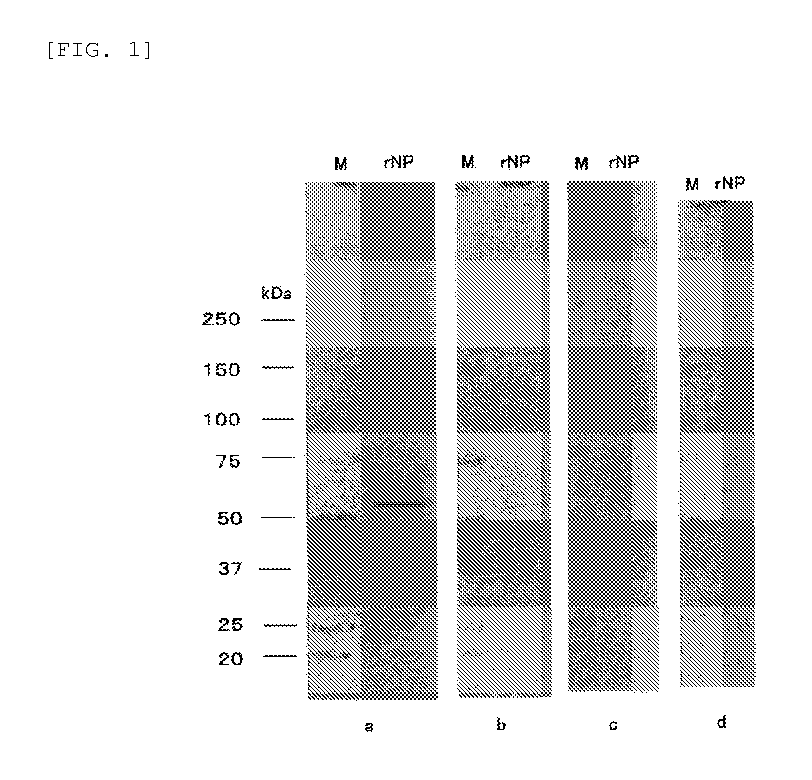
[0068]A recombinant nuclear protein produced based on the amino acid sequence of a nuclear protein derived from influenza A virus A / Puerto Rico / 8 / 34 (H1N1) strain (DDBJ / GenBank database, Accession No. V01084) was used as the immunogen. An equal parts of complete Freund's adjuvant was added into and completely mixed with the immunogen, and then a BALB / c mouse was immunized 4 times in total at 2-week intervals. Spleen cells were collected from the immunized mouse three days after the last immunization, and hybridomas were produced by the fusion of the spleen cells with myeloma cells (P3U1) using a known standard technique. 10 to 15 days after the production of the hybridomas, the screening for an antibody that is specific to an influenza A virus nuclear protein was performed using a culture supernatant of the hybridoma.

[0069]In the primary screening, an antibody causing an antigen-antibody reaction with the influenza A virus...

example 2

Production of Test Kit by Immunochromatography

(1) Production of Diluent for Capture Antibody

[0074]Isopropyl alcohol was mixed with 50 mM phosphate buffer solution (pH 7.4) so as to be diluted to 5%, and thus a diluent for the first antibody was prepared.

(2) Production of Determination Site on Chromatography Medium

[0075]One antibody, or two antibodies in combination were selected among the antibodies 1C6, 6F7, and 10G5, and diluted with a diluent for a capture antibody so as to have the total antibody concentration of 1.0 mg / mL. The antibody solution was applied on a nitrocellulose membrane (manufactured by Millipore) having a size of 25×2.5 cm using an applicator (manufactured by BioDot), and dried at 50° C. for 5 minutes, then further dried at room temperature for 1 hour, as a result, a determination site was prepared on a chromatography medium.

(3) Production of Labeling Antibody Solution

[0076]A gold colloidal suspension (manufactured by TANAKA KIKINZOKU KOGYO K.K.: average particl...

example 3

Measurement of Influenza A Virus

[0079]Using a test kit produced in the above, a reactivity test with influenza A virus was performed according to the following method, and thus the performance of the test kit of the present invention was examined.

[0080]Nasal mucus was collected from a subject who had been determined to be negative in the infection test of influenza A virus (H3N2) using a PCR method. The collection of nasal mucus was performed as follows: one tube of a suction trap was inserted to the inner part of the nasal cavity of a subject, and the other tube was connected to a suction pump, and the suction pump was set to negative pressure to suck up the nasal mucus. The nasal mucus was diluted 20 fold with a developer, and thus an influenza A virus negative sample was prepared. An inactivated influenza A virus A / Panama / 2007 / 99 (H3N2) was added to the negative sample, and thus an influenza A virus positive sample was prepared.

[0081]150 μL of each of a positive sample and a nega...


PUM

PropertyMeasurementUnit
molecular weightaaaaaaaaaa
pHaaaaaaaaaa
pHaaaaaaaaaa

Abstract

The present invention is a test kit for rapidly diagnosing influenza according to the principles of immunochromatography, and the purpose thereof is to provide a test kit for the influenza A virus in which the sensitivity in detecting the influenza A virus is greater than in conventional test kits, and a determination of “positive” is obtained stably and with high precision at an earlier time during the onset of influenza symptoms. The present invention pertains to a kit for detecting influenza A virus, in which an antibody that is in solid phase in the chromatographic medium enters into an antigen-antibody reaction with native nuclear proteins of the influenza A virus, but in Western blots the antibody does not enter into antigen-antibody reactions with full-length nuclear proteins of the influenza A virus that have been separated using SDS-polyacrylamide gel electrophoresis.

Description

TECHNICAL FIELD[0001]The present invention relates to a test kit by immunochromatography for detecting influenza A virus, using an antibody specifically causing an antigen-antibody reaction with an influenza A virus nuclear protein but not causing an antigen-antibody reaction by Western blotting with a full-length influenza A virus nuclear protein separated using SDS (sodium dodecyl sulfate)-polyacrylamide gel electrophoresis.BACKGROUND ART[0002]Influenza means an infectious disease caused by influenza virus. It is known that as the typical symptom, fever, headache, physical weariness, myalgia, arthralgia, and the like suddenly appear, and cough, nasal discharge, and the like follow in tandem, and it is said that these symptoms subside in around a week. As compared with other so-called cold syndromes, characteristics of influenza are the severe systemic symptoms. In order to make an accurate diagnosis, virological support is required. In the time when influenza is epidemic, it is im...

Claims


Application Information

Patent Timeline
Patent Type & Authority Applications(United States)
IPC IPC(8): G01N33/569C07K16/10
CPCG01N33/56983G01N2333/11C07K16/1018
Inventor IWAMOTO, HISAHIKOKAWAMOTO, HIROKOKATO, SHINICHI
Owner TANAKA PRECIOUS METAL IND
Why Eureka
Try Eureka