HPV Detection in Urine

a technology of hpv and detection method, applied in combinational chemistry, biochemistry apparatus and processes, library screening, etc., can solve the problems of no processing of urine samples into cell-containing and cell-free fractions, or soluble and insoluble fractions

Inactive Publication Date: 2015-12-31
TROVAGENE
View PDF3 Cites 3 Cited by
  • Summary
  • Abstract
  • Description
  • Claims
  • Application Information

AI Technical Summary

Benefits of technology

[0005]The disclosure relates to methods for the detection of human papillomavirus (HPV) nucleic acid molecules in urine samples from human subjects. The disclosed methods do not fractionate urine into cell-free and cell-containing fractions, or soluble and insoluble fractions, and surprisingly provide sensitivities that are higher than thos

Problems solved by technology

The disclosed methods do not fractionate urine into cell-free and cell-containing fractions, or soluble and insoluble fractions, and surprisingly provide sensitivities that are higher than those reported for cellular pellets.
The lack of fractionat

Method used


Image

  • HPV Detection in Urine
  • HPV Detection in Urine

Examples


example 1

Clinical Samples

[0081]154 urine samples were obtained from a cohort of women who were referred to a colposcopy clinic. The subjects were identified based on one or more abnormal Papanicolaou tests (Pap smears). The women were not pregnant, had not been treated previously for cervical intraepithelial neoplasia (CIN), and did not undergo a hysterectomy. The population of subjects was expected to have a high prevalence (˜80%) of HPV (all types).

[0082]The samples were treated with addition of EDTA to approximately 40-70 mM final concentration before storage at reduced temperature. Some samples were frozen while others were stored at about 4° C.

example 2

Sample Preparation

[0083]An aliquot of 500 μl of each urine sample was transferred to a tube. An equal amount of lysis buffer containing GITC (such as QIAGEN AL buffer) with about 11.2 μl / ml of carrier RNA is added to the sample followed by addition of an aliquot of protease. Proteolysis was allowed to proceed at approximately 56° C. for about 15 minutes. Ethanol (600 μl of 96-100%) was added for about 5 minutes at room temperature to enhance nucleic acid recovery.

[0084]The tube's contents were applied to a silica-based nucleic acid affinity column (such as the QIAmp MinElute column) and washed at least twice with ethanol rinses. Bound nucleic acids were eluted with 10 mM Tris, pH 8.0, optionally containing a preservative such as 0.04% azide.

example 3

Sample Analysis (HPV Detection)

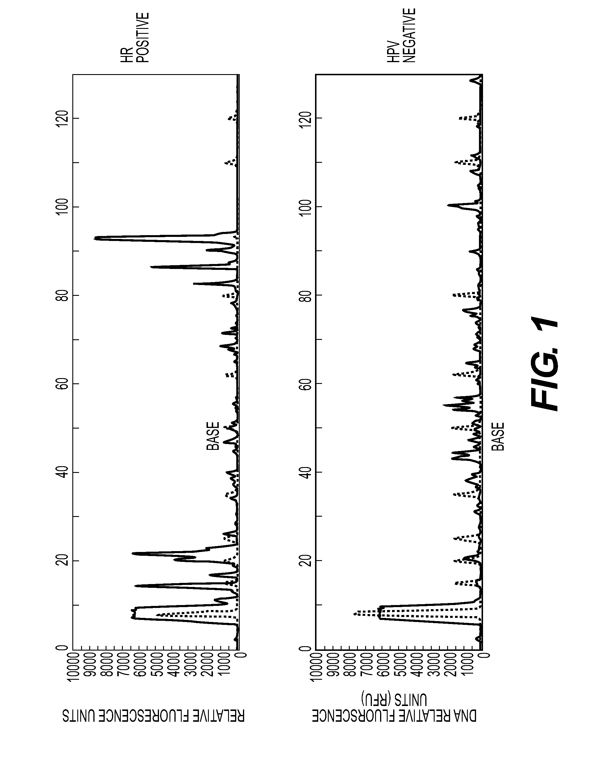
[0085]Prepared nucleic acids from each sample were PCR amplified with a forward and reverse primer pair (SEQ ID Nos: 30 and 31) followed by capillary electrophoresis to detect the presence of an amplicon of 93-96 bp—the expected size of a high-risk HPV PCR product. FIG. 1 shows exemplary traces from a “positive” (HPV 16) sample and a “negative” (HPV 54) sample. Positive and negative refer to whether the primer pair is complementary to, and so would amplify, an HPV sequence and as confirmed by sequencing described below. The trace represents FAM-labeled products, including any amplified material from the forward primer and unincorporated forward primer (large peak at approximately 15 bp).

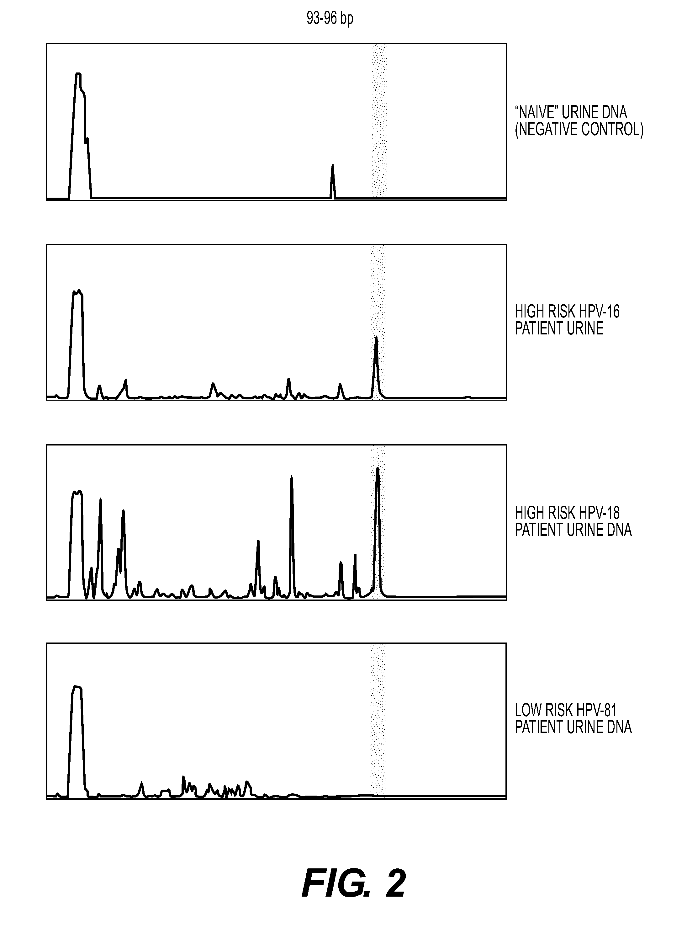
[0086]FIG. 2 shows exemplary traces from a negative control urine and two high risk urine samples (HPV 16 and 18) and one low risk sample (HPV 81). The identification of the HPV types was confirmed by sequencing as described below.

[0087]For fragment analysis, the instru...


PUM

PropertyMeasurementUnit
Fractionaaaaaaaaaa
Temperatureaaaaaaaaaa
Concentrationaaaaaaaaaa

Abstract

The disclosure provides compositions and methods for the detection of human papillomavirus (HPV) nucleic acids in a urine sample from a human subject. The nucleic acids may be from one or more high risk forms of HPV. The urine sample is minimally processed, without fractionation into cell-free and cell-containing portions, before preparation of the nucleic acids for detection of HPV sequences.

Description

FIELD OF THE DISCLOSURE[0001]This disclosure relates to the fields of medicine and clinical diagnostics based on molecular techniques. The techniques include methods for detecting human papillomavirus (HPV) nucleic acid molecules in urine samples from human subjects as an indicator of HPV infection in the subject. The detection methods include type-specific detection of HPV infections and the detection of HPV genotypes.BACKGROUND OF THE DISCLOSURE[0002]As recently as 2011, the detection of human papillomavirus (HPV) infections by use of a urine sample included a concern over the presence of soluble PCR inhibitors in urine (see page 1745, right column, in Bissett et al.). This concern led Bissett et al. to test three different protocols for the processing of urine samples as part of extraction of HPV nucleic acids. One protocol, which was reported as having the better sensitivity among the three, was centrifugation of urine (1 ml) at 13,000 rpm for 20 minutes, removal of the resultin...

Claims


Application Information

Patent Timeline
IPC IPC(8): C12Q1/70
CPCC12Q1/708C12Q2600/118C12Q2600/156C12Q2600/158
Inventor VIBAT, CECILE ROSE
Owner TROVAGENE
Why Eureka
Try Eureka