Three-Dimensional Scaffold Functionalized with Micro-Tissues for Tissue Regeneration

a tissue regeneration and micro-tissue technology, applied in the field of biomaterials, can solve the problems of unsatisfactory bone regeneration processes, current approaches that do not allow sufficiently robust bone regeneration, and limited thickness of implants, so as to achieve efficient inducing bone regeneration in vivo and enhance the

Inactive Publication Date: 2016-05-19
INST NAT DE LA SANTE & DE LA RECHERCHE MEDICALE (INSERM) +1
View PDF0 Cites 15 Cited by
  • Summary
  • Abstract
  • Description
  • Claims
  • Application Information

AI Technical Summary

Benefits of technology

[0012]It was discovered that such scaffolds allow for accelerated regeneration of living cells deposited thereon. Also, the tissue regeneration was shown to proceed in depth, up to the core of a scaffold. Tissue generation upon such biomaterials thus does not require the presence of a growth factor.
[0013]It has further been demonstrated that such functionalized nanofibrous scaffolds are capable of efficiently inducing bone regeneration in vivo.
[0014]The biomaterials according to the present invention represent a new generation of biomaterials for regenerative nanomedicine. Indeed, the strategy of functionalizing the scaffold by microtissues affects cell proliferation and thereby allows for complete colonisation even of thick scaffolds, which enhances the efficacy compared to scaffolds functionalized with living cells used in the clinic today. It is presently thought that this improvement is due to the replacement of the bidimensional cell monolayer formed by living cells on the scaffold by a further three-dimensional environment for the cells formed by the microtissues.
[0035]The term “nanofibrous scaffold” refers to a specific three-dimensional scaffold formed by fibers, notably polymeric fibers, whose diameter is less than 1 μm. Due to the presence of fibers, nanofibrous scaffolds not only mimic the three-dimensional structure of a tissue, but also facilitate adhesion and spreading of cells.
[0060]The biomaterials functionalized with microtissues according to the invention are particularly interesting because they allow for easy and fast in-depth cell colonisation of the scaffold. Such colonisation allows the spontaneous migration of cells throughout the nanofibrouse three-dimensional scaffold without use of external forces such as a pulsatile flow or an injection under pressure. Therefore, the invention is particularly advantageous for the preparation of thick biomaterials, i.e. when the scaffold has a thickness of above 50 μm, advantageously up to 50 mm, and most preferred from 0.1 to 20, and in particular from 0.5 to 10 mm.
[0069]According to an embodiment of the present invention, the three-dimensional scaffold may further also comprise a therapeutic molecule other than a growth factor and thus also serve as a reservoir for the therapeutic molecule. Such a scaffold further functionalized with a therapeutic molecule, allows for sustained release of said therapeutic molecule at the site of implantation of the biomaterial according to the invention.

Problems solved by technology

Current bone-regeneration processes, including the free fibula vascularised graft, autologous bone graft, allograft implantation, and use of growth factors, scaffolds and osteo-progenitor cells are unsatisfactory as they yield insufficient quantities of bone.
Current approaches do not allow sufficiently robust bone regeneration.
However, it appears to be limited to implants having a thickness of up to 50 μm, as cell colonisation and bone induction are not as satisfactory for thicker implants.

Method used


Image

  • Three-Dimensional Scaffold Functionalized with Micro-Tissues for Tissue Regeneration
  • Three-Dimensional Scaffold Functionalized with Micro-Tissues for Tissue Regeneration
  • Three-Dimensional Scaffold Functionalized with Micro-Tissues for Tissue Regeneration

Examples


example 1

Material and Methods

[0103]Chemicals.

[0104]Poly(ε-caprolactone) (PCL), Capa 6800 (Mw=80000) analytical grade, was obtained from Perstorp (Industriparken, Sweden). PCL was dissolved in a mixture of dichloromethane / dimethylformamide (DCM / DMF 40 / 60 vol / vol) at 27% wt / vol and was stirred overnight before use to ensure good polymer solubilisation.

[0105]Electrospinning

[0106]A standard electrospinning set-up Apparatus EC-DIG purchased from IME Technologies (Eindhoven, Netherlands) was used to fabricate the PCL three-dimensional nanofibrous scaffolds. The PCL solution was poured into a 5 mL syringe and ejected through a 21G needle of 0.8 mm diameter at a flow rate of 1.2 mUh thanks to a programmable pump (ProSense). The electrospun hjet was focused thjanks to the use of a poly(methyl metacrylate) (PMMA) plate of 2.5 mm thick pierced with a hole (25 mm in diameter) placed over the conductive collector. The collector was placed at a distance from the needle of 16 cm. A voltage of +15 kV was ap...

example 2

Scaffold Functionalized with Microtissues (In Vitro)

[0136]The comparative Example was repeated, except that the scaffold was seeded with living human osteoblasts microtissues instead of living human osteoblasts single cells.

[0137]The microtissues were prepared according to the protocol indicated above, using a GravityPLUS™ plate for hanging-drop cell culture from InSphero AG (Zurich, Switzerland).

[0138]The incorporation, cell colonization, proliferation and bone induction by the living human osteoblasts microtissues into the scaffold was studied.

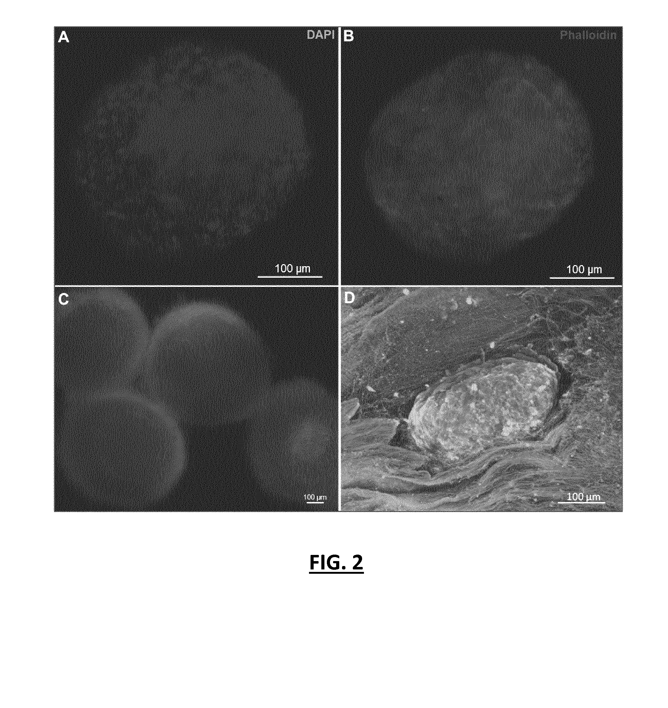
[0139]In particular, the behavior of the cells in contact with the scaffold observed by fluorescence microscopy showed that the microtissues remain viable after incorporation into the scaffold (FIG. 2D). The behavior of the osteoblasts microtissues and the capability of the functionalized scaffold to induce bone regeneration after colonization and proliferation were studied by SEM. The behavior of the microtissues with time after the deep co...

example 3

Scaffold Functionalized with Microtissues (In Vivo)

[0143]Using Thick scaffolds functionalized with osteoblasts microtissues prepared in Comparative Example and in Example 2, subcutaneous and calvaria implantations were realized in nude mice (FIG. 5A-D).

[0144]The results for the scaffolds functionalized with the living microtissues showed that, after 4 weeks, cells have migrated into the scaffold, colonizing the scaffold even deep within, and also show evidence that bone induction has occurred (FIGS. 5B and 5D).

[0145]It is further noted that bone induction is faster for scaffolds functionalized with microtissues in comparison to scaffolds functionalized with osteoblast single cells (FIGS. 5C and 5D). Indeed, the comparison of the amount of mineralized matrix yields 8% mineralized area for the scaffold functionalized with osteoblast single cells against 22% mineralized area for the scaffold functionalized with microtissues.

[0146]Further, mice calvaria implantations have been carried o...


PUM

PropertyMeasurementUnit
thicknessaaaaaaaaaa
sizeaaaaaaaaaa
thicknessaaaaaaaaaa

Abstract

The present invention concerns a biomaterial devoid of a growth factor, comprising: —a three-dimensional scaffold made of a biocompatible polymer; and—living cells, wherein said living cells are in form of microtissues and the nanofibrous three-dimensional scaffold is a nanofibrous scaffold. It further concerns a method for manufacturing such a biomaterial. Finally, it concerns such a biomaterial for use in the treatment of a bone and / or cartilage defect.

Description

TECHNICAL FIELD OF THE INVENTION[0001]The present invention concerns biomaterials comprising a three-dimensional functionalized scaffold and living cells useful for tissue generation. It further concerns a method for producing such biomaterials.[0002]It also concerns methods of treating bone or cartilage defects using such biomaterials and biomaterials for use in the treatment of bone or cartilage defects, in particular as implants.BACKGROUND OF THE INVENTION[0003]Bone regeneration is a complex, well-orchestrated physiological process of bone formation, which occurs during normal fracture healing, and is involved in continuous remodelling throughout adult life. In the clinic, bone regeneration may be required in large quantity, such as for skeletal reconstruction of large bone defects created by trauma, tumour resection, or cases in which the regenerative process is compromised (non-unions, osteoporosis). Current bone-regeneration processes, including the free fibula vascularised gr...

Claims


Application Information

Patent Timeline
Patent Type & Authority Applications(United States)
IPC IPC(8): A61L27/38A61L27/54A61L27/52A61L27/18
CPCA61L27/3821A61L27/3817A61L27/18A61L27/54A61L2430/06A61L2300/64A61L2300/412A61L2430/02A61L27/52A61L27/56A61L2430/24C12N5/0068C12N5/0654C12N2533/40
Inventor BENKIRANE-JESSEL, NADIAEAP, SANDYKELLER, LAETITIA
Owner INST NAT DE LA SANTE & DE LA RECHERCHE MEDICALE (INSERM)
Why Eureka
Try Eureka