
Method for preparing an animal decellularized tissue matrix material and a decellularized tissue matrix material prepared thereby

a tissue matrix material and decellularized technology, applied in the field of biological tissue treatment and the manufacture of tissue matrix materials, can solve the problems of tissue matrix prone to torn when used, complex process procedure for tissue matrix and organ manufacturing, and insufficient physical method to achieve complete decellularization

Active Publication Date: 2016-07-14
BEIJING RUIJIAN GAOKE BIOTECH
View PDF7 Cites 12 Cited by
  • Summary
  • Abstract
  • Description
  • Claims
  • Application Information

AI Technical Summary

Benefits of technology

The method described in this patent text uses an enzymatic process to remove cell components and antigens that can cause inflammation and rejection in the body when using animal tissue scaffolds. The method also improves the flexibility of the scaffold by removing DNA. This helps to create a safer and more effective scaffold for use in regenerative medicine.

Problems solved by technology

A process procedure for manufacturing the matrix of the tissue and organ is very complex, including processes such as collection, preservation, washing, disinfection, decellularization, antigenicity reduction, virus inactivation, and terminal sterilization of the tissue and organ, and the like.
The physical method itself is generally not sufficient to achieve complete decellularization.
If the rate of cooling and heating is too rapid or not uniform within the tissues, tiny cracks can occur in local regions of the tissues and the tissue matrix is prone to be tore when used.
After the treatment using these solutions, the tissue matrix is damaged to different extent, especially with the effects of sodium hydroxide, sodium hypochlorite, and iodine solution being greater.
The terminal sterilization treatment of the tissue product is often one of the most destructive steps for the tissue material.

Method used

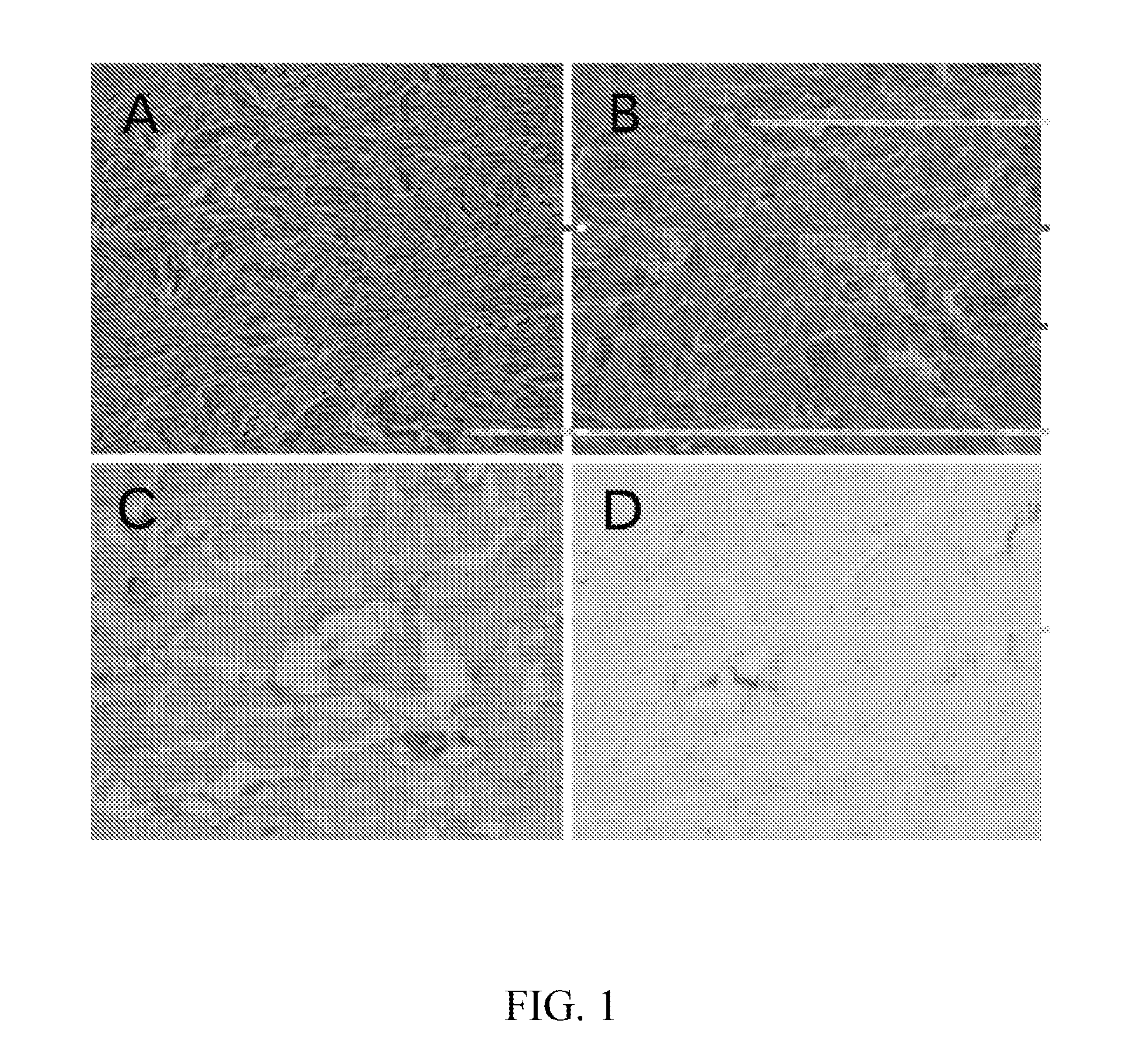
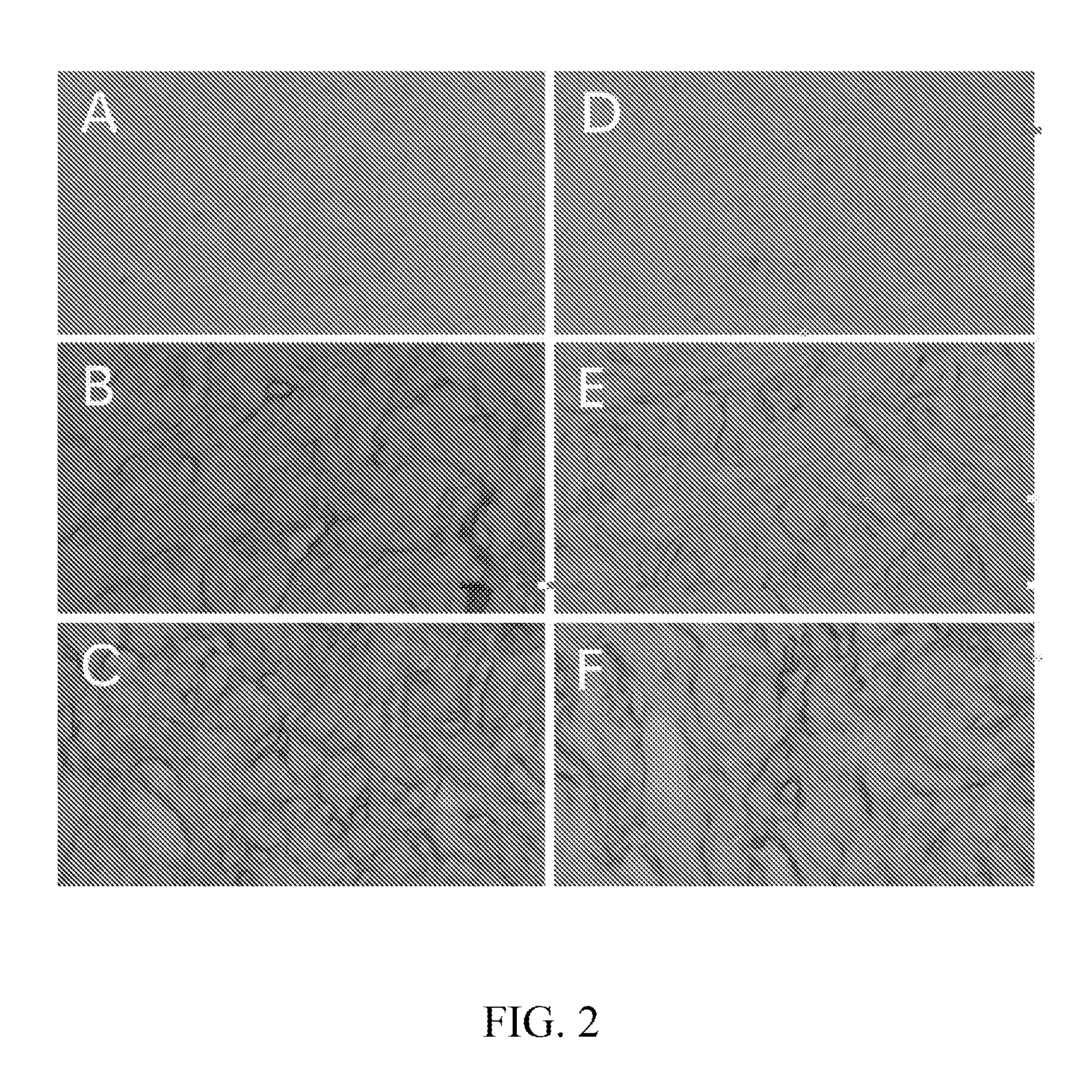

Image

  • Method for preparing an animal decellularized tissue matrix material and a decellularized tissue matrix material prepared thereby
  • Method for preparing an animal decellularized tissue matrix material and a decellularized tissue matrix material prepared thereby
  • Method for preparing an animal decellularized tissue matrix material and a decellularized tissue matrix material prepared thereby

Examples


example 1

Manufacture And Performance Detection of an Animal Acellular Tissue Matrix Material

[0055]1. Manufacture

[0056](1) Collection and Preservation of a Tissue and Organ

[0057]Fresh porcine hide was collected from a newly slaughtered pig, and temporarily preserved in a refrigerator at 4° C. After the porcine hide was dehaired mechanically, the porcine hide was split into a dermis layer having a thickness of about 1.0 mm, which was cryopreserved at −20° C.

[0058](2) Decellularization

[0059]After the dermis was thawed, it was firstly flushed with a normal saline twice, each time for 30 minutes. The flushed porcine dermis was soaked in a saline solution containing 100 mg of gentamicin per litre, and 2.0 millimole concentration of calcium chloride, 2.0 millimole concentration of magnesium chloride, and 150 units per litre of neutral dispase were further added to the solution, and the dermis is treated at 37° C. for 24 hours.

[0060](3) Washing

[0061]After being soaked in gentamicin, the dermis was w...

example 2

Determination of Appropriate Conditions for Washing and Disinfecting the Dermis

[0070]Fresh dermis was collected from a porcine body, and the fresh porcine dermis was treated in sodium hydroxide solutions with pH of 10.6, 11.5 and 11.8 at 37° C., respectively. Each kilogram of the porcine hide was in 4 litre of sodium hydroxide solution, with the control of phosphate buffer. After 24 hours, colony-forming unit per milliliter solution was determined. The phosphate buffer contained 10.3±1.3 logarithmic colony-forming unit (LogCFU)(N=3); and the logarithmic colony-forming unit in the solution with pH of 10.6, 11.5 and 11.8 was 2.1±0.1, 0 and 0, respectively. As could be seen, disinfection and sterilization effect in moderate alkaline solution was significant. It was demonstrated by using differential scanning calorimeter that the tissue matrix was damaged with pH of 11.5 or more, and the stability of protein in the tissue matrix was significantly reduced. The damage of high pH on the ti...

example 3

Manufacture and Performance Detection of an Animal Acellular Tissue Matrix Material

[0071]1. Manufacture

[0072](1) Collection and Preservation of a Tissue and Organ

[0073]Fresh porcine hide was collected from a newly slaughtered pig, and temporarily preserved in a refrigerator at 4° C. After the porcine hide was dehaired mechanically, the porcine hide was split into a dermis layer having a thickness of about 1.0 mm.

[0074](2) Collection and Washing

[0075]After collection and washing (see example 1), the porcine dermis with a thickness of 1.0 mm was temporarily preserved in a refrigerator at −80° C.

[0076](3) Decellularization

[0077]After being thawed, the dermis was flushed with 5 mM of hydroxyethylpiperazine ethane sulfonic acid solution (pH 7.4), and was then treated at 37° C. for 18 hours after adding 2.0 mM of calcium chloride and 0.2 unit per milliliter of neutral dispase.

[0078](4) Washing

[0079]The dermis was washed with 1.0% sodium deoxycholate solution at 37° C. for 20 hours.

[0080](...


PUM

PropertyMeasurementUnit
Temperatureaaaaaaaaaa

Abstract

A method for manufacturing an animal acellular tissue matrix material and a tissue matrix material manufactured by the same. The tissue matrix material manufactured by the method retains an original basic scaffold structure of a tissue extracellular matrix, with an antigen causing immunological rejection in a human body being effectively removed from the animal tissue. An animal dermal matrix manufactured by the method retains the biological integrity of a natural dermal tissue matrix and can be used for restoration and repair of lesion and missing tissues.

Description

TECHNICAL FIELD[0001]The present invention relates to the technical field of treatment of biological tissues and manufacture of tissue matrix materials, and in particular relates to a method for manufacturing an animal acellular tissue matrix material and an acellular tissue matrix material manufactured by the same.BACKGROUND OF THE RELATED ART[0002]There are great similarity and homology in an extracellular matrix of a tissue and organ of a human body and many animals. A biological matrix material manufactured by decellularization of an allogeneic or xenogenic tissue and organ has been successfully used for the repair and restoration of human tissues in clinical medicine. The decellularized tissue and organ matrix is also widely used for various studies in tissue engineering and regenerative medicine, for example, removing original cell components of a tissue and organ of animals, and re-cellularizing and functionalizing the matrix of the tissue and organ having a three-dimensional...

Claims


Application Information

Patent Timeline
IPC IPC(8): A61L27/36
CPCA61L27/362A61L27/3691A61L2430/40A61L2300/252A61L2430/34A61L27/3687A61L27/3683A61L27/58
Inventor LIU, ZHI-GANGLIU, XINHUA
Owner BEIJING RUIJIAN GAOKE BIOTECH
Learn More