Preparation method for therapeutic agent of bead-type chondrocyte
- Summary
- Abstract
- Description
- Claims
- Application Information
AI Technical Summary
Benefits of technology
Problems solved by technology
Method used
Image
Examples
examples 1
Isolation and Proliferation of Chondrocytes
[0065]Costal cartilage tissues were washed with phosphate-buffered saline (PBS) containing antibiotics 3-5 times to remove blood and contaminants. The cartilages were minced into 1-2 mm3, and extracellular matrix was then digested by the treatment of 0.5% pronase and 0.2% type II collagenase to isolate cells. After the inoculation of isolated cells into culture dishes at a cell density of 2-4×10⁴ cells / cm2, a culture medium for cell proliferation (mesenchymal stem cell growth medium [MSCGM] containing 1 ng / ml of FGF-2) was added thereto, and the cells were then cultured until being confluent in a 37° C., 5% CO2 incubator. Cells detached from the culture dishes with a trypsin-EDTA solution were inoculated into culture dishes at a cell density of 1-2×10⁴ cells / cm2, and cultured to passages 6 to 8.
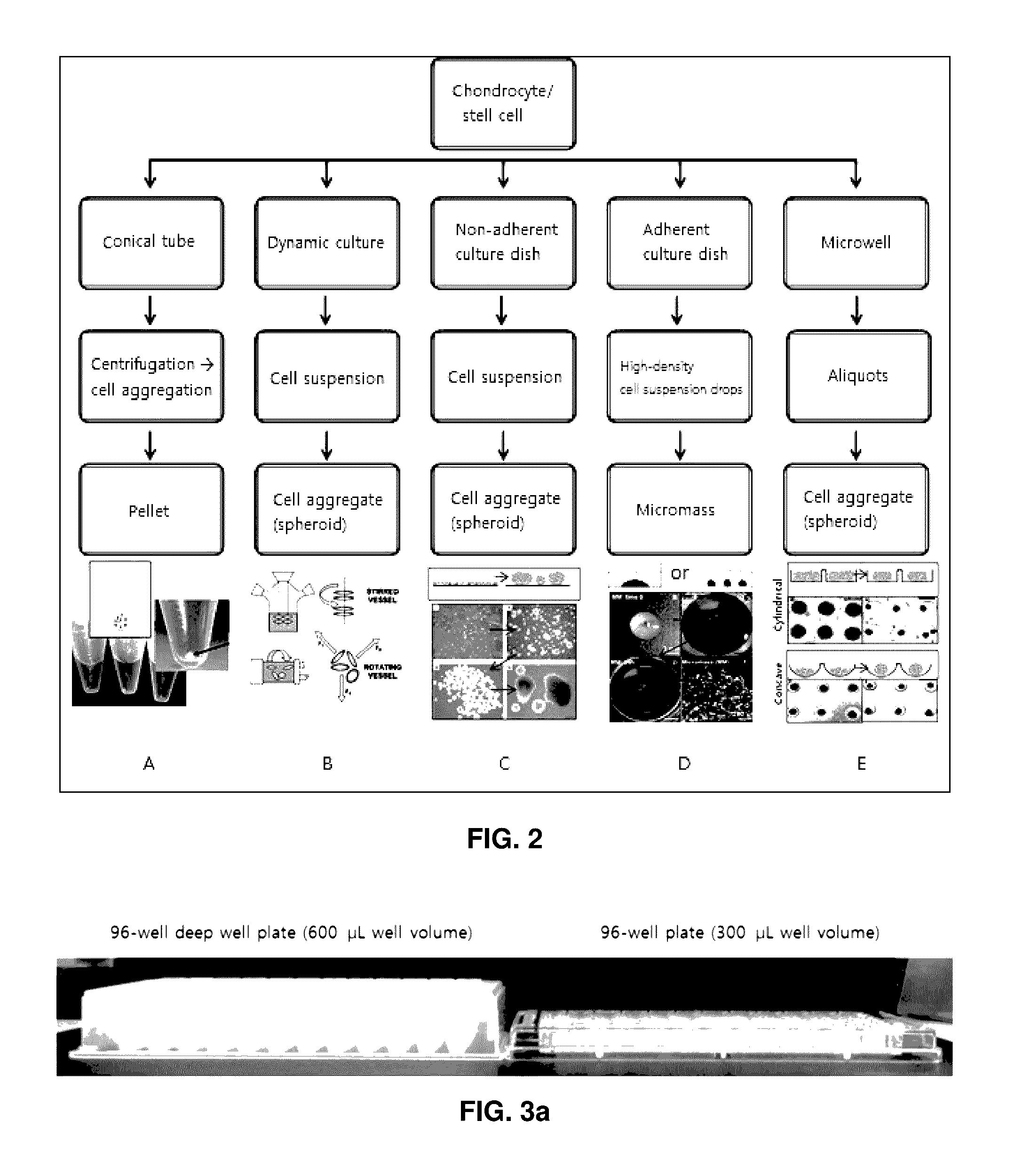
[0066]Three-Dimensional Pellet Culture of Chondrocytes at Various Kinds of 96-Well Plates or 96-Well Deep Well Plates
[0067]Proliferation completed c...
example 2
Preparation of Bead-Type Three-Dimensional Cartilage Tissue
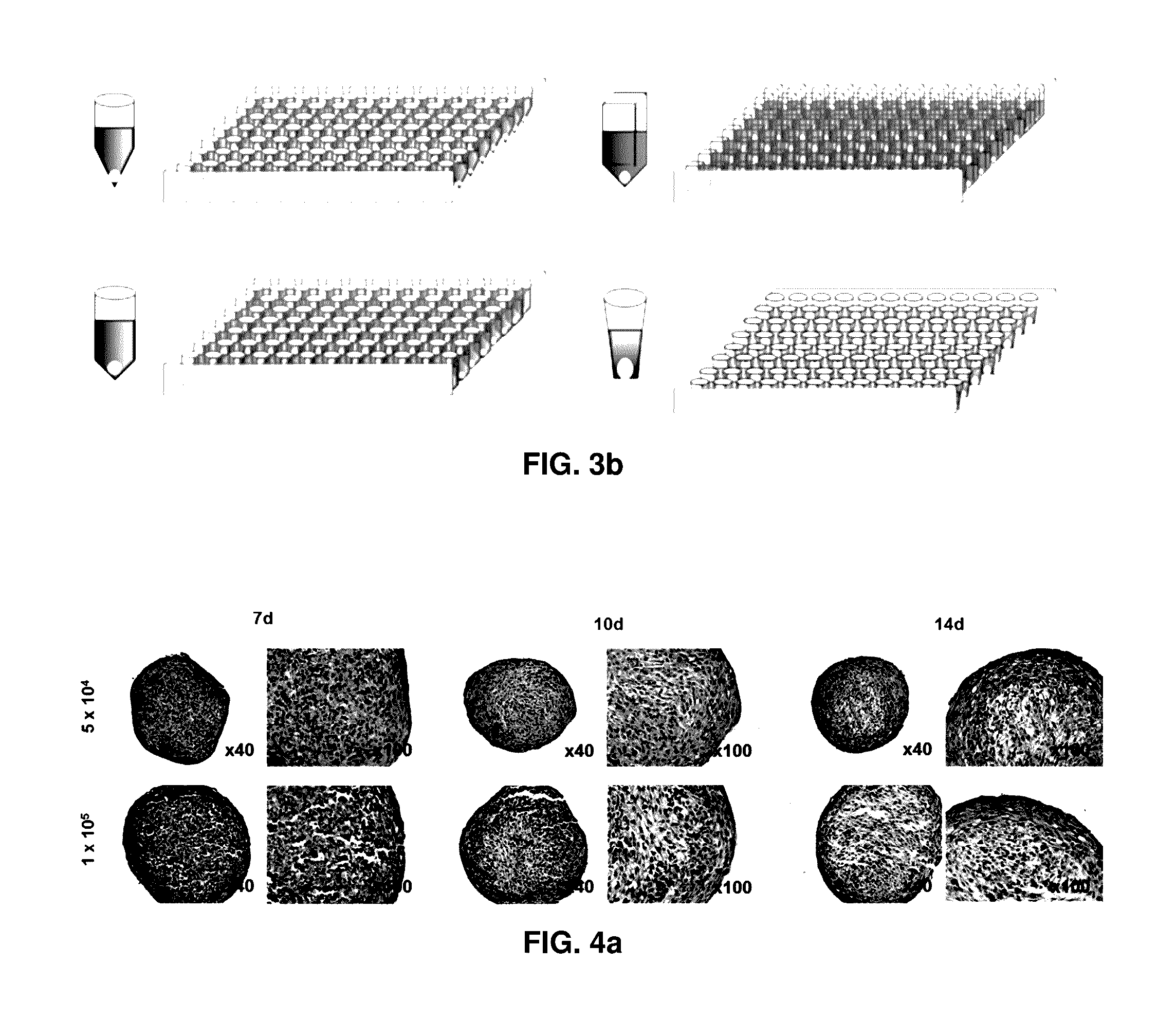
[0070]Rabbit chondrocytes cultured in a culture medium for cell proliferation to passages 6 to 8 were pellet cultured in a 96-well deep well plate having V-shaped bottom with 0.5×10⁵ cells / 400 μl / well, 1.0×10⁵ cells / 400 μl / well or 2.0×10⁵ cells / 400 μl / well and evaluated for 28 days.
[0071]Evaluation Methods for Properties of Bead-Type Three-Dimensional Cartilage Tissue
[0072](1) Appearance and Size
[0073]In the course of three-dimensional culture, pellets were collected from each well and their properties were evaluated. First of all, appearance was evaluated with the naked eye, and a pellet was mounted on a slide glass and a photograph was taken with a digital camera installed on a microscope with 40× magnifications to measure the area.
[0074](2) DNA Amount
[0075]To measure the DNA amount in pellets, 125 μg / mL of papain was added to pellets and homogenized. After overnight treatment at 65° C., the prepared sample was mixed with ...
example 3
[0103]Rabbit costal chondrocytes cultured in a culture medium for cell proliferation to passages 6 to 8 were suspended in a culture medium for differentiation and dispensed into a 96-well deep well plate having V-shaped bottom with 1.0×10⁵ cells / 400 μl / well. The plates were centrifuged at 1,200 rpm for 5 minutes, and then pellet cultured in a 37° C., 5% CO2 incubator for 10 days with exchanging culture media at an interval of 3 or 4 days to prepare a bead-type cartilage tissue without a scaffold for transplanting to articular cartilage damage. After culturing, the bead-type cartilage tissue was collected by the use of portable pellet collection apparatus (FIG. 8c), and properties were evaluated according to the methods described in Example 2.
[0104]As a result, semitranslucent bead-type three-dimensional structures having white and smooth surface with the naked eye and 1.0 to 1.5 mm of diameter were observed (FIG. 6a). Their plane sizes were in the range of 1.6 mm2±20%, and the forma...
PUM
 Abstract
Description
Claims
Application Information
 - R&D
- Intellectual Property
- Life Sciences
- Materials
- Tech Scout
- Unparalleled Data Quality
- Higher Quality Content
- 60% Fewer Hallucinations
Browse by: Latest US Patents, China's latest patents, Technical Efficacy Thesaurus, Application Domain, Technology Topic, Popular Technical Reports.
© 2025 PatSnap. All rights reserved.Legal|Privacy policy|Modern Slavery Act Transparency Statement|Sitemap|About US| Contact US: help@patsnap.com


