Soluble Extracellular Matrix Composition and Method for Intravascular Delivery

a technology of soluble extracellular matrix and composition, which is applied in the direction of drug compositions, peptide/protein ingredients, prosthesis, etc., can solve the problems of inability to fully soluble or transparent colloids, inability to deliver intravascular/infusion-type intravascular/infusion-type therapy, and decellularized hydrogels, etc., to increase viable tissue mass, induce new vascular formation, and increase blood flow

Pending Publication Date: 2021-12-09
RGT UNIV OF CALIFORNIA
View PDF0 Cites 0 Cited by
  • Summary
  • Abstract
  • Description
  • Claims
  • Application Information

AI Technical Summary

Benefits of technology

[0021]In embodiments, the invention provides that the injection or infusion of said composition repairs damage to cardiac muscle sustained by said subject, such as a myocardial infarction. In embodiments, the invention provides that the injection or infusion of said composition is used to treat muscular or neurological damage caused by disease, trauma, stroke and / or ischemia in said subject. In embodiments, the invention provides that said effective amount is an amount that increases blood flow, increases viable tissue mass, or induces new vascular formation in the area of the injection or infusion of the subject. In embodiments, the invention provides that said effective amount is an amount that promotes cell survival, reduces inflammation, and repairs damaged vasculature in the area of the injection or infusion of the subject.

Problems solved by technology

No extracellular matrix therapy is capable of intravascular / infusion delivery to a tissue of interest.
ECM hydrogels made from decellularized and digested tissue have been created, but they are not fully soluble or transparent colloids.
The current standard of care does not address this ischemic damage.
A number of stem cell and growth factor therapies have reached clinical trials; however, these therapies have displayed poor efficacy, likely due to the inadequate retention of non-encapsulated therapies.
Current delivery of the MM is limited to transendocardial catheter injections, as it not amenable to intracoronary infusion because it contains submicron particles that are too large to pass through the leaky coronary vasculature into the infarct.
However, this material did not show significant improvements in cardiac function, possibly due to the limited bioactivity of alginate [10].

Method used


Image

  • Soluble Extracellular Matrix Composition and Method for Intravascular Delivery
  • Soluble Extracellular Matrix Composition and Method for Intravascular Delivery
  • Soluble Extracellular Matrix Composition and Method for Intravascular Delivery

Examples


experiment 1

tion and Characterization

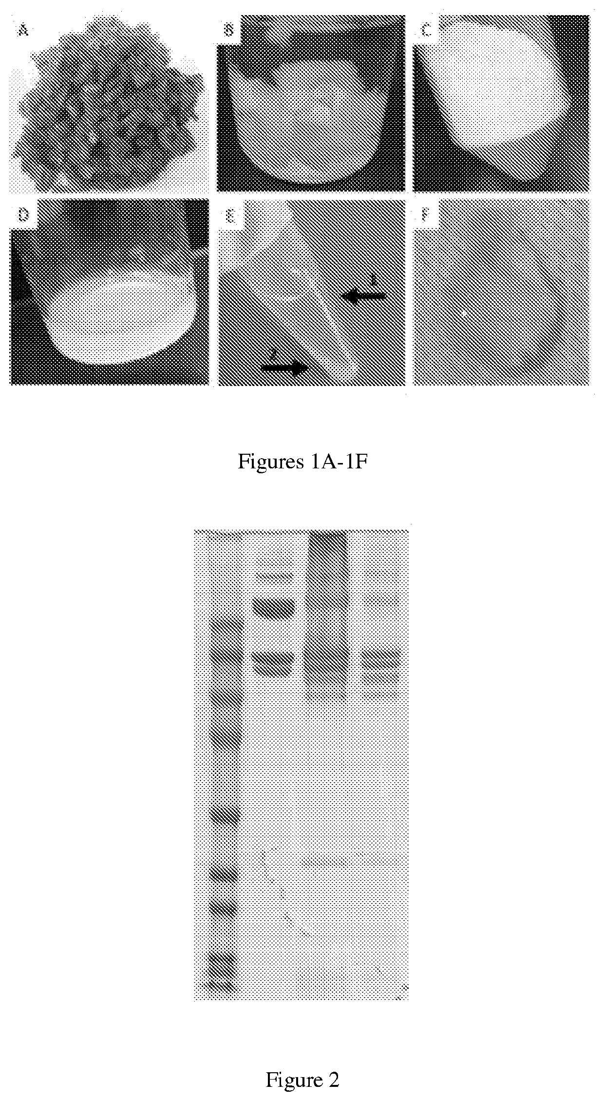
[0088]The formulation of myocardial matrix (MM) can be generated based on previously described protocols (FIG. 1) [3]. In brief, fresh hearts are harvested from pigs (approx. 30-45 kg) and the LV myocardium is isolated. Major vessels and connective tissue are removed, and the remaining tissue will be cut into pieces less than 5 mm3 (FIG. 1A). Tissue is decellularized in 1% (w / v) sodium dodecyl sulfate (SDS) for 4-5 days until the tissue is completely white, followed by an additional day of water rinsing to remove residual SDS (FIG. 1B). The material is lyophilized and milled into a fine powder (FIG. 1C) and subsequently partially enzymatically digested for 48 hours. The material is then neutralized and buffered to match in vivo conditions, yielding MM (FIG. 1D), capable of thermally induced gelation.

[0089]Next, the MM is centrifuged at 15,000 RCF at 4° C. to separate the soluble and insoluble fractions (FIG. 1E). The supernatant is isolated from the insolubl...

experiment 2

ility of the SolMM with Human Blood

[0090]The interaction between SolMM and human blood samples (n=4) is assessed at different dilutions (1:1, 1:2, 1:10) of SolMM to whole human blood or platelet rich plasma. 1:1 represents the highest possible ratio between blood and SolMM, whereas 1:10 represents a physiologically relevant dilution based on the volumetric flow rate of the coronary vasculature and intended infusion rate (1 ml / min). Hemocompatibility is assessed as previously described for MM [4]. Red blood cell aggregation will be performed within 4 hours of sampling on a Myrenne aggregonometer (Myrenne GmbH) after adjusting hematocrit to 45% with autologous plasma. Aggregation is assessed following stasis (M0) or a low shear rate (3 Hz; M1) while absorbance (800 nm) is measured for 5 seconds. Similarly, platelet aggregation is measured with isolated platelet rich plasma on a lumi-aggregonometer (Chrono-log). Using the same dilutions as above for sample to platelet rich plasma, high...

experiment 3

, Retention, and Efficacy in a Small Animal Ischemia-Reperfusion Model

[0092]Using a Sprague Dawley rat (225-250 g) ischemia-reperfusion model of MI, the left coronary artery is occluded for 45 minutes, followed by reperfusion. Within 5 minutes following reperfusion, the aorta is clamped for approximately 15 seconds to simulate intracoronary infusion, and 200 μl of SolMM is injected in to the LV lumen at a concentration of 6, 10, or 14 mg / mL. This will force the material into the coronary arteries and then distribute into the infarcted myocardium [12]. Hearts (n=2 per concentration) are isolated 60-minutes post-injection to determine if the material will initially distribute and then be retained in the heart, as non-gelling materials are cleared from the heart within an hour [5]. SolMM is conjugated with Alexa Fluor™ 568 N-hydroxysuccinimidyl ester (Invitrogen) to allow for fluorescent detection and analysis. Retention of the material is tested with the optimal concentration at the t...


PUM

PropertyMeasurementUnit
size exclusionaaaaaaaaaa
sizeaaaaaaaaaa
sizeaaaaaaaaaa

Abstract

Compositions and methods for their manufacture and use are provided comprising a soluble extracellular matrix fraction for intravascular delivery which forms a gel or coating in situ for treatment of myocardial infarction and ischemia in a variety of tissues and endothelial injury/dysfunction.

Description

CROSS REFERENCE TO RELATED APPLICATIONS[0001]This application claims the priority benefit of U.S. Provisional Application No. 62 / 750,303, filed Oct. 25, 2018, which application is incorporated herein by reference.GOVERNMENT SPONSORSHIP[0002]This invention was made with government support under grant No. HL113468 awarded by the National Institutes of Health (NIH). The government has certain rights in the invention.TECHNICAL FIELD[0003]The present invention relates to an infusable soluble extracellular matrix composition and therapy for minimally invasive delivery to tissues / organs / cells, including ischemic or injured heart, brain, skeletal muscle, blood vessels, and endothelial cells.BACKGROUND[0004]Extracellular matrix therapies include native tissue components providing a scaffold for tissue regeneration. Current decellularized extracellular matrix therapies are restricted to patches or direct injections. No extracellular matrix therapy is capable of intravascular / infusion delivery...

Claims


Application Information

Patent Timeline
Patent Type & Authority Applications(United States)
IPC IPC(8): A61L27/36A61L27/54A61K38/01
CPCA61L27/3633A61L27/3683A61L2400/06A61K38/014A61L27/54A61K35/22A61K35/30A61K35/38A61K35/34A61K38/012A61K9/0019A61P9/00A61K9/0024A61K47/26A61L2430/20
Inventor CHRISTMAN, KARENSPANG, MARTINGROVER, GREGORY
Owner RGT UNIV OF CALIFORNIA
Why Eureka
Try Eureka