Preparation method for therapeutic agent of bead-type chondrocyte

a technology of chondrocytes and therapeutic agents, which is applied in the field of preparation of bead-type chondrocyte therapeutic agents, can solve the problems of inability to reproduce the structural features or constitutions of normal articular cartilage, death of interior cells, and limited spontaneous regeneration, so as to achieve the effect of repairing damage and facilitating and stably preparing uniform, high-quality cartilage tissues

Active Publication Date: 2018-05-01
MCTT BIO
View PDF5 Cites 0 Cited by
  • Summary
  • Abstract
  • Description
  • Claims
  • Application Information

AI Technical Summary

Benefits of technology

[0017]The present invention uses a 96-well deep well plate having V-shaped bottom in the preparation of three-dimensional cartilage tissue without a scaffold, thereby being capable of easily and stably preparing uniform, high-quality cartilage tissues with repetitive reproducibility in large quantities; being capable of automation; and being capable of preparing a therapeutic agent of bead-type chondrocyte which has effectiveness in repairing damage by simply being implanted into cartilage damage of patients by injection, as compared with conventional culture methods.

Problems solved by technology

Because cartilage tissue is avascular tissue and the ratio of cells in the tissue is very low, spontaneous regeneration is very limited.
However, in cases of elderly patients and large-size damage, structural features or constitutions of normal articular cartilage cannot be reproduced.
However, in view of clinical application, xenogeneic and allogeneic natural materials may cause immune reaction, and in the case of synthetic materials safety problems may be caused due to the harmful degradation product.
Furthermore, when chondrocytes are inoculated to a scaffold, most of them distribute to the outer part of the scaffold, and extracellular matrixes which are synthesized and secreted by chondrocytes form shell at the outer part of the scaffold to hinder diffusion and exchange of nutrients, wastes and gases, resulting in death of interior cells.
However, there are still many unresolved problems such as interaction between cells and biomaterials, irregular degradability of biomaterials, biocompatibility, uneven cellular distribution, lack of linkage (bonding) between tissue-engineered cartilage and peripheral cartilage and the like (Sittinger et al., 1996; Grande et al., 1997; Nehrer et al., 1998; Sims et al., 1998; Hutmacher et al., 2000; Ochi et al., 2001; Naumann et al., 2004; Park et al., 2006; and Wolf et al., 2008).
Studies about methods for the preparation of three-dimensional cartilage tissue without the use of scaffold have been continually carried out, but it has been reported that such methods are very limited in direct clinical applications since tissues are formed depending on only cells and ECM synthesis capacity of cells so that it is difficult to prepare tissues suitable for the size of damage where implantation is needed (Adkisson et al., 2001; Grogan et al., 2003; and Marlovits et al., 2003).
In addition, the shape and depth of damaged cartilage are not uniform (FIG. 1A).
Tissue-engineered cartilages developed up to now are implanted in such a manner, but they cannot be adjusted to the thickness of damage.
However, for development as a therapeutic agent, a technique for preparing uniform cartilaginous tissues with repetitive reproducibility is necessary, and a large-scale culture system for preparing considerably large number of cartilaginous tissues is required for using in wide damaged area.
However, evaluation of applicability of cartilaginous structure prepared by pellet culture as a cell-therapy product has not been carried out since a pellet system is a useful method for preparing high quality cartilaginous tissues but it has been regarded being difficult to apply to regeneration of damaged cartilage for the problem of difficulty in preparing sufficient pellet size.
As a result, it is difficult to apply this method to large-scale culture (FIG. 2A).
However, this culture method cannot adjust the number of cells which produce one cell aggregate, and there is a disadvantage of not being standardized as a tissue-engineered / cell therapy product since the size of each cartilaginous tissue and chondrification vary due to the possibility of fusion between formed cartilaginous tissues.
However, it cannot be guaranteed to stably obtain uniform cartilaginous tissues since the capacity of forming cell aggregate spontaneously is different depending on cell condition.
However, because they are also methods to induce spontaneous cellular aggregation, it cannot be guaranteed that they stably obtain uniform cartilaginous tissues.
In addition, because the size of prepared cell aggregates is too small, physical strength is weak and handling is difficult, so there is a limit in it being used as a therapeutic agent of three-dimensional chondrocyte.
As such, methods known up to now as those for preparing three-dimensional cartilage tissue without the use of scaffold have problems such that sufficient pellet size is not formed, it is not suitable for large-scale preparation, uniform cartilage tissue with repetitive reproducibility is not formed, the size of formed cell aggregate is too small, and strength is low.
Therefore, such methods are inappropriate for the preparation of a therapeutic agent of bead-type chondrocyte without a scaffold

Method used


Image

  • Preparation method for therapeutic agent of bead-type chondrocyte
  • Preparation method for therapeutic agent of bead-type chondrocyte
  • Preparation method for therapeutic agent of bead-type chondrocyte

Examples


examples 1

[0067]Isolation and Proliferation of Chondrocytes

[0068]Costal cartilage tissues were washed with phosphate-buffered saline (PBS) containing antibiotics 3-5 times to remove blood and contaminants. The cartilages were minced into 1-2 mm3, and extracellular matrix was then digested by the treatment of 0.5% pronase and 0.2% type II collagenase to isolate cells. After the inoculation of isolated cells into culture dishes at a cell density of 2-4×10⁴ cells / cm2, a culture medium for cell proliferation (mesenchymal stem cell growth medium [MSCGM] containing 1 ng / ml of FGF-2) was added thereto, and the cells were then cultured until being confluent in a 37° C., 5% CO2 incubator. Cells detached from the culture dishes with a trypsin-EDTA solution were inoculated into culture dishes at a cell density of 1-2×10⁴ cells / cm2, and cultured to passages 6 to 8.

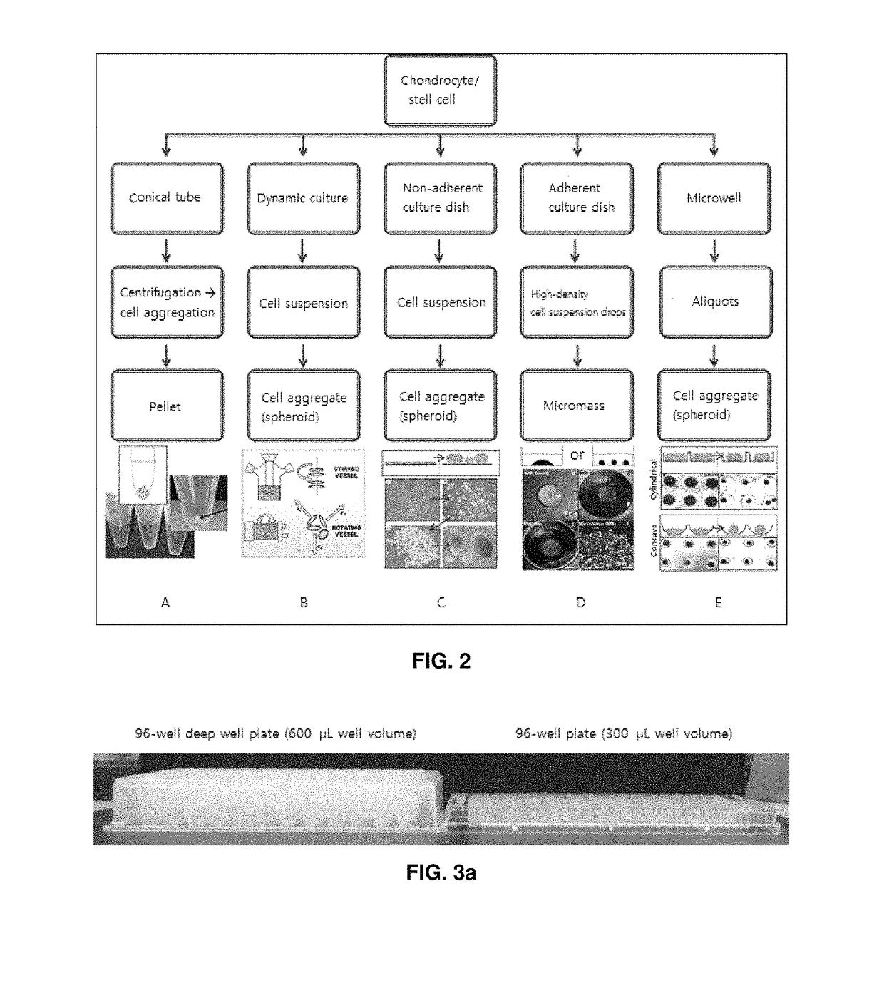
[0069]Three-dimensional Pellet Culture of Chondrocytes at Various Kinds of 96-well Plates or 96-well Deep well Plates

[0070]Proliferation compl...

example 2

[0073]Preparation of Bead-type Three-dimensional Cartilage Tissue

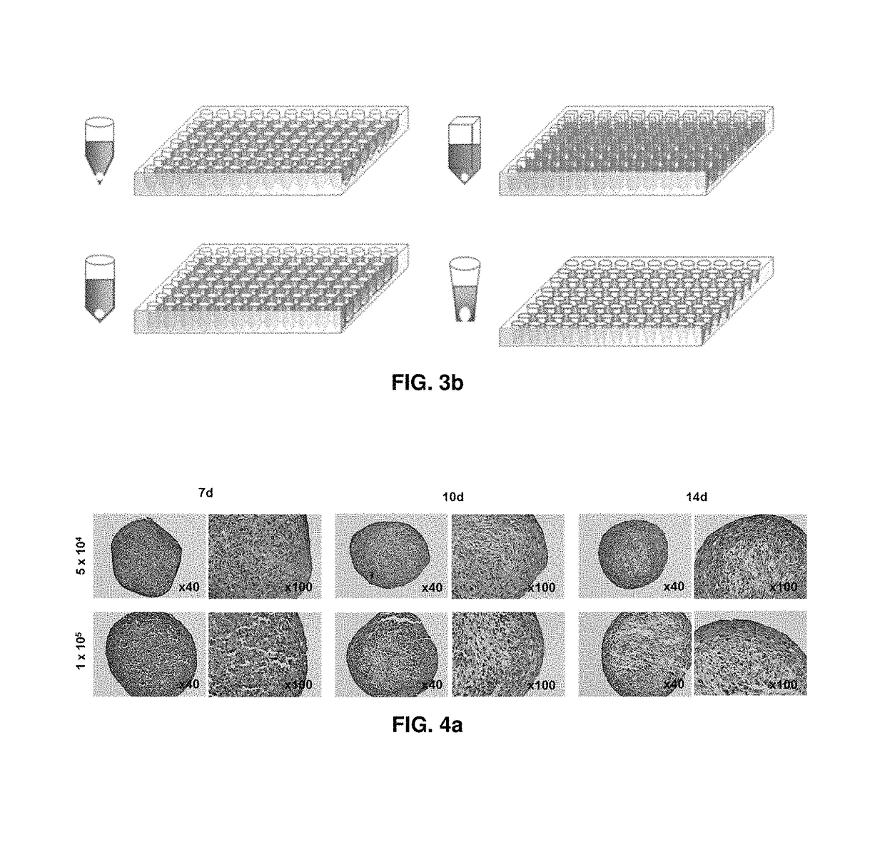
[0074]Rabbit chondrocytes cultured in a culture medium for cell proliferation to passages 6 to 8 were pellet cultured in a 96-well deep well plate having V-shaped bottom with 0.5×10⁵ cells / 400 μl / well, 1.0×10⁵ cells / 400 μl / well or 2.0×10⁵ cells / 400 μl / well and evaluated for 28 days.

[0075]Evaluation Methods for Properties of Bead-type Three-dimensional Cartilage Tissue

[0076](1) Appearance and Size

[0077]In the course of three-dimensional culture, pellets were collected from each well and their properties were evaluated. First of all, appearance was evaluated with the naked eye, and a pellet was mounted on a slide glass and a photograph was taken with a digital camera installed on a microscope with 40× magnifications to measure the area.

[0078](2) DNA Amount

[0079]To measure the DNA amount in pellets, 125 μg / mL of papain was added to pellets and homogenized. After overnight treatment at 65° C., the prepared sample was mixed...

example 3

[0108]Rabbit costal chondrocytes cultured in a culture medium for cell proliferation to passages 6 to 8 were suspended in a culture medium for differentiation and dispensed into a 96-well deep well plate having V-shaped bottom with 1.0×10⁵ cells / 400 μl / well. The plates were centrifuged at 1,200 rpm for 5 minutes, and then pellet cultured in a 37° C., 5% CO2 incubator for 10 days with exchanging culture media at an interval of 3 or 4 days to prepare a bead-type cartilage tissue without a scaffold for transplanting to articular cartilage damage. After culturing, the bead-type cartilage tissue was collected by the use of portable pellet collection apparatus (FIG. 8c), and properties were evaluated according to the methods described in Example 2.

[0109]As a result, semitranslucent bead-type three-dimensional structures having white and smooth surface with the naked eye and 1.0 to 1.5 mm of diameter were observed (FIG. 6a). Their plane sizes were in the range of 1.6 mm2±20%, and the forma...


PUM

PropertyMeasurementUnit
sizeaaaaaaaaaa
diametersaaaaaaaaaa
volumeaaaaaaaaaa

Abstract

The present invention relates to a preparation method for a therapeutic agent of a bead-type chondrocyte and, more specifically, to: a preparation method for a therapeutic agent of a bead-type chondrocyte without a support, comprising the steps of: a) dispensing chondrocytes and / or cells having chondrogenic differentiation capability in a 96-well deep well plate having a V-shaped bottom; b) centrifuging the plate; c) three-dimensionally culturing the plate in an incubator; and d) recovering pellets from each well, thereby being capable of easily and stably preparing cartilage tissues of a uniform quality, in large quantities; and a therapeutic agent of a bead-type chondrocyte without a support, prepared by the method, having effectiveness to repair damage by simply being implanted into cartilage damage by injection.

Description

CROSS-REFERENCE TO RELATED APPLICATIONS[0001]This application is a national phase application of PCT Application No. PCT / KR2014 / 006680, filed on Jul. 23, 2014, which claims benefit of Korean Patent Application 10-2013-0087533, filed on Jul. 24, 2013. The entire disclosure of the application identified in this paragraph is incorporated herein by reference.FIELD[0002]The present invention relates to a method for preparing a therapeutic agent of bead-type chondrocyte. More specifically, the present invention relates to a method for preparing a therapeutic agent of bead-type chondrocyte without a scaffold which comprises the steps of: a) dispensing chondrocyte and / or cell having chondrogenic potential into a 96-well deep well plate having V-shaped bottom; b) centrifuging the plate; c) three-dimensionally culturing the plate in an incubator; and d) collecting a pellet from each well, thereby being able to easily and stably prepare uniform, high-quality cartilage tissues with repetitive r...

Claims


Application Information

Patent Timeline
Patent Type & Authority Patents(United States)
IPC IPC(8): A61K35/32C12N5/077C12N5/00A61K35/28
CPCA61K35/32A61K35/28C12N5/0655C12N5/0062C12N2513/00C12N2500/90A61P19/00A61P19/02A61P43/00
Inventor LEE, JUNG SUNLEE, JIN YEONCHAE, BYUNG CHULSON, YOUNG SOOKCHANG, SONG SUN
Owner MCTT BIO
Why Eureka
Try Eureka