
Circulating tumour cell typing and identification kit

a tumour cell and identification kit technology, applied in the field of molecular biology, can solve the problems of increased mortality risk of malignant tumour patients, false positive or false negative detection results, and neoadjuvant chemotherapy did not eliminate the ctcs in which emt had, so as to reduce false negative results, reduce false positive results, and improve detection sensitivity.

Active Publication Date: 2017-09-28
SUREXAM BIO TECH
View PDF1 Cites 3 Cited by
  • Summary
  • Abstract
  • Description
  • Claims
  • Application Information

AI Technical Summary

Benefits of technology

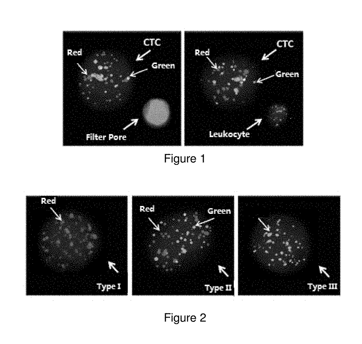
The patent aims to provide a kit and method for detecting and typing circulating tumour cells with high specificity and sensitivity. The marker genes used in the kit were chosen through extensive experimentation and statistical analysis. The identification method uses a multiple RNA probe that can label a plurality of genes simultaneously and identify different cell types, reducing false-negative results. The probes show good specificity and high signal to noise ratio. A novel RNA in situ hybridization method is also used with signal amplification, improving the sensitivity of the detection process. The detection process is completed in 8 hours and the sensitivity of the method is significantly improved.

Problems solved by technology

After entering into peripheral blood, most CTCs ended in apoptosis or being devoured, while a few may escape and anchor to develop into a metastatic lesion, resulting in increased mortality risk of malignant tumour patients.
They found that neoadjuvant chemotherapy did not eliminate these CTCs in which EMT had occurred.
False positive or false-negative detection results may occur due to the possible presence of a number of non-neoplastic epithelial cells in peripheral blood, potential contamination in blood samples by normal epithelial cells during blood sampling, and that some CTCs cannot be detected due to loss of epithelial antigens during EMT.
However, drawbacks exist as RT-PCR process has high requirements for environments and operation, mRNA is prone to degradation which disables CTC cell typing, and the like.

Method used


Image

  • Circulating tumour cell typing and identification kit
  • Circulating tumour cell typing and identification kit
  • Circulating tumour cell typing and identification kit

Examples


example 1

[0039]The circulating tumour cell typing and identification kit disclosed in this example can take two forms, either with or without a labeled probe.

[0040]A circulating tumour cell typing and identification kit A with a labeled probe mainly comprises:

[0041]I. Capture Probe

[0042]A capture probe consists of three components from 5′-terminal to 3′-terminal in sequence: a sequence P1 which complementarily pairs with corresponding marker gene mRNA, a spacer arm sequence, and a P2 sequence that is able to complementarily pair with a P3 sequence of a corresponding amplification probe, wherein the same P2 sequence is used in capture probes for marker genes of the same type. The spacer arm separates the P2 sequence of a capture probe from the target mRNA. A spacer arm sequence of appropriate length is usually provided within a probe to reduce steric hindrance and improve hybridization efficiency and specificity. The spacer arm of a capture probe of the present disclosure is preferably 5-10T,...

example 2

Detection of Circulating Tumour Cells in Peripheral Blood of Tumour Patients Using the Kit A According to Example 1

[0052]The formulations of the various solutions are listed below:

Name of solutionFormulationPreserving16.6 g NH4Cl, 2 g KHCO3, 8 ml 0.5M EDTA, filled to 1 L with ultrapuresolutionwaterFixing agent10% Neutral formalinPBS2.9 g Na2HPO4•12H2O, 0.3 g NaH2PO4•2H2O, 8.06 g NaCl, 0.2 g KCl,filled to 1000 ml with ultrapure water, pH7.4Permeabilization0.25% TritonX-100agentDigestive enyzme1 ug / ml proteinase KProbe buffer100 μg / ml denatured salmon sperm DNA, 100 mM LiCl, 0.1% sodiumsolutiondodecyl sulfate, 9 mM EDTA, 50 mM HEPES (pH 7.5), 0.05% ProClin300Amplification6 mM Tris-HCl (pH 8.0), 1% sodium dodecyl sulfate, 1 mL / L bovinebuffer solutionserum albumin, 0.05% ProClin 300, 0.05% sodium azideColor-developing20 mM Tris-HCl, 400 mM LiCl, 1 mL / L Tween 20, 1 mL / L bovine serumbuffer solutionalbumin, 0.05% ProClin 300Probe mixturei.e., capture probe, 0.75 fmol / ul / genesolutionAmplifi...

example 3

Detection of Cell Lines Using the Kit A According to Example 1

[0106]I. Selection of Cell Lines

[0107]In the kit of present disclosure, the epithelial cell marker genes are two or more selected from the group consisting of EPCAM, E-cadherin, CEA, KRT5, KRT7, KRT17, and KRT20; the mesenchymal cell marker genes are two or more selected from the group consisting of VIMENTIN, N-cadherin, TWIST1, AKT2, ZEB2, ZEB1, FOXC1, FOXC2, SNAI1, and SNAI2; and the leukocyte marker gene is CD45. The epithelial marker genes, mesenchymal marker genes, and leukocyte marker genes provided in this disclosure are genes selected by the inventors through numerous experiments and statistical analysis that specifically express on CTCs, with good specificity and accuracy for identification and typing of CTCs.

[0108]In this example, an epithelial type cell line MCF-10A, a mesenchymal type tumour cell line U118, and a mixed epithelial-mesenchymal type lung cancer cell line PC-9 were used for experiments, while CCRF...


PUM

PropertyMeasurementUnit
Magnetic fieldaaaaaaaaaa
Temperatureaaaaaaaaaa
Volumeaaaaaaaaaa

Abstract

This disclosure relates to a circulating tumour cell typing and identification kit, comprising a capture probe, an amplification probe, and a labeled probe for each marker gene mRNA, wherein the marker gene mRNA comprises the following two types: at least two epithelial cell marker gene mRNAs selected from the group consisting of EPCAM, E-cadherin, CEA, KRT5, KRT7, KRT17, and KRT20 mRNAs; and, at least two mesenchymal cell marker gene mRNAs selected from the group consisting of VIMENTIN, N-cadherin, TWIST1, AKT2, ZEB2, ZEB1, FOXC1, FOXC2, SNAI1 and SNAI2 mRNAs. This disclosure prevents false-positive results caused by, for example, possible presence of a number of non-neoplastic epithelial cells in peripheral blood, introduction of normal epithelial cells during blood sampling, and the like. Accordingly, it may be assured that cells detected with epithelial cell marker genes and / or mesenchymal cell marker genes are indeed circulating tumour cells, further improving accuracy and reliability of the detection results.

Description

TECHNICAL FIELD[0001]The present disclosure belongs to the field of molecular biology, and relates to the medical sciences and biological technologies. More particularly, it relates to a circulating tumour cell typing and identification kit.BACKGROUND[0002]Circulating tumour cell (CTC) refers to collectively all types of tumour cells occurring in peripheral blood, which was detached from a solid tumour lesion (primary lesion or metastatic lesion) spontaneously or due to medical operations. After entering into peripheral blood, most CTCs ended in apoptosis or being devoured, while a few may escape and anchor to develop into a metastatic lesion, resulting in increased mortality risk of malignant tumour patients.[0003]Research in recent years has revealed that tumour cells, during the process of entering peripheral blood (PB) circulation, may go through epithelial-mesenchymal transition (EMT). Tumour cells gone through epithelial-mesenchymal transition (EMT) have increased migration an...

Claims


Application Information

Patent Timeline
IPC IPC(8): C12Q1/68
CPCC12Q1/6886C12Q2600/112C12Q2600/158C12Q1/6881C12Q1/6841C12Q2565/519C12Q2563/107C12Q2600/16C12Q1/682C12Q2525/197C12Q2537/143C12Q2543/10C12Q2565/102C12Q1/68C12Q1/04
Inventor XU, JIASENWU, SHIYANGLIU, SUYAN
Owner SUREXAM BIO TECH
Learn More