Spatiotemporal delivery system embedded in 3d-printing

Active Publication Date: 2018-03-29
THE TRUSTEES OF COLUMBIA UNIV IN THE CITY OF NEW YORK
View PDF4 Cites 5 Cited by
  • Summary
  • Abstract
  • Description
  • Claims
  • Application Information

AI Technical Summary

Benefits of technology

[0006]Among the various aspects of the present disclosure is the provision of a 3D-Printing delivery system and

Problems solved by technology

While conventional scaffolds can include transplanted cells from a subject (which can reduce immunologic complications), commercialization of these cell-seeded scaffolds can be problematic with respect to ex vivo cell culture, packing and s

Method used


Image

  • Spatiotemporal delivery system embedded in 3d-printing
  • Spatiotemporal delivery system embedded in 3d-printing
  • Spatiotemporal delivery system embedded in 3d-printing

Examples


example 1

3D Printed Biocompatible Scaffold for Multi-Tissue Interface

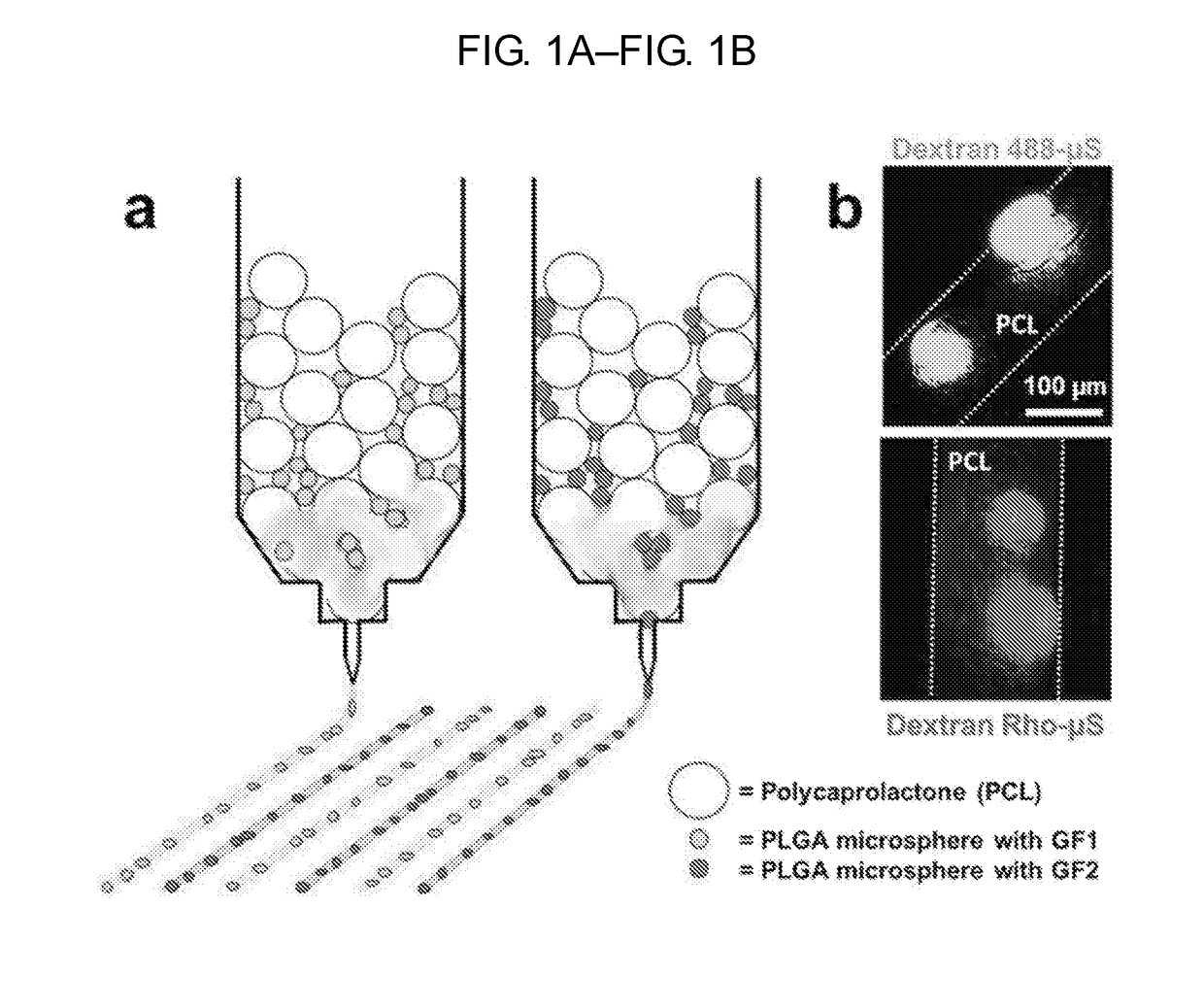
[0271]The following example provides for a 3D printed biocompatible scaffold with spatiotemporal delivery of microsphere encapsulated CTGF and TGFβ3.

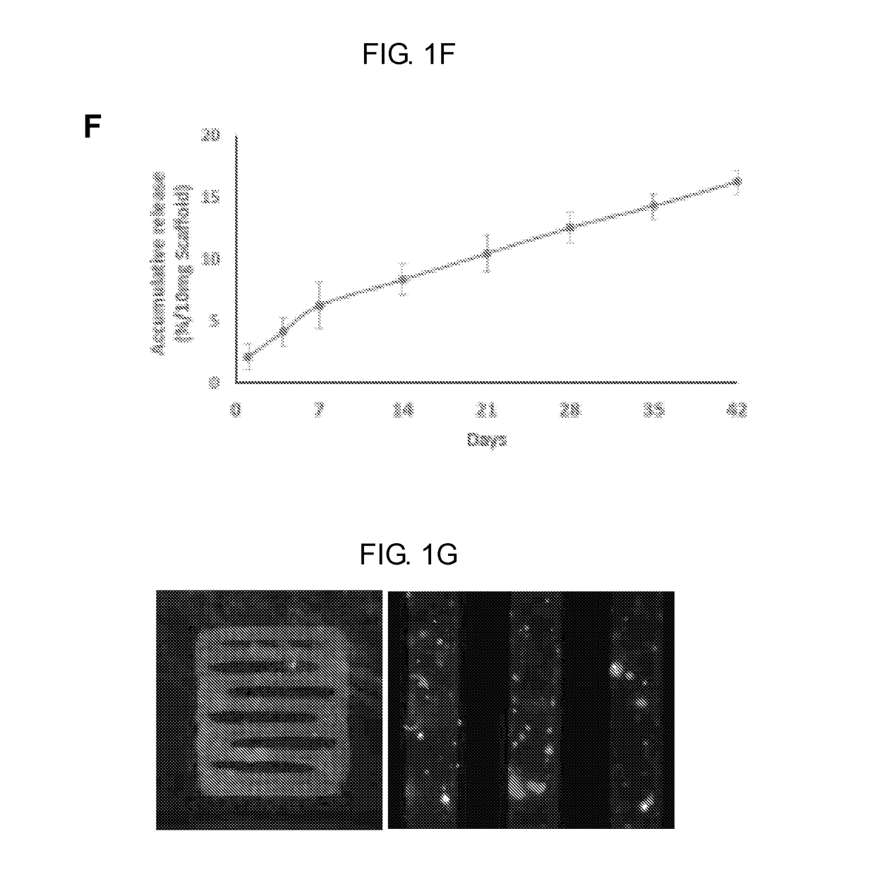
[0272]Selected growth factors (GF) were encapsulated in poly(lactic-co-glycolic acids) (PLGA) microspheres (μS) for controlled release. Poly(lactic-co-glycolic acid) (PLGA) microspheres (10-400 μm) encapsulating CTGF, TGFβ3, and BMP growth factors, were prepared by double-emulsion technique.

[0273]Then 3D scaffolds with custom-designed microstructure and outer shape were constructed using layer-by-layer deposition of continuous PCL microfibers. PCL pellets and growth factor-encapsulated microspheres were mixed in dispensing cartilages of 3D Bioplotter® (EnvisionTec, Germany) and heated up to 100° C., selected from heating diffusion analysis (see e.g., FIG. 1A). Material was dispensed through a fine stainless steel needle (inner diameter 50 μm to 400 μm), creating micro-sized gro...

example 2

Growth Factor Delivery in 3D Printed Multi-Tissue Interface Scaffold

[0281]The following Example demonstrates formation of multi-tissue interfaces in 3D-printed scaffold with spatiotemporal delivered CTGF and TGFβ3. Methods are according to Example 1 except as indicated otherwise.

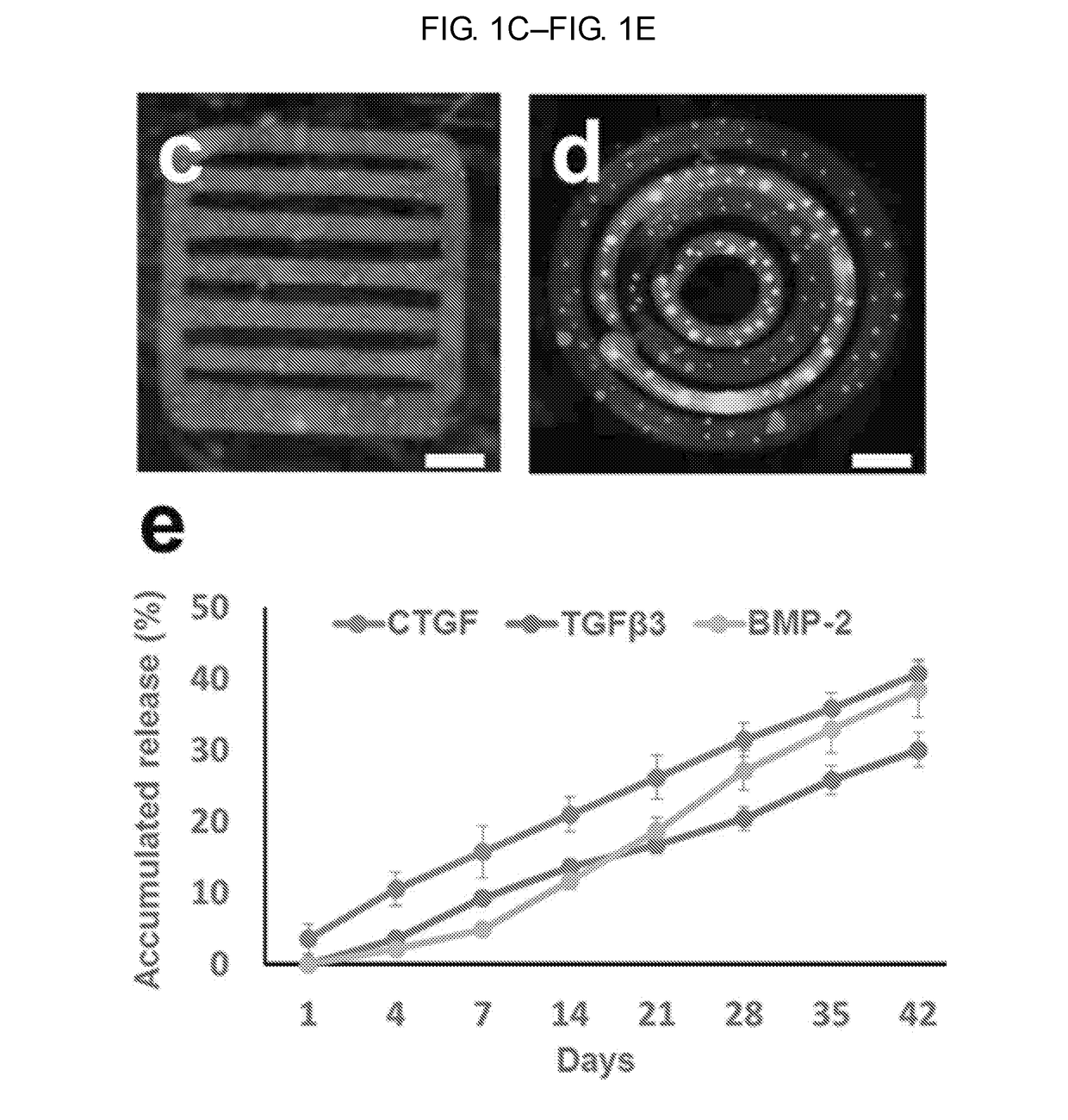
[0282]A single layered rectangular structure was constructed (see e.g., FIG. 3A) with alternative PCL microfibers-embedding CTGF-μS and TGFβ3-μS (see e.g., FIG. 3B). The size of microfibers and interfibers space was 100 μm.

[0283]Then human bone marrow derived mesenchymal stem / progenitor cells (MSCs) were delivered in the scaffold's inter-fiber space via fibrin gel. After 4 weeks culture in vitro, alternative depositions of COL-I+ and COL-II+ matrices, corresponding to the pattern of growth factor delivery, forming an integrated interface within the 100-200 μm interfiber zones (see e.g., FIG. 3C).

[0284]This demonstrated PCL scaffolds with spatiotemporal delivery of CTGF and TGFβ3 successfully formed native-li...

example 3

Spatiotemporal Delivery of Growth Factors Embedded in 3D Printed Multi-Tissue Scaffolds

[0285]The following example will test the efficacy of the spatiotemporal delivery of growth factors (GFs) embedded in 3D printed scaffolds (see e.g., Example 1) for regeneration of multi-tissue complexes.

[0286]Scaffolds are first cultured with multiple stem / progenitor cell populations in vitro (see e.g., Example 5, Example 6, and Example 7), followed by in vivo implantation in a relevant animal model (see e.g., Example 5).

[0287]Scaffolds are designed with spatiotemporal delivery of growth factors. An anterior cruciate ligament-fibrocartilage-bone interfaces (see e.g., FIG. 4A) and supraspinatus tendon-fibrocartilage (unmineralized and mineralized)-bone interfaces (see e.g., FIG. 4B) are reconstructed in 3D-printed scaffolds with spatiotemporally delivered growth factors.

[0288]In a similar way, a cementum (CM)-periodontal ligament (PDL)-alveolar bone (AB) complex is reconstructed (e.g., periodontiu...


PUM

PropertyMeasurementUnit
Temperatureaaaaaaaaaa
Lengthaaaaaaaaaa

Abstract

Provided herein is a 3D printing system and related compositions, and method of using such, that can produce a polymeric microfiber having embedded microspheres encapsulating an active agent with micron precision and high spatial and temporal resolution. One aspect of the present disclosure provides a method of forming a biocompatible scaffold. Another aspect provides a method of forming a polymeric fiber having a microencapsulated agent distributed in the polymeric fiber. Another aspect provides a composition including a polymeric microfiber produced by 3D printing.

Description

CROSS-REFERENCE TO RELATED APPLICATIONS[0001]The present application claims the benefit of U.S. Provisional Application Ser. No. 62 / 303,843 filed 4 Mar. 2016; U.S. Provisional Application Ser. No. 62 / 174,232 filed 11 Jun. 2015; U.S. Provisional Application Ser. No. 62 / 148,074 filed 15 Apr. 2015; and U.S. Provisional Application Ser. No. 62 / 144,890 filed 8 Apr. 2015; each of which are incorporated herein by reference in their entireties.STATEMENT REGARDING FEDERALLY SPONSORED RESEARCH OR DEVELOPMENT[0002]This invention was made with government support under grant number 5K12DE023583-02 (C.H.L) awarded by NIH / NIDCR. The government has certain rights in the invention.MATERIAL INCORPORATED-BY-REFERENCE[0003]Not Applicable.BACKGROUND OF THE INVENTION[0004]Tissue engineering provides for medical applications, biosensors, experimental drug testing, or other non-therapeutic applications. Biocompatible scaffolds are generally necessary to support growth for tissue generation to occur. While ...

Claims


Application Information

Patent Timeline
IPC IPC(8): A61L27/44A61L27/18A61L27/54A61L27/38A61L27/36A61L27/56B33Y70/00B29C64/112B33Y10/00B33Y80/00
CPCA61L27/44A61L27/18A61L27/54A61L27/3847A61L27/3834A61L27/3608A61L27/56B33Y70/00B29C64/112B33Y10/00B33Y80/00A61L2300/414A61L2300/622A61L2430/02A61L2430/06A61L2430/10A61L2430/38A61L2300/602B29L2031/7532B29K2067/04B29K2995/006B29K2995/0056A61L27/50C08L67/04D01D5/0007
Inventor LEE, CHANG HUN
Owner THE TRUSTEES OF COLUMBIA UNIV IN THE CITY OF NEW YORK
Why Eureka
Try Eureka