Method and system for determining a risk of cardiac conduction abnormalities

a risk and cardiac conduction technology, applied in the field of preoperative planning of transcatheter structural heart interventions, can solve the problems of paravalvular aortic regurgitation, paraventricular aortic regurgitation, and 6% in-hospital mortality of over 65 year old patients, and achieve the effect of higher peak pressure or strain and higher risk

Active Publication Date: 2018-10-11
FEOPS NV
View PDF0 Cites 4 Cited by
  • Summary
  • Abstract
  • Description
  • Claims
  • Application Information

AI Technical Summary

Benefits of technology

[0025]Optionally, determining the measure of the mechanical interaction includes defining a predetermined region of the patient-specific anatomical model, and determining a total contact force or total strain within the predetermined region. The total contact force in the predetermined region can be the determined contact pressure integrated over the surface area of the predetermined region. A larger total force or strain in the predetermined region can indicate a higher risk.
[0026]Optionally, determining the measure of the mechanical interaction includes defining a predetermined region of the patient-specific anatomical model, and determining a peak pressure or peak strain of the determined pressure within the predetermined region. A higher peak pressure or strain can indicate a higher risk.

Problems solved by technology

This invasive procedure requires total anesthesia, sternotomy (open-heart surgery) and cardiopulmonary bypass (the blood is pumped and oxygenated using an external machine), and is associated with about 6% in-hospital mortality for over 65 year old patients.
Moreover, at least one-third of the patients with severe aortic stenosis are denied valve surgery as the risks associated with surgery are too high.
Undersizing of a valve implant may lead to paravalvular aortic regurgitation, while oversizing may result in a rupture of the aortic annulus or in a suboptimal functional behavior of the implant.
A problem with these tools is that they do not provide preoperative insights into the interaction between a certain implant device and the specific patient anatomy, and can thus not be used to predict complications such as regurgitation.
Another problem is that is difficult to reconstruct native leaflets from e.g. CT images.
In the currently deployed methods, an incomplete leaflet image is obtained, comprising gaps whereby the gaps represent a lack of data.
US 2011 / 0153286 A1 does not provide a prediction of the mechanical behavior and interaction of the patient-specific aortic root, ascending aorta and aortic valve leaflets with the deployment of a valve implant.
Neither does it provide a means to study the hemodynamic performance of an implant deployed in the aortic valve.
Balloon-expandable devices whose deployment is based on permanent plastic deformations of the metal cannot be modeled.
Problem is that the aortic annulus is not circular, that the aortic annulus may deform and that calcium deposits may deform a valve frame.
Another problem is that the aortic root visualised with Computed Tomography (CT) imaging changes in shape and size after TAVI.
On the one hand this gives higher costs for the health system.
If the incorrect device / size of the TAV is chosen, the first TAVI procedure may fail and additional treatments, including a second TAVI procedure (valve-in-valve), SAVR, or rehospitalization may be necessary, with a considerable increase of the costs per patient.
On the other hand this leads to a lower prognosis.
Sub-optimal treatment planning may result in peri-procedural complications, which affect both the life quality and the life expectancy of the patient.

Method used


Image

  • Method and system for determining a risk of cardiac conduction abnormalities
  • Method and system for determining a risk of cardiac conduction abnormalities
  • Method and system for determining a risk of cardiac conduction abnormalities

Examples


Embodiment Construction

[0052]Left bundle branch block (LBBB) after a transcatheter aortic valve implantation (TAVI) procedure is a frequent complication. LBBB after TAVI may occur in as much as 20 to 50% patients. This can result in increased mortality after one year. The underlying cause is to date still subject of speculation. Using the present technology, however, a predictor for the occurrence of LBBB or other conduction abnormalities can be given.

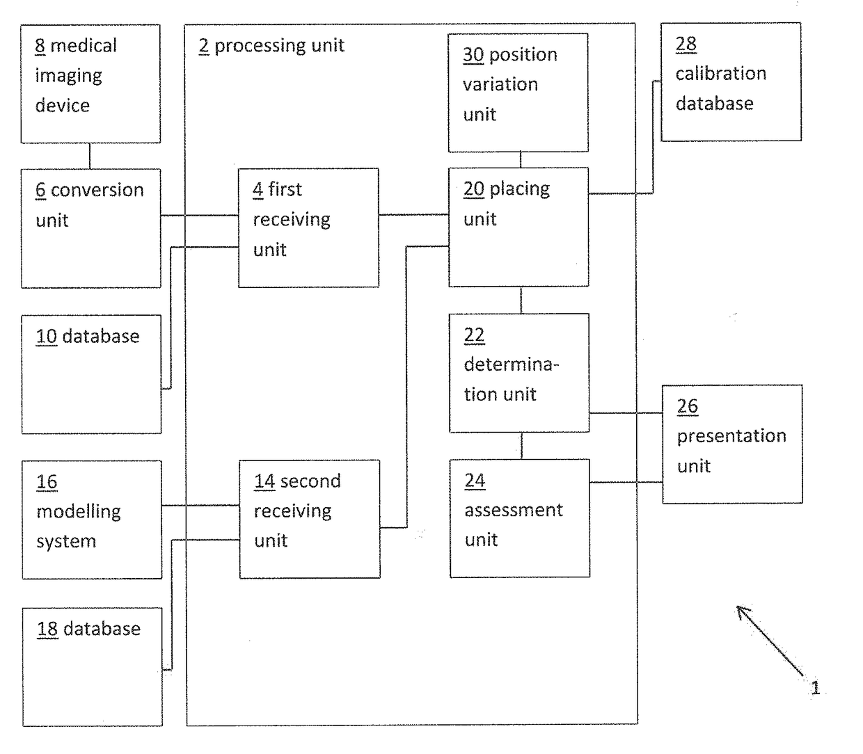
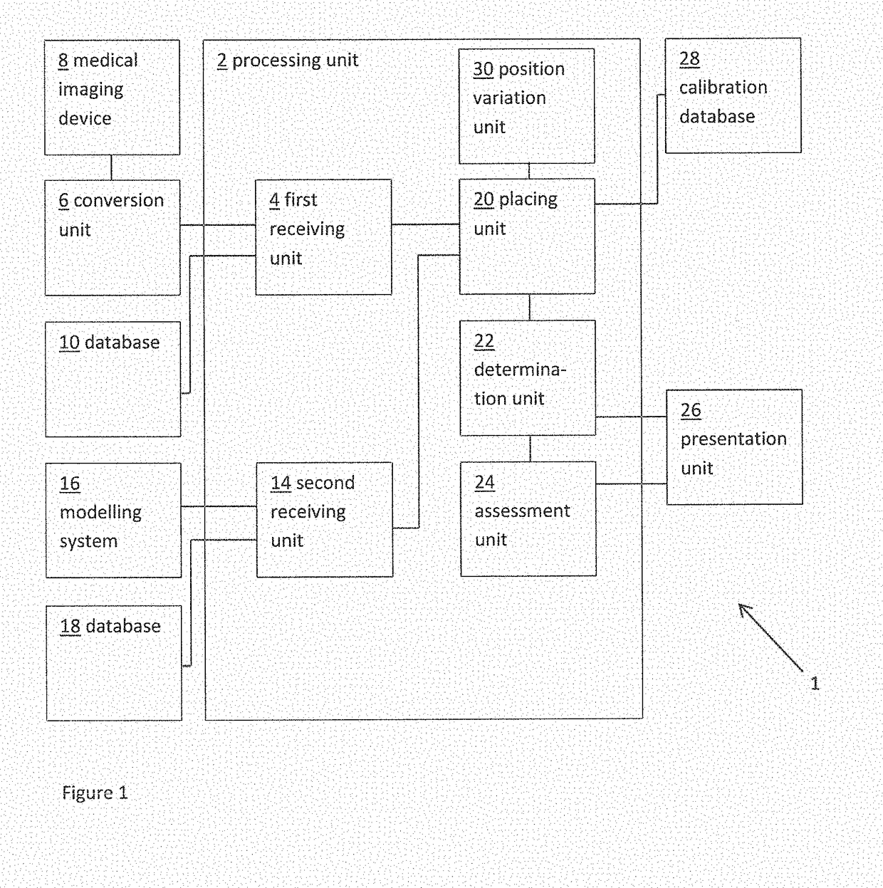
[0053]FIG. 1 shows a schematic example of a system 1 for determining a measure of a risk of a patient developing cardiac conduction abnormalities as a result of transcatheter cardiac valve treatment. The system includes a processing unit 2. The processing unit 2 includes a first receiving unit 4 for receiving a patient-specific anatomical model. Here the patient-specific anatomical model represents a patient-specific cardiac valve region. In this example, the patient-specific anatomical model is provided as a three dimensional (3D) finite element model compr...


PUM


Abstract

A method and system for determining a measure of a risk of a patient developing cardiac conduction abnormalities as a result of transcatheter cardiac treatment. The method includes providing a patient-specific anatomical model representing cardiac region and an implant model representing a finite element representation of a cardiac implant. The method includes virtually placing said implant model into said patient-specific anatomical model. A measure of mechanical interaction between the implant model and the patient-specific anatomical model is determined and a measure of risk of the patient developing cardiac conduction abnormalities is determined on the basis of the determined mechanical interaction.

Description

FIELD OF THE INVENTION[0001]The present invention relates to the field of pre-operative planning of transcatheter structural heart interventions, e.g. valve treatment, such as valve implantation and / or repair. More in particular, the invention relates to pre-operative prediction of the risk a patient developing cardiac conduction abnormalities as a result of transcatheter valve treatment.BACKGROUND TO THE INVENTION[0002]The left ventricle of the heart pumps the blood to the aorta through the aortic valve. Aortic (valve) stenosis is a pathology occurring when the aortic valve does not open fully because the leaflets calcify, thicken and stiffen and, as a result, the blood flow going from the heart to the systemic circulation decreases. Aortic stenosis manifests itself in elderly people, with a prevalence going from 1.3% in over 65 and 4% in over 85 year old people. Currently it is the most common valvular heart disease in the Western world and its prevalence is increasing with the ag...

Claims


Application Information

Patent Timeline
Patent Type & Authority Applications(United States)
IPC IPC(8): A61B34/10A61F2/24G06T7/00G06T17/20G16H50/30G16H50/50
CPCA61B34/10A61F2/2427G06T7/0012G06T17/20G16H50/30G16H50/50A61B2034/104A61B2034/105G06T2207/30048G06F17/5018A61F2/24G06T2210/41G06T19/00G16H50/20A61B2017/00243A61B2017/00783G06F30/23
Inventor MORTIER, PETER EDDY JDE SANTIS, GIANLUCADE BEULE, MATTHIEU ROBERT ANNA FIRMIN
Owner FEOPS NV
Why Eureka
Try Eureka