METASTATIC CANCER DIAGNOSIS VIA DETECTING pH-DEPENDENT ACTIVATION OF AUTOPHAGY IN INVASIVE CANCER CELLS

Inactive Publication Date: 2018-11-01
NANO HESGARSAZAN SALAMAT ARYA
View PDF0 Cites 0 Cited by
  • Summary
  • Abstract
  • Description
  • Claims
  • Application Information

AI Technical Summary

Benefits of technology

[0007]In one general aspect, the present disclosure describes an exemplary method for detecting a metastasis state of biological cells. The method may include seeding a plurality of biological cells onto an array of electrodes of an electrical cell-substrate impedance sensor (ECIS) by dropping a cell suspension including the plurality of biological cells in a cell culture medium onto the array of electrodes, forming a plurality of cultured biological cells attached onto the array of electrodes by maintaining the ECIS in an incubator, reducing pH value of an extracellular media around the plurality of cultured biological cells to a pH value between 6.2 and 6.7 by dropping an acidic solution onto the array of electrodes, activating an intra

Problems solved by technology
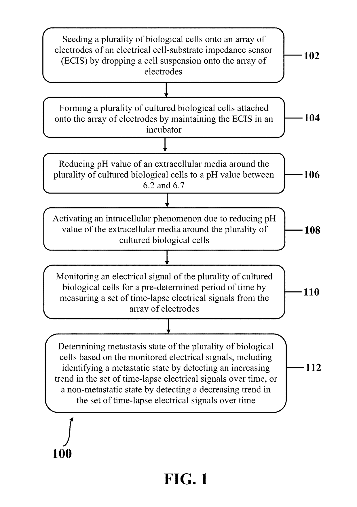
Such systems are so complicated and expensive that they may not be used before the appearance of clinical signatures of the patient-reducing adv

Method used


Image

  • METASTATIC CANCER DIAGNOSIS VIA DETECTING pH-DEPENDENT ACTIVATION OF AUTOPHAGY IN INVASIVE CANCER CELLS
  • METASTATIC CANCER DIAGNOSIS VIA DETECTING pH-DEPENDENT ACTIVATION OF AUTOPHAGY IN INVASIVE CANCER CELLS
  • METASTATIC CANCER DIAGNOSIS VIA DETECTING pH-DEPENDENT ACTIVATION OF AUTOPHAGY IN INVASIVE CANCER CELLS

Examples


Example

Example 1: Fabricati
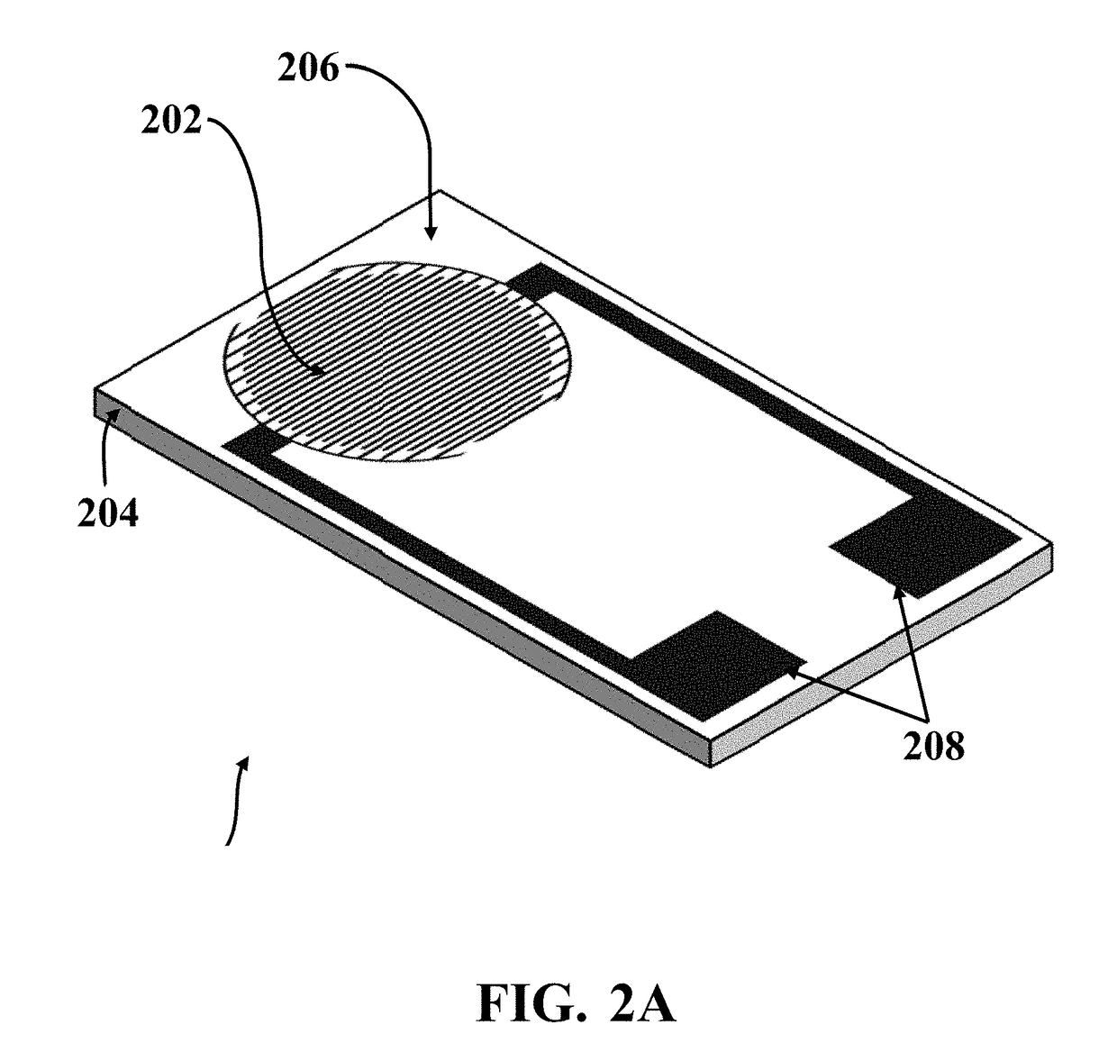
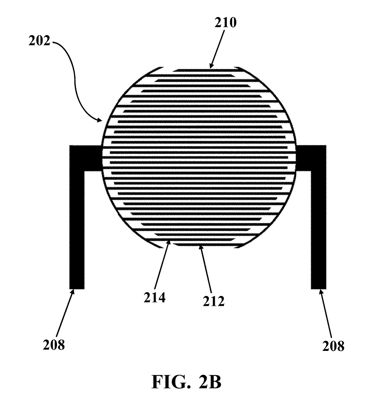
on of SiNWs Impedance Sensor (SiNW-ECIS)

[0086]In this example, exemplary ECISs with SiNWs electrodes array (SiNW-ECIS) similar to exemplary ECIS 200 were fabricated. Silicon wafers, which were used as substrates, were cleaned through standard RCA#1 cleaning method (NH4OH:H2O2:H2O solution and volume ratio of 1:1:5, respectively). Subsequently, a thin layer of SiO2 with a thickness of about 300 nm was grown on the substrate by wet oxidation furnace. A thin layer of gold (Au) with a thickness of about 10 nm was coated on SiO2 layer by a sputtering system at a pressure of about 20 mTorr as a catalyst layer. The gold layer was patterned and the design of the sensor transferred to the substrate. Au-covered samples were located in a low pressure chemical vapor deposition (LPCVD) system with a quartz tube chamber. The growth of silicon nanowires (SiNWs) was a two-step process named as graining and growth. During the graining, a thermal annealing at 450° C.-550° C. for a...

Example

Example 2: Cell Culture and Seeding

[0088]In this example, normal cell line (MCF10 non-cancerous breast epithelial cell line), primary cancerous cell line (MCF7 human breast cancer cell line), and metastatic cell line (MDA-MB-468 human breast cancer cell line) were obtained from a standard cell bank, which were isolated from normal sites, grades I and IV of human breast tumors, respectively. The cell lines were held at about 37° C. in an incubator (about 5% CO2, about 95% air) in RPMI-1640 medium supplemented with about 5% fetal bovine serum, and about 1% penicillin / streptomycin for MCF7 and MDA-MB468 cells and in DMEM-F12 supplemented with about 10% horse serum, about 1% antibiotic / antimitotic solution, about 0.2% NaHCO3, insulin (about 5 μg / ml), EGF (about 10 ng / ml) and hydrocortisone (about 1 μg / ml) for MCF10 cells. The fresh medium was replaced every other day. Standard cell culture methods were used for cell propagation. The cells were counted and suspended in about 100 μl of re...

Example

Example 3: PH Dependent Electrical Impedance of the Cells Measured by SiNW-ECIS

[0093]In this example, the electrical impedances of normal and malignant cells attached onto the SiNWs of exemplary fabricated SiNW-ECIS were measured by an exemplary impedance meter board similar to electrical readout board 304 that was designed and prepared in-house for impedance measurement purposes. Exemplary Impedance meter board was connected through coaxial wires to exemplary SiNW-ECIS including the attached normal and malignant cells onto the SiNWs. Measurements were performed with an applied voltage of about 200 mV. The real time measurements of cellular bioelectrical functions were recorded at desired frequencies (about 0.4 kHz-400 kHz) in the known interval of times. DMEM (including Na+, K+, Mg2+, Ca2+ and etc. ions and amino acid, glucose and etc. buffers) was used as cells' carrier solution. The pH was controlled by low concentrations of HCl with the assistance of a pH meter. The electrical r...


PUM


Abstract

A method for detecting a metastasis state of biological cells is disclosed. The method includes seeding a plurality of biological cells onto an array of electrodes of an electrical cell-substrate impedance sensor (ECIS) by dropping a cell suspension including the plurality of biological cells in a cell culture medium onto the array of electrodes, forming a plurality of cultured biological cells attached onto the array of electrodes by maintaining the ECIS in an incubator, reducing pH value of an extracellular media around the plurality of cultured biological cells to a pH value between 6.2 and 6.7 by dropping an acidic solution onto the array of electrodes, activating an intracellular phenomenon due to reducing pH value of the extracellular media around the plurality of cultured biological cells, monitoring an electrical signal of the plurality of cultured biological cells for a pre-determined period of time, and determining metastasis state of the plurality of biological cells based on the monitored electrical signals. The intracellular phenomenon includes one of autophagy phenomenon in metastatic cells, or cell's proliferation reduction and/or apoptosis in non-metastatic cells. Monitoring the electrical signal of the plurality of cultured biological cells includes applying an electrical voltage to the array of electrodes and extracting a set of time-lapse electrical signals from the array of electrodes. Determining metastasis state of the plurality of biological cells includes identifying a metastatic state for the plurality of biological cells by detecting an increasing trend in the set of time-lapse electrical signals over time, where the increasing trend occurs responsive to activation of the autophagy phenomenon. Determining metastasis state of the plurality of biological cells further includes identifying a non-metastatic state for the plurality of biological cells by detecting a decreasing trend in the set of time-lapse electrical signals over time, where the decreasing trend occurs responsive to activation of the cell's proliferation reduction and/or apoptosis phenomenon.

Description

CROSS-REFERENCE TO RELATED APPLICATION[0001]This application claims the benefit of priority from pending U.S. Provisional Patent Application Ser. No. 62 / 530,300 filed on Jul. 10, 2017, and entitled “TRACING THE PH DEPENDENT ACTIVATION OF AUTOPHAGY IN CANCER CELLS”, which is incorporated herein by reference in its entirety.SPONSORSHIP STATEMENT[0002]This application has been sponsored by Iran Patent Office, which does not have any rights in this application.TECHNICAL FIELD[0003]The present disclosure generally relates to cancer diagnosis, and particularly, to distinguishing invasive tumor cells from non-invasive tumor and / or healthy cells by monitoring the electrical impedance change of the cells due to acidity changes of extracellular media.BACKGROUND[0004]In contrast to normal cells, cancer cells resist against acidic stress by upregulating autophagy as a survival mechanism to maintain their vital functions. Autophagy activates the acidic stress ion channels (ASIC) in cancer cells....

Claims


Application Information

Patent Timeline
IPC IPC(8): G01N33/483G01N27/02
CPCG01N33/4836G01N27/026G01N33/48735G01N2033/4977
Inventor ABDOLAHAD, MOHAMMADALIKHANI, ALIREZAGHAROONI, MILADABIRI, HAMEDFAROKHMANESH, FATEMEH
Owner NANO HESGARSAZAN SALAMAT ARYA
Why Eureka
Try Eureka