Unlock instant, AI-driven research and patent intelligence for your innovation.

Three-Dimensional (3D) Tissue Scaffold with Cell Alignment

a tissue scaffold and three-dimensional technology, applied in the field of three-dimensional tissue scaffolds, can solve the problems of limiting the cell templating of holes, unfavorable cell viability of 3d sheets, and limiting the depth of the device, so as to facilitate the transport of nutrients and waste products throughout the device, and the spread of template cells

Pending Publication Date: 2018-12-06
THE TRUSTEES FOR PRINCETON UNIV
View PDF0 Cites 0 Cited by
  • Summary
  • Abstract
  • Description
  • Claims
  • Application Information

AI Technical Summary

Benefits of technology

The patent describes a method for creating a scaffold model that can recapitulate native tissue properties. The method involves patterning a polymer substrate with a cell-adhesive organometallic layer and a phosphonic acid cell-binding molecule. The substrate is then perforated with a laser to create holes in the substrate. Cells are then attached to the substrate and align with the patterned layer. The resulting 2D construct can be stacked to create a 3D construct that supports cell attachment and alignment. The invention provides a way to create a scaffold model that can recapitulate native tissue properties and has potential applications in regenerative medicine and tissue engineering.

Problems solved by technology

Stacking such sheets in a 3D construct could, however, be unfavorable for cell viability in the depths of the device, i.e., multiple layers down.
This is because lateral transport of oxygen and nutrients from the device periphery to growing cells toward the device's center, and removal of waste products from the center of the device, could be limiting.
If perforations or holes were to be present in each 2D-patterned layer of a 3D construct, transport of nutrients and waste products throughout the device would be facilitated; yet such holes could compromise cell templating.

Method used


Image

  • Three-Dimensional (3D) Tissue Scaffold with Cell Alignment
  • Three-Dimensional (3D) Tissue Scaffold with Cell Alignment
  • Three-Dimensional (3D) Tissue Scaffold with Cell Alignment

Examples


embodiments

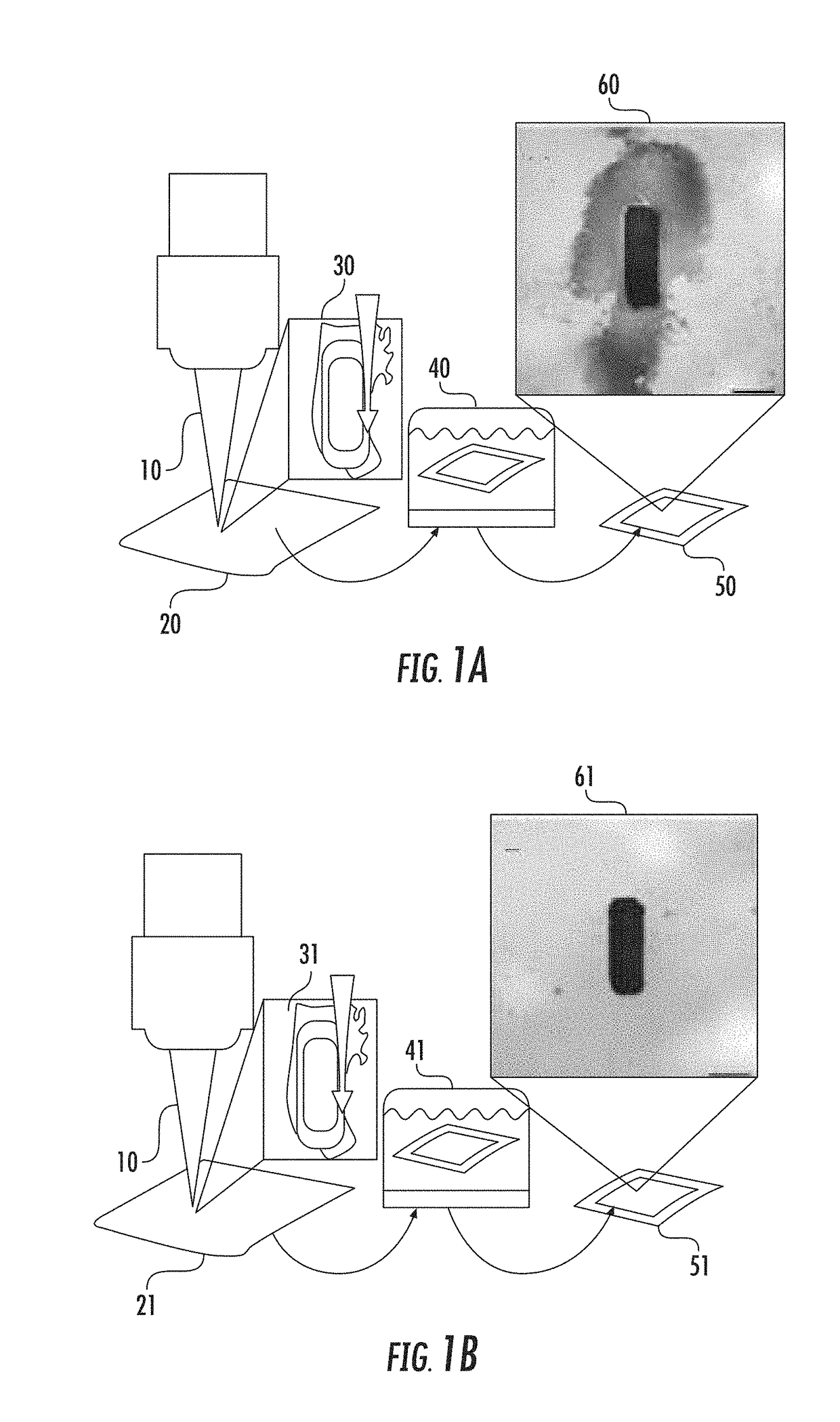
[0019]As disclosed herein, polymeric substrates, such as those in sheet-form, can be perforated by ablation or another method that provides holes through the substrate, where the holes are approximately cell-sized or slightly larger, e.g. about 20-40 microns, up to about 130 microns, and are uncompromised by a debris field when the substrates are first treated with a thin protective layer of photoresist. Preferably the ablation is laser ablation, which cleanly provides holes of controlled size. Polymer substrates, e.g. sheets, perforated with holes comprising 5%, 10%, 20% up to about 30% of the nominal surface area are then patterned in stripes by photolithography, which is followed by synthesis in exposed regions of a cell-adhesive organometallic / α,ω-diphosphonate adduct interface. Preferably the α,ω-diphosphonate is a C3-C16 α,ω-diphosphonate, more preferably 1,4-butanediphosphonic acid or 1,2-dodecanediphosphonic acid. Microscopic and SEM analyses following removal of unexposed p...

example 1

DS Analysis

[0039]Perforated polymer substrates were characterized with an FEI Quanta 200 Environmental-SEM equipped with an Oxford INCA Synergy 450 energy-dispersive X-ray microanalysis system with an X-Max 80 large area analytical silicon drift detector (SDD) at an acceleration voltage of 5 keV. SEM and EDS were performed in low-vacuum mode (0.53 torr) to avoid melting of the polymer by the electron beam.

example 2

on of Polymer Films for Laser Ablation

[0040]PEEK films were cut into 1 cm×1 cm coupons and cleaned by sonication in ethanol for 15 minutes. Films were rinsed with isopropanol and baked at 95° C. to remove moisture. Both sides of the PEEK films were spin-coated with a layer of AZ-5214E photoresist to protect the native polymer surfaces from debris created during the laser ablation process. The photoresist was cured by heating at 95° C. for 45 seconds.


PUM

PropertyMeasurementUnit
Fractionaaaaaaaaaa
Adhesivityaaaaaaaaaa
Surface areaaaaaaaaaaa

Abstract

Polymeric substrates, optionally in sheet form, treated with a thin layer of photoresist are perforated by laser ablation. Following removal of the photoresist the polymer substrates perforated with holes are patterned in stripes by photolithography, which is followed by synthesis of a cell-adhesive organometallic / self-assembled monolayer of phosphonate (SAMP) interface in the exposed regions, providing well-aligned continuous stripes for various levels of perforation. Cells plated on each of these 2-dimensional (2D) perforated surfaces attach to the interface and spread in alignment with pattern fidelity that is as high as that measured on a non-perforated, patterned substrate. A stack of such 2D patterned polymers yields a 3-dimensional (3D) device which facilitates cell growth and viability via the perforations.

Description

CROSS-REFERENCE TO RELATED APPLICATIONS[0001]This application claims the benefit under 35 USC § 119(e) of U.S. Provisional Application No. 62 / 512,257 filed on May 30, 2017, the entire disclosure of which is incorporated herein by reference.STATEMENT REGARDING FEDERALLY SPONSORED RESEARCH OR DEVELOPMENT[0002]The present invention was made with government support under Grant No. DMR-1420541 awarded by the NSF MRSEC. The government has certain rights in the invention.FIELD OF THE INVENTION[0003]The present invention is related to the field of three-dimensional tissue scaffolds capable of supporting cell alignment and growth, suitable for various medical applications.BACKGROUND[0004]A challenge for tissue engineering is to direct cell organization throughout a three-dimensional (3D) space in a 3D device. Ideally such a device should induce cells to adopt the specific, native-like organization characteristic of a particular tissue type that is required for its proper function. Among such...

Claims


Application Information

Patent Timeline
IPC IPC(8): C12M1/12C12N5/00
CPCC12M25/14C12N5/0068A61L27/3891C12N2533/40A61F2002/0086C12M33/00A61L27/18A61L27/3804A61L2400/18C12N2535/10C12N2535/00C08L71/00
Inventor BANDINI, STEPHEN B.SPECHLER, JOSHUAARNOLD, CRAIGSCHWARTZ, JEFFREY
Owner THE TRUSTEES FOR PRINCETON UNIV
Features
  • R&D
  • Intellectual Property
  • Life Sciences
  • Materials
  • Tech Scout
  • Unparalleled Data Quality
  • Higher Quality Content
  • 60% Fewer Hallucinations
Learn More