
Decellularised cell wall structures from plants and fungus and use thereof as scaffold materials

a cell wall structure and plant or fungus technology, applied in the field of scaffold materials, can solve the problems of affecting the quality of the finished product, the risk of disease transmission, and the inability to maintain shape, so as to achieve the effect of reducing the risk of disease transmission, and reducing the cost of the end user

Active Publication Date: 2019-02-28
UNIVERSITY OF OTTAWA
View PDF0 Cites 10 Cited by
  • Summary
  • Abstract
  • Description
  • Claims
  • Application Information

AI Technical Summary

Benefits of technology

This patent describes a biomaterial that is low-cost, efficient, and can be used to produce complex structures. It has the ability to maintain shape, is highly biocompatible, and induces rapid vascularization. The biomaterial can be derived from plants or fungi and is considered organic and biodegradable. It can be produced from food waste, offering an alternative route for discarded produce. The scaffold biomaterial can be implanted at the spinal cord and promote repair or regeneration after spinal cord injury.

Problems solved by technology

Commercial biomaterials often require complicated and time consuming production methods, which leads to a high cost to the end user, even if they are not approved for human use.
In addition, most commercial biomaterials are derived from human / animal origin, resulting in potential rejection by the body and / or adverse immune responses and / or risk of disease transmission.
The source materials can also have negative environmental impact, and can also lead to problems with unethical sourcing.
Also, some commercial biomaterials lose their shape after implantation, which can result in reduced success of the tissue repair / replacement.
However, these approaches are not without some disadvantages [48].
Importantly, resorbable biomaterials suffer from the fact that regenerated tissues often collapse and become deformed due to the loss of structure [58-62].

Method used


Image

  • Decellularised cell wall structures from plants and fungus and use thereof as scaffold materials
  • Decellularised cell wall structures from plants and fungus and use thereof as scaffold materials
  • Decellularised cell wall structures from plants and fungus and use thereof as scaffold materials

Examples


example 1

Experimental Protocol Examples for Scaffold Biomaterial Production

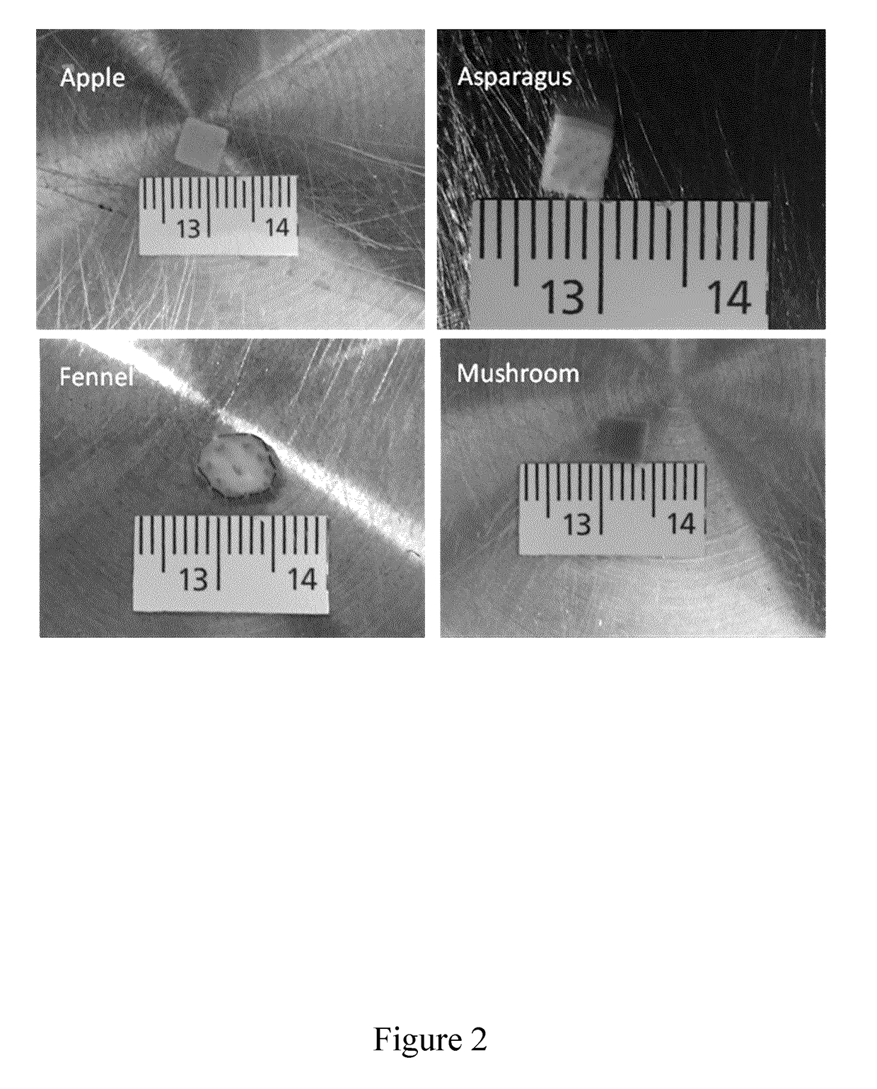
[0176]In this Example, two experimental protocols are described for preparing scaffold biomaterials as described herein from an apple hypanthium tissue (Malus pumila). It will be understood that these protocols are provided as illustrative and non-limiting examples intended for the person of skill in the art. The skilled person having regard to the teachings herein will be aware of various modifications, additions, substitutions, and / or other changes which may be made to these exemplary protocols.

[0177]The initial experimental protocol described below was successfully used for preparing scaffold biomaterials. This protocol, however, took many weeks to provide full cell infiltration under the conditions tested. A modified protocol was, therefore, subsequently developed, which includes the use of a calcium chloride wash (CaCl2), which gave similar results to scaffold biomaterials prepared by the first protocol, but with...

example 2

Mouse Implantation

[0240]In vivo mouse implantation studies were performed to study in vivo effects of scaffold biomaterial embodiments as described herein.

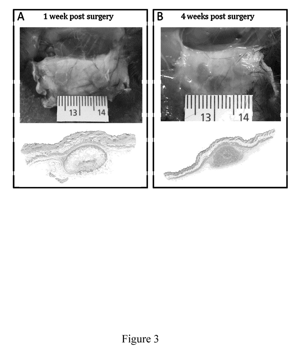
[0241]Results indicate that, following subcutaneous implantation in a mouse model, full cell infiltration was observed (See FIGS. 7; 1, 4 and 8 weeks after implantation), with collagen deposition (FIG. 4A) and, importantly, angiogenesis with functional blood vessel formation within 4 weeks post-implantation (FIGS. 4B and 8). When scaffolds were implanted in vivo, the minimum footprint promoted cell infiltration, angiogenesis and tissue repair and only a minimal inflammatory response (mainly produced by the surgery itself rather than the scaffold). Plant / fungus derived scaffolds were fully biocompatible in vivo in these studies. These scaffolds were also fully compatible with in vitro studies as shown in (FIG. 5).

[0242]A Non-Biodegradable Biomaterial:

[0243]The field has been primarily focused on biodegradable materials; however, th...

example 3

In Vivo Biocompatibility of Scaffold Biomaterials

[0250]To address the question of in vivo biocompatibility of the scaffold biomaterials, the response of the body to apple-derived cellulose scaffolds has been characterized. Macroscopic (˜25 mm3) cell-free cellulose biomaterials were produced and subcutaneously implanted in a mouse model for 1, 4 and 8 weeks. Here, the immunological response of immunocompetent mice, deposition of extracellular matrix on the scaffolds and evidence of angiogenesis (vascularization) in the implanted cellulose biomaterials was assessed. Notably, although a foreign body response was observed immediately post-implantation, as expected for a surgical procedure, only a low immunological response was observed with no fatalities or noticeable infections whatsoever in all animal groups by the completion of the study. Surrounding cells were also found to invade the scaffold, mainly activated fibroblasts, and deposit a new extracellular matrix. As well, the scaffo...


PUM


Abstract

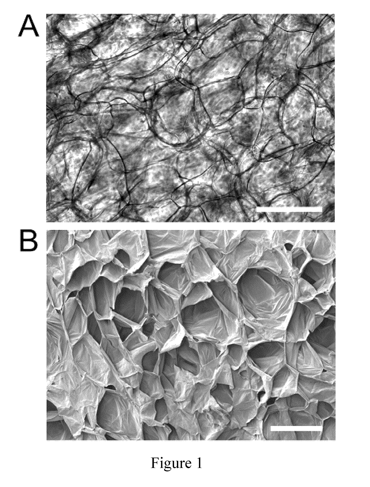
Provided herein are scaffold biomaterials comprising a decellularised plant or fungal tissue from which cellular materials and nucleic acids of the tissue are removed, the decellularised plant or fungal tissue comprising a cellulose- or chitin-based 3-dimensional porous structure. Methods for preparing such scaffold biomaterials, as well as uses thereof as an implantable scaffold for supporting animal cell growth, for promoting tissue regeneration, for promoting angiogenesis, for a tissue replacement procedure, and / or as a structural implant for cosmetic surgery are also provided. Therapeutic treatment and / or cosmetic methods employing such scaffolds are additionally described.

Description

FIELD OF INVENTION[0001]The present invention generally relates to scaffold biomaterials and uses thereof. More specifically, the present invention relates to decellularised plant or fungus tissue, and uses thereof as scaffold biomaterials.BACKGROUND[0002]The biomaterials industry is estimated to have a market value of $90 Billion USD and is driven by novel materials derived from natural sources, synthetic polymers, metals, and ceramics. These materials can form three dimensional high porosity scaffolds possessing nano / microscale structures that are biocompatible and promote the growth of living cells. There is intense interest in novel biomaterials which support the invasion and proliferation of living cells for potential applications in tissue engineering and regenerative medicine, for example.[0003]Biomaterial scaffolds have applications in multiple sectors, including dental and cosmetic surgery, clinical and medical therapies (such as regenerative medicine, wound healing, tissue...

Claims


Application Information

Patent Timeline
IPC IPC(8): A61L27/36A61L27/54A61L27/38
CPCA61L27/3637A61L27/3687A61L27/54A61L27/38A61L2300/414A61L27/3683A61L27/507A61L27/52A61L27/60A61L2430/02A61L2430/16A61L2430/32A61L2430/40A61P27/02A61P43/00A61K8/97C12N5/0062C12N5/0068
Inventor PELLING, ANDREW EDWARDCUERRIER, CHARLES MICHELMODULEVSKY, DANIEL J.HICKEY, RYAN JOSEPH
Owner UNIVERSITY OF OTTAWA
Learn More