

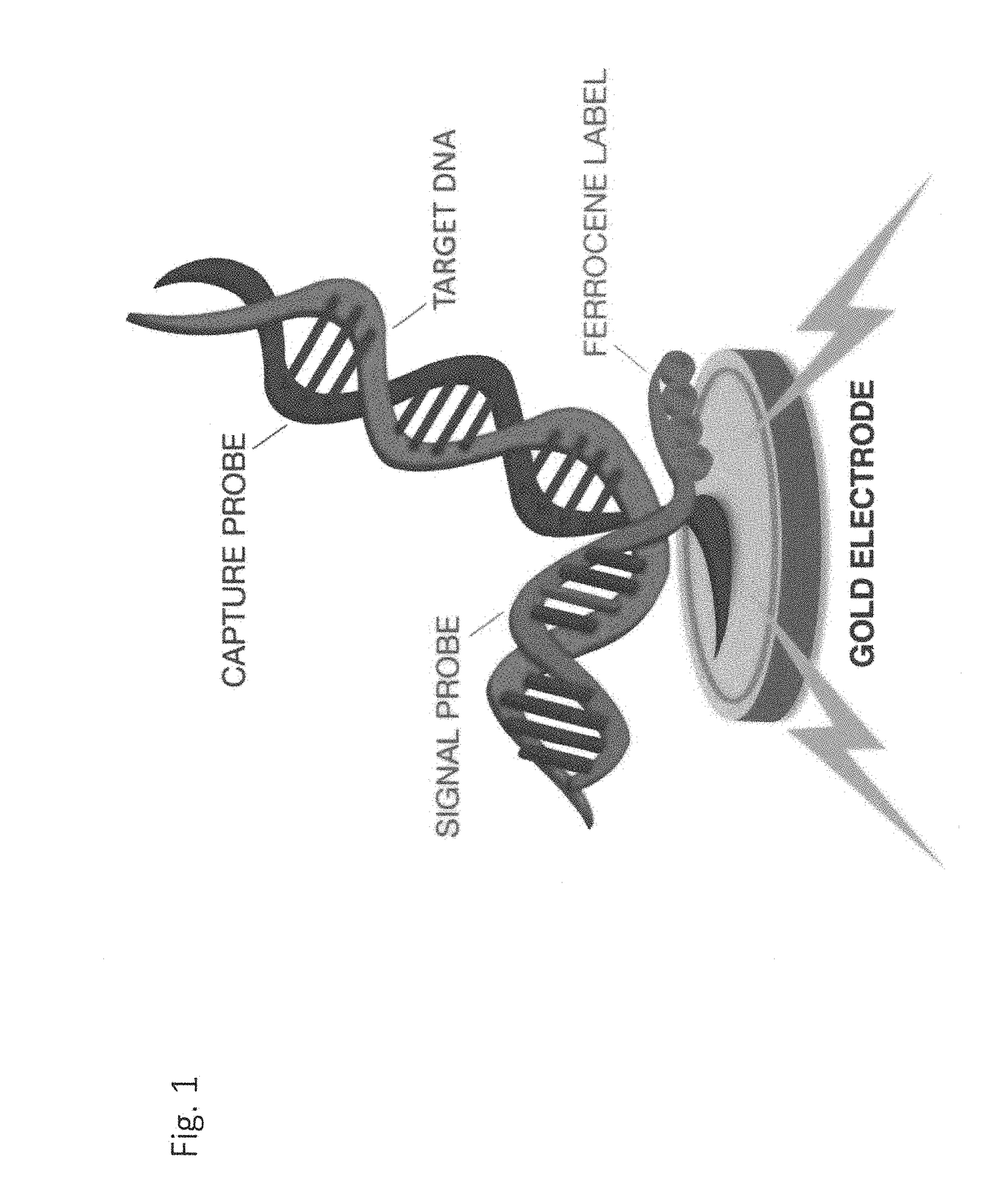
Electrochemical detection of bacterial and/or fungal infections
a technology of bacterial and fungal infections and detection methods, applied in the field of electrochemical detection of bacterial and/or fungal infections, can solve the problems of higher death rate, associated fungal infections, and significant methods, and achieve the effects of improving patient care outcomes, detecting infection, and ensuring the safety of the environmen
- Summary
- Abstract
- Description
- Claims
- Application Information
AI Technical Summary
Benefits of technology
Problems solved by technology
Method used
Image

Examples
example 1
tive Panel, Blood Culture Contamination
[0371]False positive Enterococcus faecalis, Pan-GN and Pan-candida signals were observed when Applicants ran the sLRM GP assay. Applicants investigated whether the blood culture matrix was the source of the contamination.
[0372]Desired blood culture bottles were collected. The rubber sealer of each blood culture bottle was cleaned with ethanol before puncturing it with a needle. 75 uL from each bottle was aspirated. sLRM was performed (Bead beater sample- to lyse cell, add 300 uL lysis buffer, 500 uL binding buffer-wait 2 min, and wash with 150 uL wash buffer). 100% of the washed magnetic beads were loaded onto the cartridge. H1 and H3 (annealing heaters) were run at 61.5° C.
[0373]A preliminary test of NTC sLRMs (bottle matrix with no blood or bacterial targets) showed high false positive signals for Enterococcus faecalis, Pan-candida, and Pan-GN but buffer alone runs did not (see FIG. 4). As a result it was determined that contamination is comi...
example 4
tive, Contamination Mitigation
[0387]Next Applicants evaluated 37-cycle PCR for all targets to reduce or eliminate contamination from blood matrix bottles. Three types of bottles were tested (Bactec Pediatric Plus / F, Bactec Aerobic Plus / F, Bactec Anaerobic Lytic / 10) with and without blood.
[0388]Surprisingly, when PCR cycles are reduced from 40 to 37, most blood matrix contamination is eliminated. FIG. 13.
example 5
tive, Detuning to Eliminate Blood Culture Contamination
[0389]In light of the above experiments, Applicants reduced all cycling to 35 or 30 cycles. Even with the reduction in cycles, false positives were still detected. For example, Corynebacterium was reduced from 40 to 35 and then to 30 cycles but false positives persisted. Applicants then dropped the primer concentration by 50% to 250 nM and the false positives were eliminated. Enterococccus false positives were eliminated when PCR cycles were dropped from 40 to 35 cycles and primer concentration was reduced by 50% to 250 nM. S anginosus false positives were eliminated when PCR cycles were dropped from 40 to 35 cycles and primer concentration was reduced by 75% to 250 nM. The primer concentration for the other targets ranges from 125 to 1000 nM. As summarized in the table below, Applicants were surprisingly able to make their BCID-GP assay less sensitive, to eliminate or reduce detection of contaminants in the sample by “detuning”...
PUM
| Property | Measurement | Unit |
|---|---|---|
| temperature | aaaaa | aaaaa |
| temperatures | aaaaa | aaaaa |
| AC voltage | aaaaa | aaaaa |
Abstract
Description
Claims
Application Information