Self-assembled nanoparticle releasing soluble microneedle structure and preparation method therefor

a nanoparticle and soluble technology, applied in the direction of prosthesis, genetic material ingredients, antibody medical ingredients, etc., can solve the problems of pain and inflammation, difficulty in injection, and difficulty in delivering the needle for direct hypodermic injection, so as to improve the efficiency of simultaneous delivery and facilitate the delivery of water-soluble. , the effect of increasing the solubility in aqueous solution

Pending Publication Date: 2019-04-25
RES & BUSINESS FOUND SUNGKYUNKWAN UNIV
View PDF2 Cites 3 Cited by
  • Summary
  • Abstract
  • Description
  • Claims
  • Application Information

AI Technical Summary

Benefits of technology

[0043]The present invention relates to a self-assembled nanoparticle-releasing soluble microneedle structure and a manufacturing method thereof, and the inventors had attempted to provide a microneedle structure for drug delivery, which can easily deliver a water-soluble or fat-soluble drug, and thereby confirmed that, when a hydrophobic drug was transdermally administered while contained in a microneedle structure without separate preparation using a biocompatible amphiphilic block copolymer having a property of dissolving in both an aqueous solution and an organic solvent, the formation of self-assembled nanoparticles results in an increase in drug solubility in an aqueous solution and activation of intracellular delivery, and thus the present invention was completed.
[0044]According to the present invention, an aqueous or hydrophobic drug can be delivered while loaded in microneedles, and particularly, a fat-soluble drug is delivered while loaded in micelle-type self-assembled nanoparticles formed by the dissolution of the structure. Accordingly, the solubility in an aqueous solution can be greatly increased, such that an existing drug with poor absorption is able to be delivered to the body through the skin, and thus it is expected to be useful for enhancing the efficiency of simultaneous delivery of a vaccine antigen and a hydrophobic vaccine adjuvant.

Problems solved by technology

Generally, one of the methods of delivering a drug, that is, a needle for direct hypodermic injection has disadvantages of pain and inflammation.
In addition, bleeding may occur, and injection may be difficult according to age or a characteristic.
However, such water-soluble microneedles were i) very limited in loading of a hydrophobic drug due to the water-soluble property of a microneedle structure, and ii) exhibited toxicity in a corresponding site due to limited diffusion caused by precipitation in a site in which dermal administration occurred.
In addition, in the case of drug-coated needles, since a drug release is adjusted by rapid dissolution and simple diffusion, it is difficult to control a biological reaction, and when a needle structure is broken after use, the structure remains and thus has a risk of infection.
Mostly, the inoculation of a vaccine is generally performed by subcutaneous, intradermal or intramuscular injection using a syringe, thereby decreasing the compliance of a patient, and the support of professional medical personnel is necessary.
However, the subunit vaccine generally has lower immunogenicity than a conventional live vaccine or killed (inactivated) vaccine, and is used by mixing an adjuvant for increasing an immune response with a vaccine antigen.
While the most generally used adjuvant, an aluminum salt, is employed in most commercially available vaccines, it has been known that the adjuvant is not suitable for cancer vaccines necessary for a cell-mediated immune response because it generally serves to induce a Th2-type immune response, has excellent antigen-mediated immune response activity, but does not have a cell-mediated immune response.
While conventional water-soluble microneedles are suitable for vaccine antigen delivery, in the case of a hydrophobic adjuvant, it was difficult to manufacture a needle structure because of the water solubility of the structure, and also difficult to deliver a vaccine antigen to immune cells due to precipitation at an administration site after transdermal delivery.

Method used


Image

  • Self-assembled nanoparticle releasing soluble microneedle structure and preparation method therefor
  • Self-assembled nanoparticle releasing soluble microneedle structure and preparation method therefor
  • Self-assembled nanoparticle releasing soluble microneedle structure and preparation method therefor

Examples


example 1

re of Microneedles

[0065]After a tri-block copolymer, Pluronic F127, was dissolved in ethanol to the final concentration of 15%, a solution in which a hydrophobic molecule was dissolved in ethanol (1,1′-dioctadecyl-3,3,3′,3′-tetramethylindodicarbocyanine iodide (DiD) or Resiquimod (R848)) was uniformly mixed, and then an ethanol solvent present in the solution was removed using a rotary evaporator.

[0066]A film was obtained, the remaining solvent was completely removed by evaporation using nitrogen, a previously prepared aqueous solution containing polyethylene glycol (PEG MW 6000) and a hydrophilic molecule, OVA, was added to the film, and the film was uniformly dispersed in the aqueous solution using a sonicator, followed by filtering the aqueous solution to remove an undissolved material.

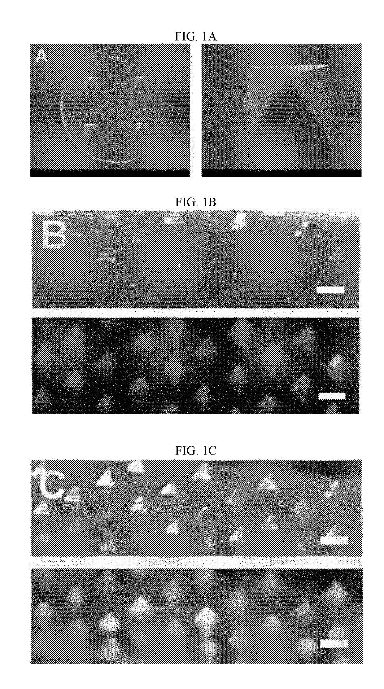
[0067]Soluble microneedles were manufactured by injecting 0.15 ml of the aqueous solution into a reusable negative-patterned template for a polydimethylsiloxane (PDMS) microneedle, which has a size...

example 2

on of Microneedles

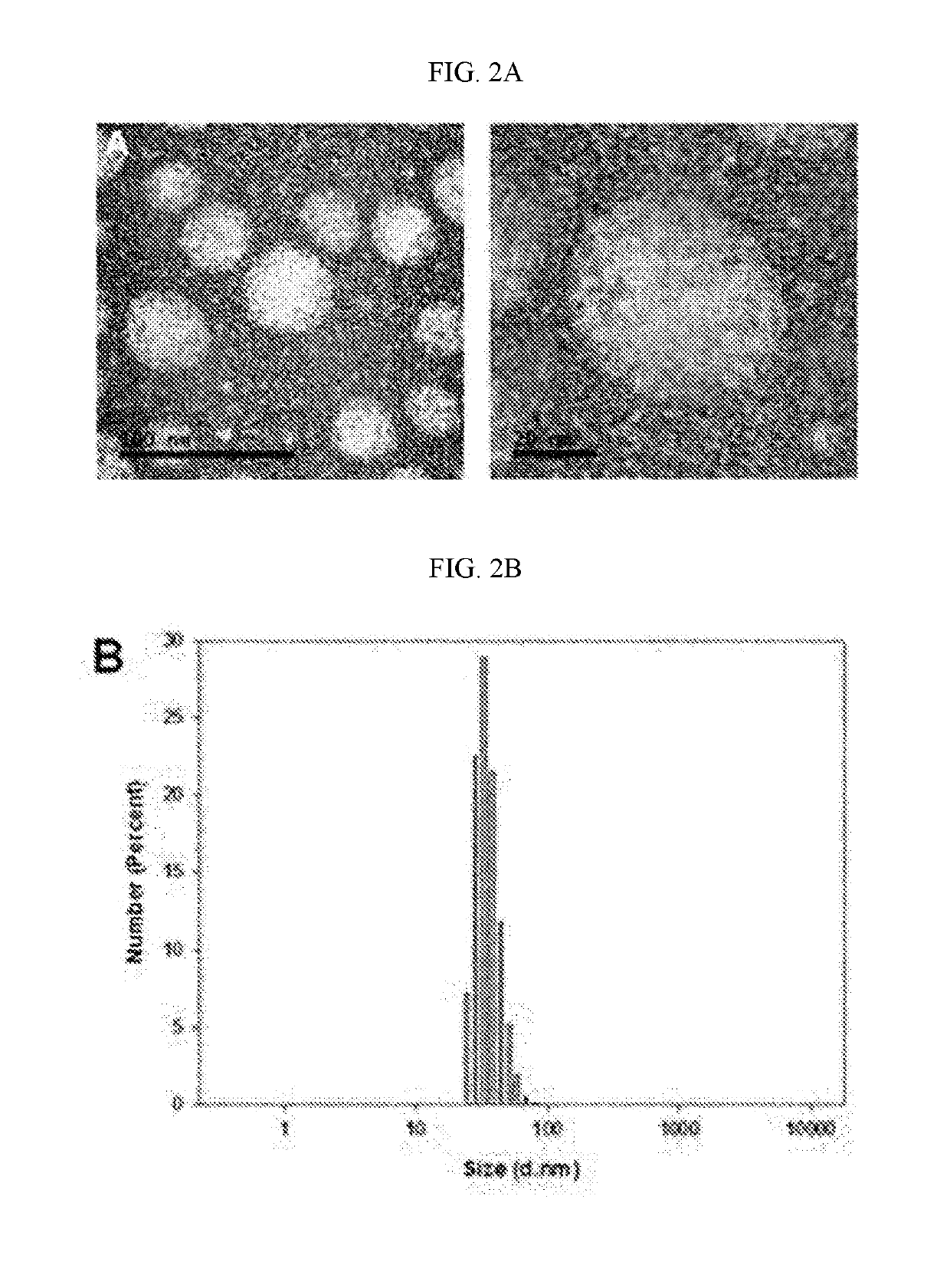
[0069]Spherical micelle-type self-assembled nanoparticles can be formed in an aqueous solution using the Pluronic F127 used in Example 1 as an amphiphilic tri-block copolymer (see the description on the polymeric micelles of FIG. 2A).

[0070]A paraffin film (Parafilm®) was placed on a Styrofoam support, microneedles were applied and then pressed vertically, and then the film and the microneedles were separated from the Styrofoam support. Afterward, the film perforated by the microneedles and the microneedles were floated on the water surface in a Petri dish (Ø=30 mm) containing 500 μl of distilled water to dissolve the microneedles, and 30 minutes later, the corresponding solution was taken and then dried on a TEM grid (formvar coated). As a result, as shown in FIG. 2A, formation of spherical particles could be confirmed using a transmission electron microscope.

[0071]In addition, as shown in FIG. 2B, it was observed that the size of a micelle in the solution analyzed...

example 3

rofiles for OVA and Resiquimod (R848)

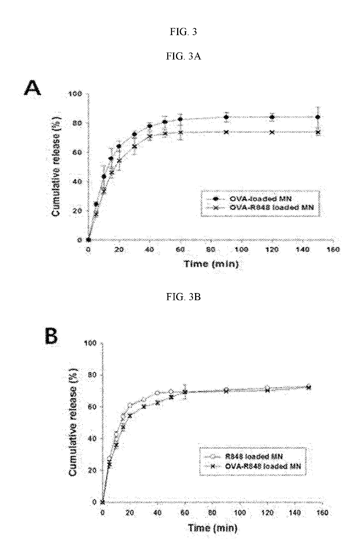
[0072]To observe the profiles of releasing OVA and Resiquimod (R848) from the soluble microneedles manufactured by the method of Example 1, the soluble microneedles were put into phosphate buffered saline (PBS, pH 7.4), stored at 37° C., and then a sample was obtained at predetermined intervals (0, 1, 2, 3, 4, 5, 10, 20, 30, 60, and 90 minutes), followed by replacement with the same volume of a new release solution.

[0073]As a result, as shown in FIG. 3A, an OVA release profile from each of OVA only-containing microneedles (OVA-loaded MN) and OVA / R848-containing microneedles (OVA-R848 loaded MN) was able to be identified by the bicinchoninic acid (BCA) assay (micro plate reader, Multiskan GO, Thermo Fisher Scientific, Vantaa, Finland) at 590 nm.

[0074]In addition, the absorbance with respect to R848 was calculated and quantified at 327 nm using a UV-Vis scanner (TECAN Infinite M500 microplate reader), confirming that, as shown in FIG. 3B, an R848 r...


PUM

PropertyMeasurementUnit
diameteraaaaaaaaaa
micelle diameteraaaaaaaaaa

Abstract

The present invention relates to a self-assembled nanoparticle releasing microneedle structure which is formed of biocompatible amphiphilic block copolymers containing a drug, and a preparation method therefor. The microneedle structure according to the present invention can deliver a water-soluble or hydrophobic drug while being carried in a microneedle. In particular, since a fat-soluble drug is delivered while being carried by micelle-type self-assembled nanoparticles which are formed as the structure is dissolved, it is possible to greatly increase the solubility in an aqueous solution. As such, existing drugs with poor absorption can be delivered through the skin of a body.

Description

TECHNICAL FIELD[0001]The present invention relates to a microneedle structure using a backbone material for a water-soluble structure capable of releasing drug-loaded self-assembled nanoparticles by dissolution, and a method of manufacturing the same.BACKGROUND ART[0002]Generally, one of the methods of delivering a drug, that is, a needle for direct hypodermic injection has disadvantages of pain and inflammation. In addition, bleeding may occur, and injection may be difficult according to age or a characteristic. Therefore, as an alternative thereto, microneedles have been actively studied because they can minimize pain, bleeding or inflammation, and enable local injection of a drug to be effectively and continuously input only to a site for injection.[0003]Recently, attempts have been made to manufacture microneedles using a harmless biodegradable material. Particularly, there were increasing attempts to manufacture soluble microneedles using a polysaccharide or water-soluble polym...

Claims


Application Information

Patent Timeline
Patent Type & Authority Applications(United States)
IPC IPC(8): A61K9/00A61K9/51A61K47/34A61K31/437A61K39/00
CPCA61K9/0021A61K9/5146A61K47/34A61K31/437A61K39/0005A61K31/4745A61K39/0011A61K2039/54A61K2039/55511A61K9/1273A61K39/395A61K48/00A61L27/26A61L27/54A61L27/58A61M37/0015A61L2400/12A61M2037/0046
Inventor JEONG, JI HOONKIM, NAK WON
Owner RES & BUSINESS FOUND SUNGKYUNKWAN UNIV
Why Eureka
Try Eureka