

A method to enhance wound healing using silk-derived protein
a technology of silk-derived protein and wound healing, which is applied in the direction of peptide/protein ingredients, prosthesis, drug compositions, etc., can solve the problems of corneal regenerative properties that cannot restore a healthy epithelial surface and cannot reconstitute critical barrier functions, etc., to promote wound healing and enhance wound healing
- Summary
- Abstract
- Description
- Claims
- Application Information
AI Technical Summary
Benefits of technology
Problems solved by technology
Method used
Image

Examples
example 1
ration and the Lawrence Stability Test
[0137]Materials.
[0138]Silkworm cocoons were obtained from Tajima Shoji Co., Ltd., Japan. Lithium bromide (LiBr) was obtained from FMC Lithium, Inc., NC. An autoclave was obtained from Tuttnauer Ltd., NY. The 3,500 Da molecular-weight cutoff (MWCO) dialysis membranes were obtained from ThermoScientific, Inc., MA. An Oakton Bromide (Br) double-junction ion-selective electrode was obtained from ISE, Oakton Instruments, IL.
[0139]Processing.
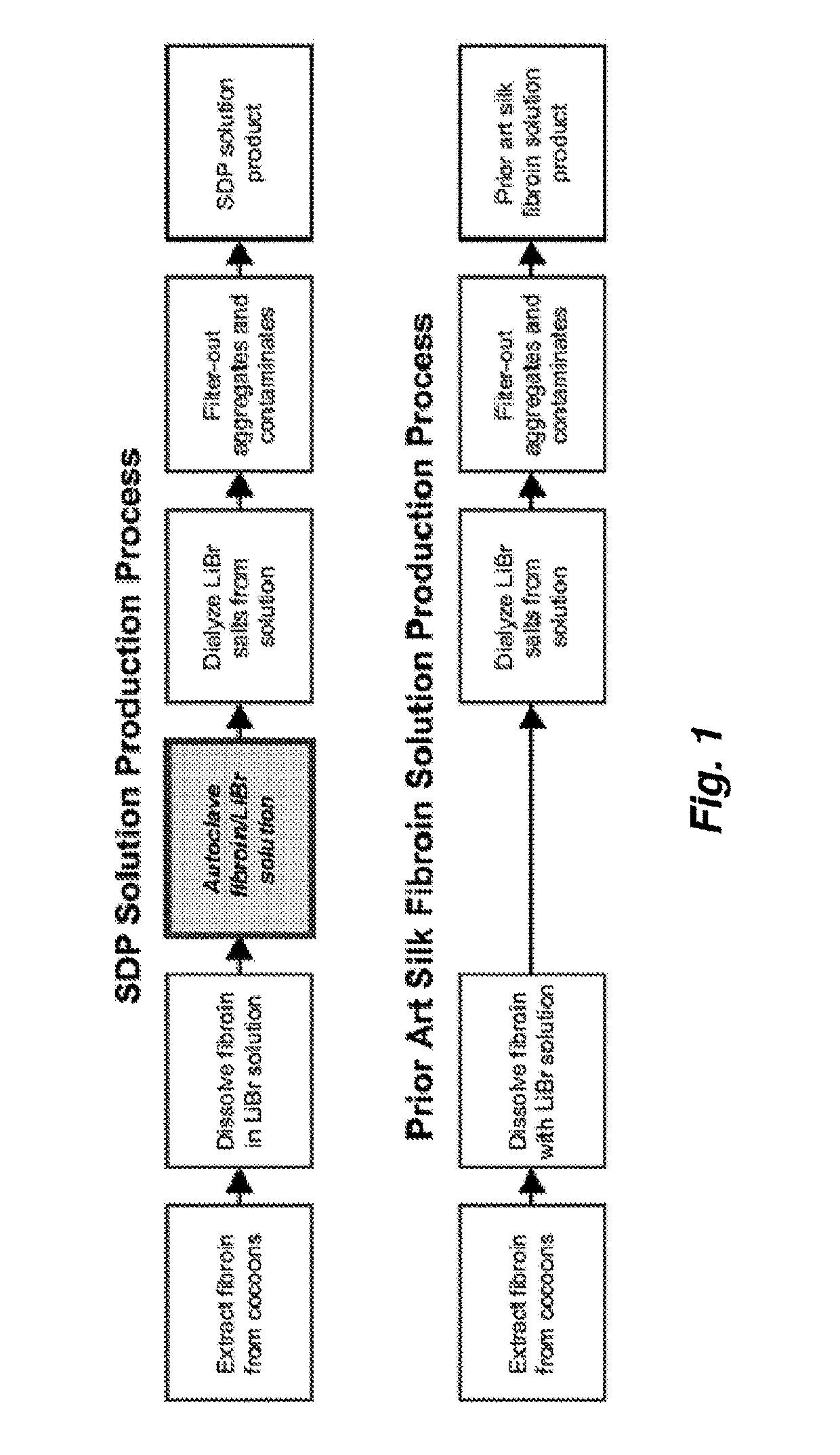
[0140]Two samples, SDP and PASF, were prepared as illustrated in FIG. 1. Briefly, SDP was produced by submerging pupae-free, cut silkworm cocoons (3-5 cuts / cocoon) into 95° C. heated, deionized water (diH2O) containing 0.3 wt % NaCO3 at 233 mL water / gram of cocoons. Cocoons were agitated in this solution for 75 minutes to dissolve sericin, thereby separating it from the silk fibers. The cocoons were subsequently washed four times in like dilutions of diH2O for 20 minutes per rinse to remove residual sericin from t...
example 2
ular Weight Characterization
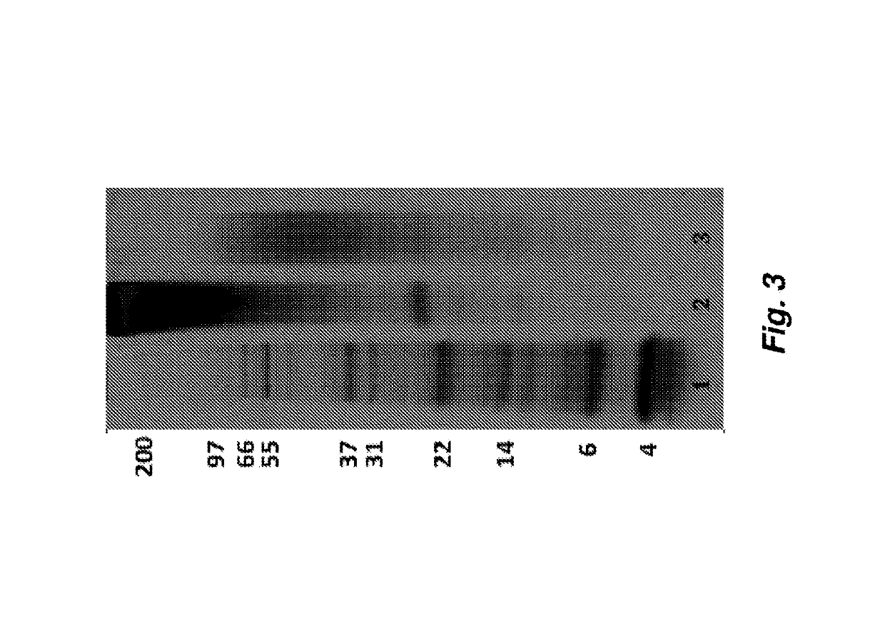
[0144]To evaluate the effect of processing on the molecular weight distribution of solubilized protein, SDP Solution and PASF Solution were subjected to polyacrylamide gel electrophoresis (PAGE), which separates proteins by molecular weight. Specifically, 15 μg of each sample was mixed with running buffer containing sodium dodecyl sulfate and dithiothreitol (Biorad Inc., CA) to remove any secondary folding structures and disulfide bonds, respectively. The mixtures were then heated to 70° C. for 5 minutes. The mixtures were loaded along with a 2.5-200 kDa molecular weight ladder (Life Technologies, CA) onto pre-cast, 4-12% polyacrylamide gradient gels containing Bis-Tris buffer salts (Life Technologies, CA), and then exposed to 120V electric field for 90 minutes on a BioRad PowerPac Power supply (BioRad Inc., CA). The gels were then removed and placed in Coomassie Blue stain for 12 hours to stain proteins, followed by 6 hours of washing in diH2O. The gels ...
example 3
lity Study
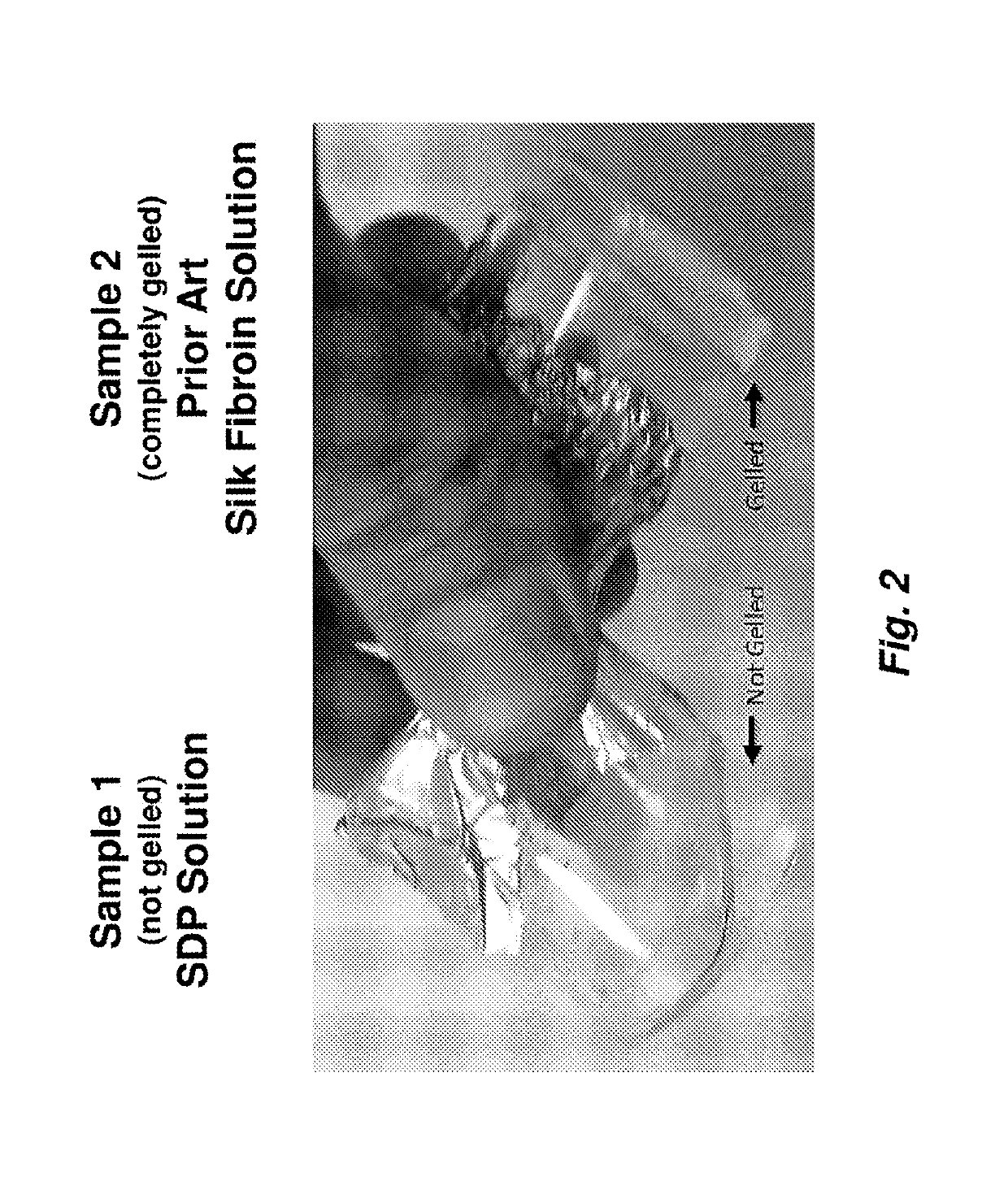
[0146]To further determine the functional impact of the autoclave process on the stability of the resulting SDP compared to the stability of prior art fibroin, the samples were analyzed using the methods of Wang et al. (Biomaterials 2008, 29(8):1054-1064) to mimic a well-characterized model of silk fibroin protein gelation. Volumes of both samples (0.5 mL, SDP and PASF) were added to 1.7 mL clear centrifuge tubes and were subjected to ultrasonication (20% amplitude, 10 Hz, 15 seconds). The clear tubes containing the solutions were then visually monitored for gel formation as a screen for gelation.
[0147]The SDP Solution samples failed to form gels, as shown in FIG. 4A. Even 3 months post-sonication, the SDP samples remained in solution and lacked protein aggregation as determined by visual inspection. The PASF Solution sample gelled rapidly (within 2 hours) following sonication. The resulting gelled PASF is shown in FIG. 4B. These results further indicate that the autoclave...
PUM
| Property | Measurement | Unit |
|---|---|---|
| Fraction | aaaaa | aaaaa |
| Fraction | aaaaa | aaaaa |
| Fraction | aaaaa | aaaaa |
Abstract
Description
Claims
Application Information