
Diagnostic methods and kits for early detection of ovarian cancer

a diagnostic method and ovarian cancer technology, applied in the field of cancer diagnosis, can solve the problems of ineffective screening programs and early detection, low sensitivity rate, and no effective improvement of survival

Pending Publication Date: 2020-01-30
TEL HASHOMER MEDICAL RES INFRASTRUCTURE & SERVICES +1
View PDF0 Cites 4 Cited by
  • Summary
  • Abstract
  • Description
  • Claims
  • Application Information

AI Technical Summary

Benefits of technology

The invention provides a method for predicting the future course of a disease or disorder, particularly ovarian cancer, by analyzing the expression of biomarker proteins. This allows for early diagnosis and personalized medicine, as treatment regimes can be determined based on the expression value of the biomarker proteins. The method requires at least two samples to be collected from the subject and analyzed. The samples can be collected at different stages of the disease and can include samples from the same patient. The method can be used for monitoring disease progression or relapse. The term "about" refers to values that may vary up to 1% higher or lower than the indicated value. The patent text also mentions the use of the phrase "consisting of" to include additional ingredients or steps as long as they do not materially alter the basic and novel characteristics of the claimed composition or method.

Problems solved by technology

This grim reality stems primarily from the lack of effective screening programs and of early stage-specific biomarkers.
A multitude of biomarkers have been proposed and tested over the years but none have shown to be effective in improving survival [2-5].
Low predictive value stems from the correlation of blood-borne proteins with tumor volume, and hence failure to diagnose the earliest, potentially curable lesions before they have disseminated beyond the ovary.
Given that these studies recruited mostly advanced-stage HGOC patients, these sensitivity rates are considered too low.
However, deep proteomic profiling of any body-fluid is challenged by the vast dynamic range of their proteomes and the masking of low abundance biomarkers by core plasma proteins, such as albumin, IgG, hemoglobin etc.

Method used


Image

  • Diagnostic methods and kits for early detection of ovarian cancer
  • Diagnostic methods and kits for early detection of ovarian cancer
  • Diagnostic methods and kits for early detection of ovarian cancer

Examples


example 1

[0392]Patients' Characteristics

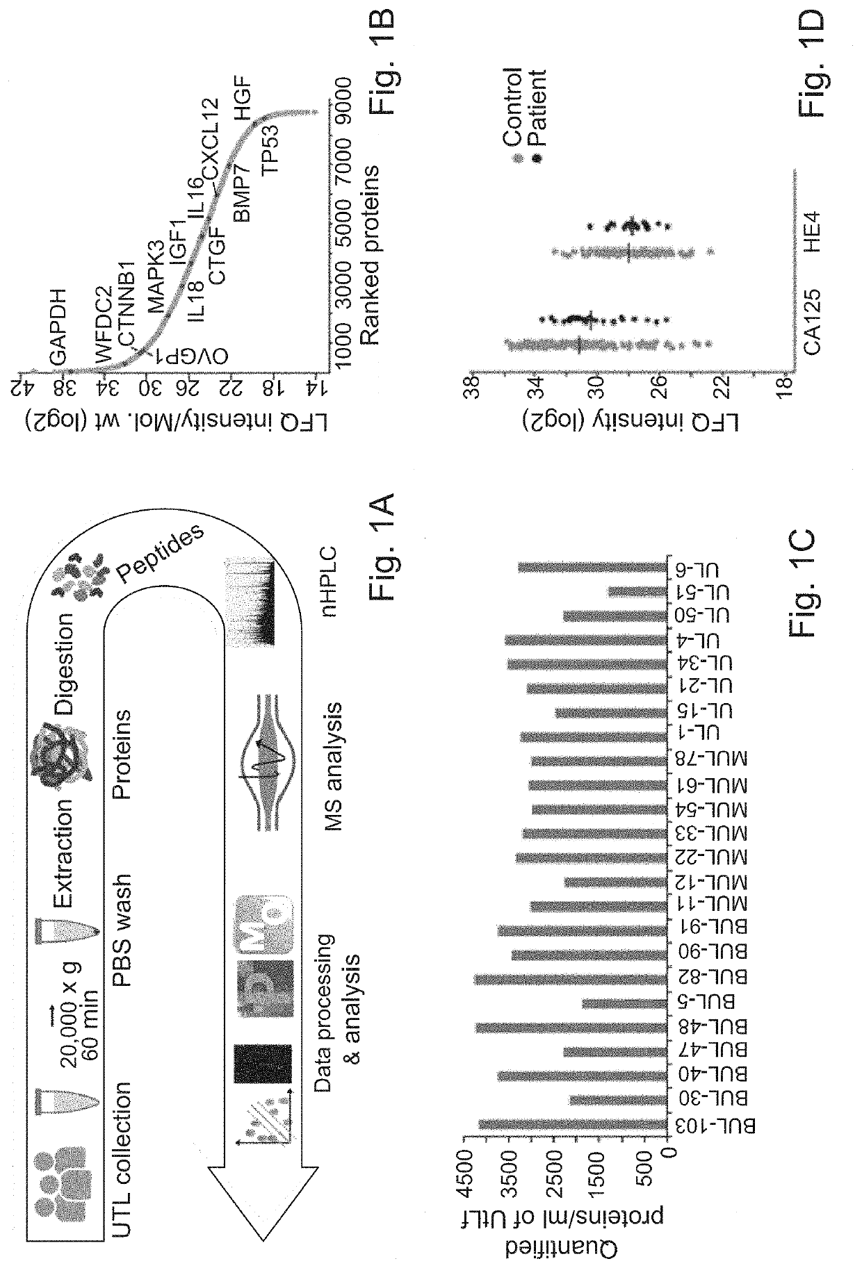
[0393]Aiming to identify early-stage biomarkers for HGOC, it was hypothesized that “localized liquid biopsy” such as UtL sampling is likely to have better sensitivity and specificity than serum biomarkers. To that end, a set of 212 UtL samples from 208 enrolled donors was analyzed (Tables 1 and 2). Eleven samples were excluded due to missing data (n=1), inappropriate ovarian tumor histological subtype (n=8), or failing the quality control measures (n=2). The discovery set (n=24) consisted of UtL samples from 12 HGOC patients and 12 representative controls from all participating medical centers, while all subsequent samples were regarded as a validation set (n=152), and analyzed independently in a blinded manner. Overall, 49 UtL samples were obtained from HGOC patients (patient cohort', average age=61.8). Of those, 27 samples were obtained at primary debulking surgery and the other 22 were obtained at interval debulking surgery, after 3 cycles of platin...

example 2

[0394]UtL microvesicle Proteomic Profiling

[0395]In order to profile the proteome of a complex body fluid and detect potential diagnostic biomarkers, the challenge inflicted by the existence of highly abundant proteins had to be overcome. Therefore the previously developed method for microparticle isolation from plasma was examined for the application to UtL samples. Therefore, microvesicles were isolated from UtL by high speed centrifugation followed by PBS wash to remove albumin contamination. The microvesicles and their protein content were denatured with urea, followed by trypsin protein digestion and LC-MS / MS analysis as illustrated by the scheme of FIG. 1A. Analysis of the entire discovery cohort identified a total of 8578 UtLF microvesicle proteins and an average number 3000 per sample (range: 1500-4000) (FIG. 1C). Among the identified proteins, known FTE / HGOC proteins were found, such as MUC16 (CA125), WFDC2 (HE4), and OVGP1 (MUC9), as well as lower abundance proteins, includ...

example 3

[0396]Identification of Protein Signature

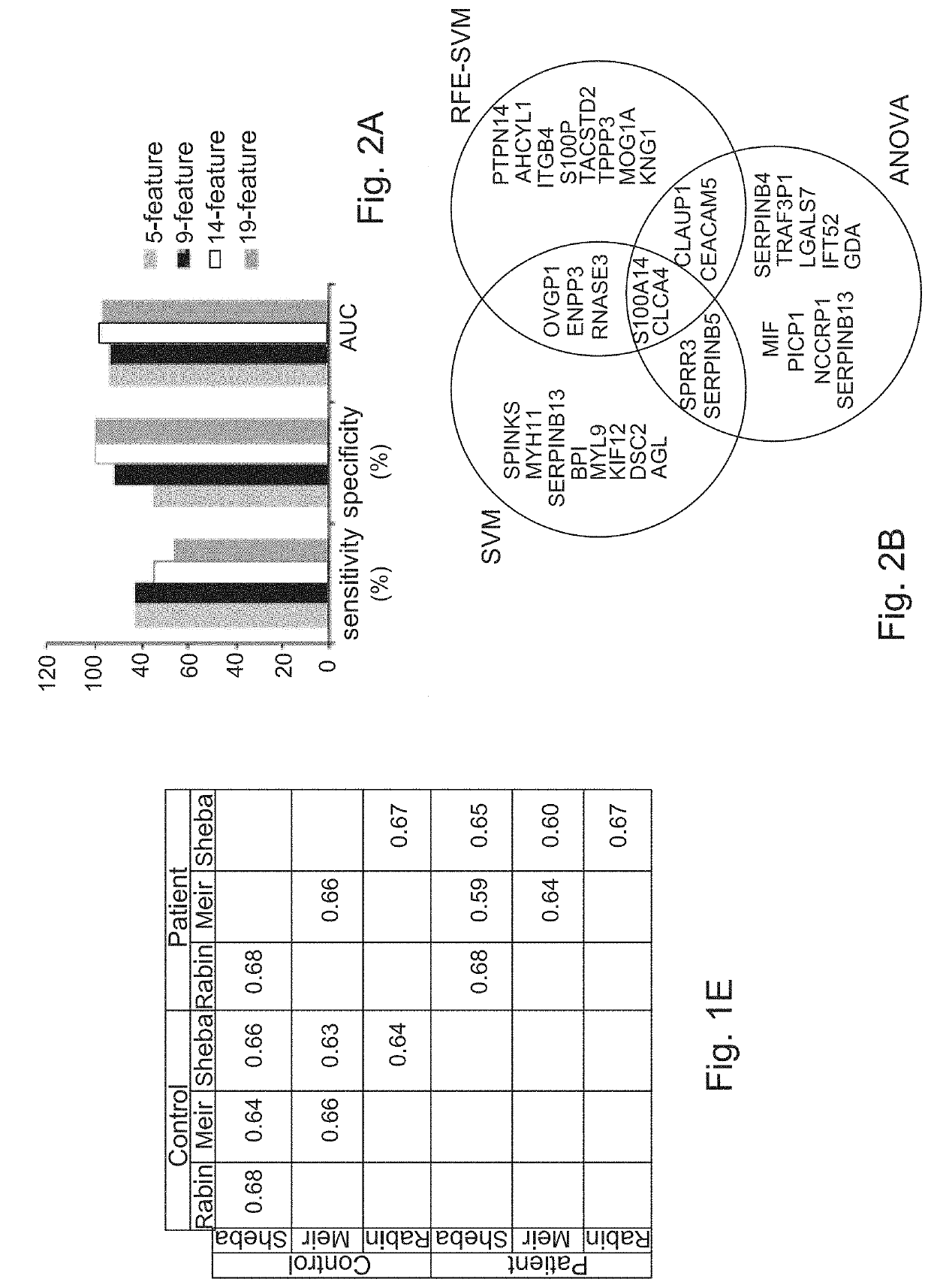
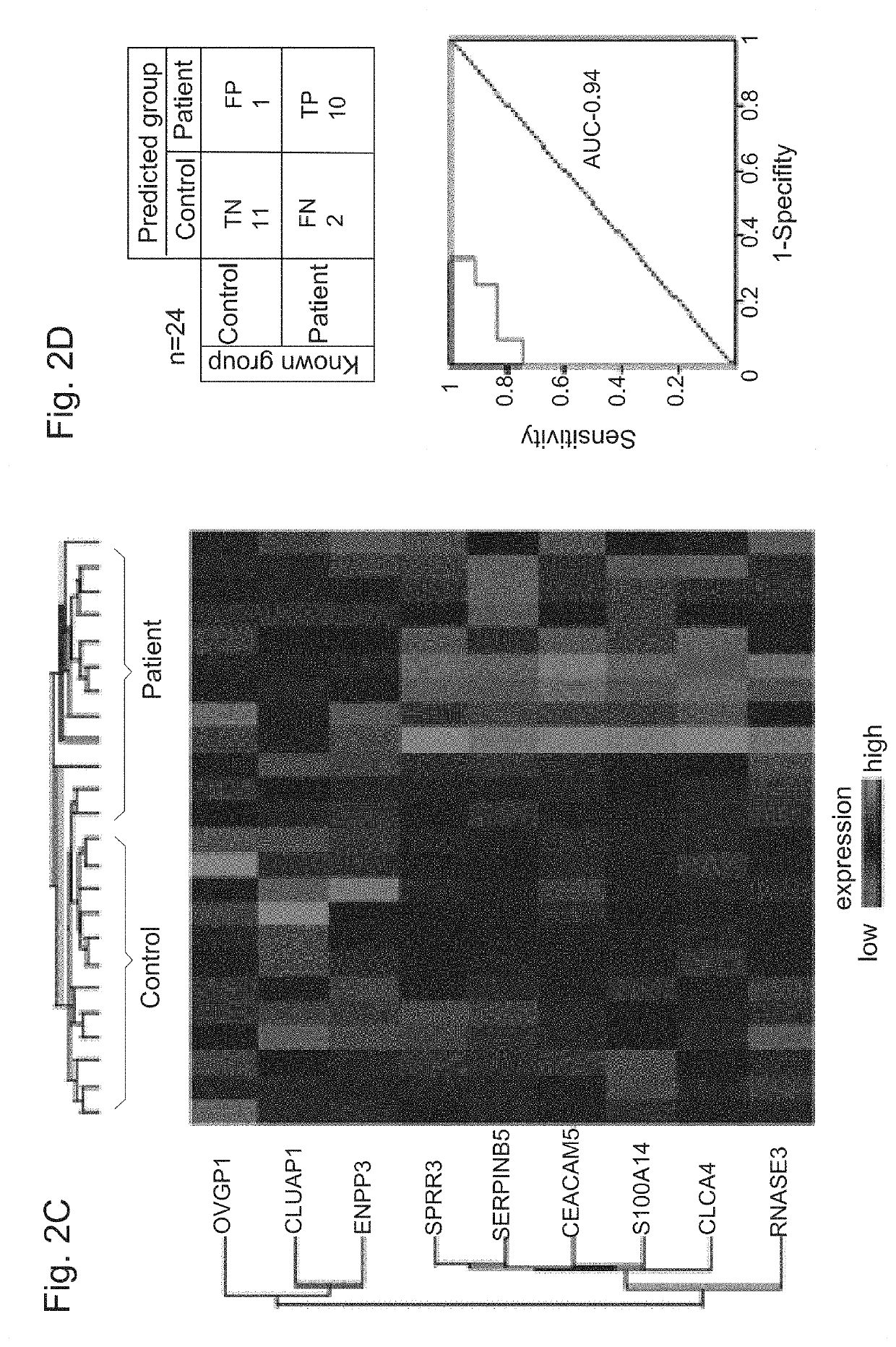
[0397]Next, the proteomic profiles of 24 patients and controls (discovery cohort') were used to construct a protein classifier for HGOC diagnosis. Support vector machine algorithm was used to classify the samples, and optimized the minimal number of features (proteins) that provide highest accuracy. For feature selection, 3 different algorithms were applied to the discovery cohort MS-datasets, SVM, RFE-SVM and ANOVA. The entire analytical workflow was embedded in a cross validation procedure to reduce over-fitting in order to identify a signature with a minimal number of proteins, a high predictive power, and a least dependence on the feature selection algorithm. The performance of several sets of top-ranked overlapping proteins, ranging in size from 5 to 19 features (FIG. 2A, 2B) was therefore examined. Optimal sensitivity, specificity, and area under the curve (AUC) of Receiver Operating Characteristic (ROC) curve of sensitivity vs. 1-speci...


PUM

PropertyMeasurementUnit
diameteraaaaaaaaaa
diameteraaaaaaaaaa
volumeaaaaaaaaaa

Abstract

The invention relates to novel biomarker signature, diagnostic methods, kits and compositions for early diagnosis of ovarian cancer, based on microvesicles prepared from body fluid sample, specifically, uterine lavage fluid (UtLF) sample.

Description

FIELD OF THE INVENTION[0001]The invention relates to diagnosis of cancer. More specifically, the present invention provides novel biomarker signature, diagnostic methods, kits and compositions for early diagnosis of ovarian cancer.BACKGROUND ART[0002]References considered to be relevant as background to the presently disclosed subject matter are listed below:[0003][1] Vaughan S, et al., Nat. Rev. Cancer 11: 719-725 (2011)[0004][2] Havrilesky L J et al., Gynecol Oncol 110:374-382 (2008).[0005][3] Kozak K R, et al., Proteomics 5:4589-4596 (2005)[0006][4] Bast Jr. R C, et al., Int J Gynecol Cancer 15 Suppl 3:274-281 (2005)[0007][5] Moore L E, et al., Cancer 118:91-100 (2012)[0008][6] Sarojini S, et al., J Oncol 2012:709049 (2012)[0009][7] Moore R G, et al., Gynecol Oncol 112:40-46 (2009)[0010][7] Freydanck M K, et al., Anticancer Res 32:2003-8 (2012)[0011][9] Lu K H, et al., Cancer 119:3454-61 (2013)[0012][10] Stukan M, et al., J Ultrasound Med 34:207-17 (2015)[0013][11] Jacobs I J, et...

Claims


Application Information

Patent Timeline
Patent Type & Authority Applications(United States)
IPC IPC(8): G01N33/574G01N33/563G01N33/543
CPCG01N33/563G01N33/54306G01N33/57449C12Q1/6886G01N33/57488C12Q1/6816C12Q2600/158C12Q2525/205
Inventor LEVANON, KERENGEIGER, TAMARBAHAR-SHANY, KERENBARNABAS, GEORGINA D.HELPMAN, LIMORKORACH, JACOBPERRI, TAMAR
Owner TEL HASHOMER MEDICAL RES INFRASTRUCTURE & SERVICES
Learn More