Nanonets and spherical particles

a macroscale nanomaterial and nanotechnology, applied in the field of macroscale macromolecule complexes comprising microns, can solve the problems of reducing peptide/protein reactivity and limited surface display systems, and achieve the effect of rapid surface presentation and effective production of bioactive 2d/3d macromolecule nanomaterials

Active Publication Date: 2020-06-18
JAMES HUTTON INSTITUTE
View PDF0 Cites 3 Cited by
  • Summary
  • Abstract
  • Description
  • Claims
  • Application Information

AI Technical Summary

Benefits of technology

[0017]Advantageously, these components or virus like particles can be obtained to high yields. Further, advantageously coat proteins (CPs) or other components (not limited to phage tail fibres, or plant virus movement proteins) can be manipulated to surface display peptides, proteins and / or enzymes which confer high catalytic / reactive activities of interest to the virion or structures surface. In a suitable advantageous embodiment, surface display of peptides / proteins / enzymes on characterised coat proteins for example, can greatly enhance the stability of the enzymes / proteins and permit provision of very dense reactive surface areas after self-assembly.
[0048]Suitably, a macroscale network may be derived from TMV coat proteins or TMV VLPs assembled from coat proteins. In embodiments the macroscale network can be formed from a microstructure comprising viral coat proteins or VLPs, suitably, wherein the viral coat proteins may comprise motifs which allow the viral coat proteins or VLPs to coalesce to form networks. The networks formed may not appear to comprise obvious virus like particles and may appear to be aggregated strings of coat proteins. These networks may be formed by at least one of insertion of amino acid sequences or motifs into the coat protein, induction of bacterial expression and protein isolation. Suitably such structures may be modified by introducing a SpyTag into the C-terminal end of the TMV CP. Advantageously, introduction of a SpyTag into the C-terminal end of the TMV-CP may provide large TMV network structures (devoid of nucleic acid), when expressed in bacteria. SpyTag at the C-terminal end of TMV CP can suitably form covalent linkages with its SpyCatcher interacting partner which may be suitably surface displayed on GFP spherical particles. Suitably networks, including SpyTag, may be decorated with surface displayed SpyCatcher, for example GFP spherical particles to provide different functional proteins (or enzymes), effectively producing bioactive 2d / 3d macroscale nanomaterials.

Problems solved by technology

However, this system for surface display is limited by the size of the peptide that can be inserted.
However, a disadvantage of chemical linkage approaches is that they may provide too much non-specific cross-linkage which may reduce peptide / protein reactivity, or alternatively / additionally there may be the requirement for additional clean up steps.

Method used


Image

  • Nanonets and spherical particles
  • Nanonets and spherical particles
  • Nanonets and spherical particles

Examples


example 1

and Plasmids

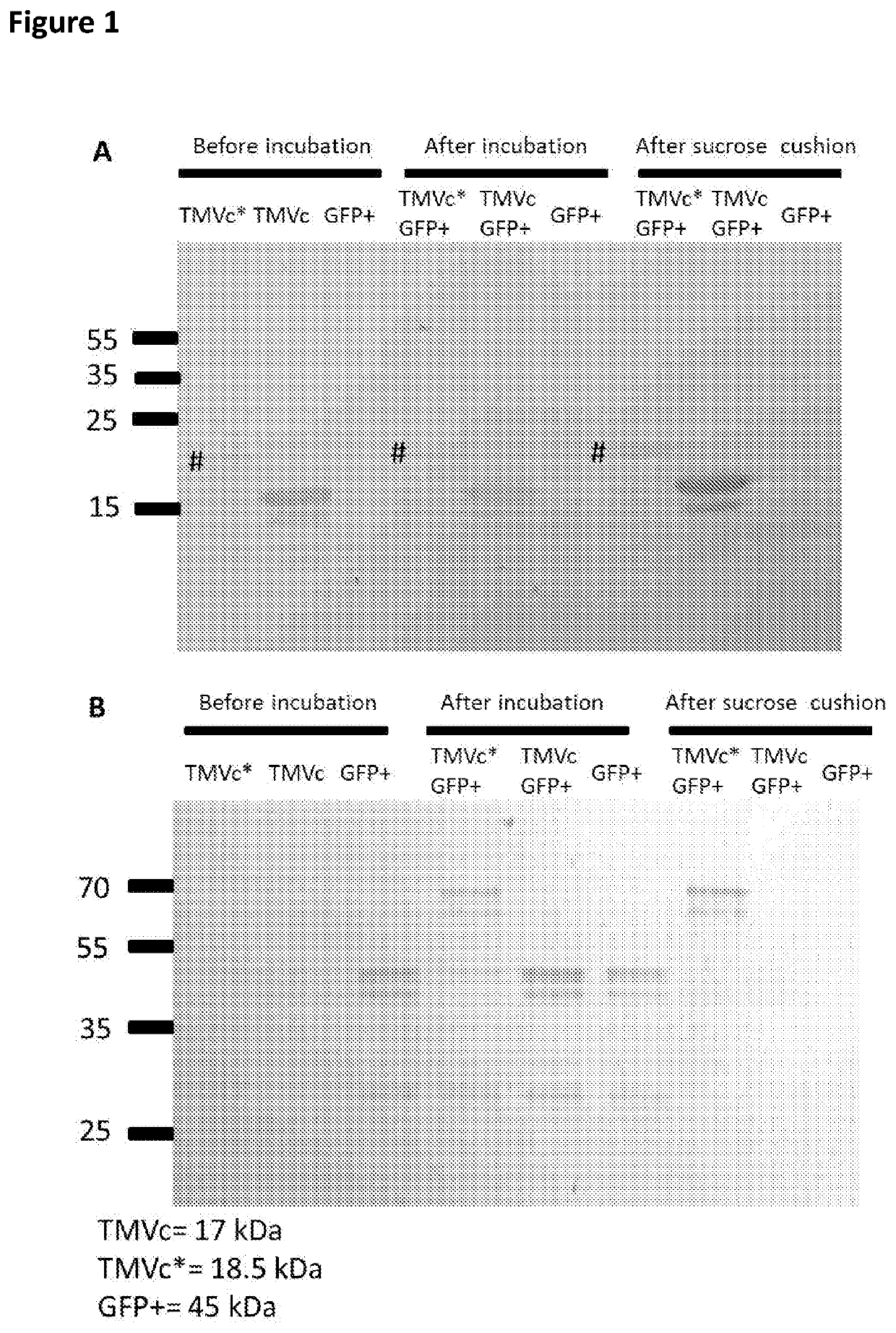
[0090]For the SpyTag-TMV (TMVc*), the TMV E50Q and D77N group modified coat protein sequence (TMVc; Brown et al., 2013) was fused at its C-terminal end to the SpyTag sequence (Long et al., 2013). For the SpyCatcher-GFP sequence (GFP+), SpyCatcher (Long et al., 2013) was fused to the N-terminal end of the eGFP sequence (GenBank: AAG27429.1). For the GalEst, this consisted of a UDP-glucose 4-epimerase sequence (GenBank: EIE36183.1) to which a 6 His and SpyTag sequence was attached at its N-terminal end. For the TMV CP-GGGGS SpyCatcher His, the TMVc* sequence had a 3x GGGGS linker-6 His-SpyCatcher fused to its C-terminal end. For the TMV CP-GGGGS-SpyTag His, a 3x GGGGS linker-SpyTag-6 His, was fused to the C-terminal end of the TMVc*. For the 11-20 SpyCatcher (SEQ ID NO: 13), a small Psoroptes ovis peptidic sequence was fused to the C-terminal of the SpyCatcher. These were codon optimized for bacterial expression and cloned into a pET-21a(+) vector by Genscript (Piscataway,...

example 2

n in Bacteria and Isolation of Proteins or Virus Structures

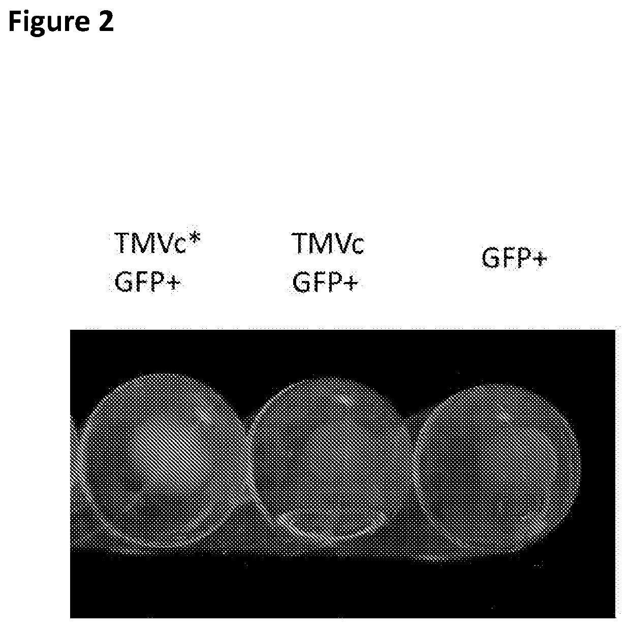
[0091]Bacteria containing the plasmids were grown at 37° C. with shaking at 200 rpm in LB media supplemented with 100 μg / ml ampicillin and 34 μg / ml chloramphenicol. Once cultures reached an OD 600 of 0.5, IPTG was added to a final concentration of 0.1 mM, and induction proceeded overnight at 20° C. with shaking at 200 rpm. Bacteria were pelleted by centrifugation at 4000 g for 15 minutes, the supernatant was removed and the cells were lysed by freeze-thawing 3 times using liquid nitrogen and a 37° C. water bath, before proceeding with protein extraction using the B-PER Complete reagent (ThermoFisher Scientific, Paisley, UK). Lysates obtained with the B-PER Complete reagent were centrifuged at 10,000 g for 20 minutes to remove debris. The lysates were combined (TMVc* or TMVc were mixed with GFP+) in equal volume and incubated at room temperature for 1 hour prior to centrifugation on a 20% sucrose cushion, after which the pell...

example 3

TMVc Purification and Buffer Exchange of GFP+ Prior to Transmission Electron Microscopy

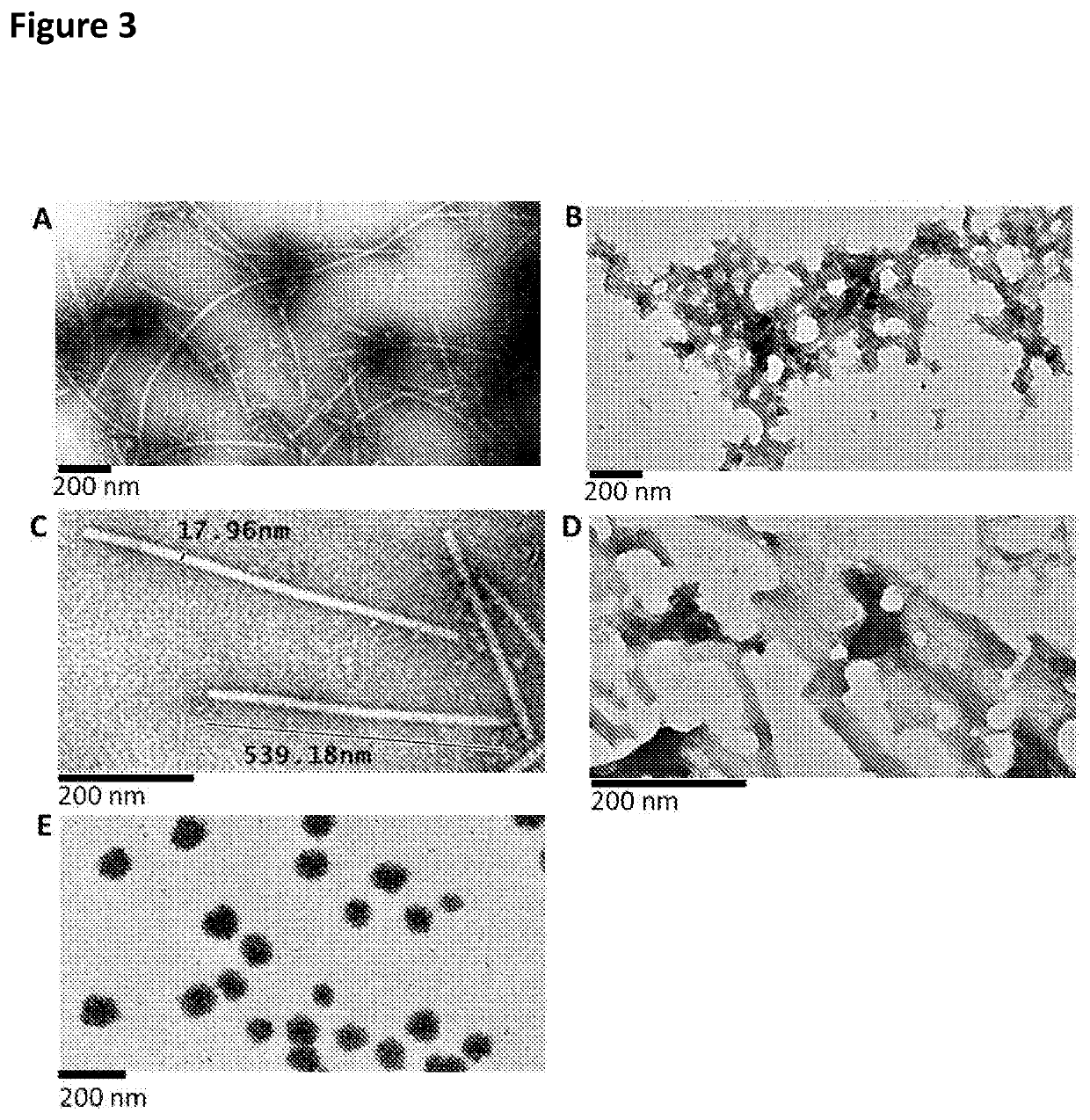
[0094]To the centrifuged TMV structure lysates, PEG 8000 and NaCl were added to a final concentration of 2% and 1% respectively, and the solutions were incubated at 4° C. overnight to ensure precipitation. These were centrifuged at 10000 g at 4° C. for 20 minutes to pellet the virus structures. The pellets were resuspended in 25 mM tris-HCl (pH 7.8), centrifuged at 10000 g to clarify and the supernatants were collected. Another two rounds of resuspension and centrifugation was carried out to obtain maximal virus structure yields, while minimizing debris. The clarified supernatants were then loaded onto 2 ml 20% sucrose 25 mM Tris-HCl (pH 7.8) cushions, which was then centrifuged in swing out rotors (SW41 Beckman Coulter; Beckman Coulter, California, USA) at 32000 rpm for 2 hours at 4° C. Pellets were resuspended in 0.01 M Tris-HCl (pH 7.8), ultracentrifuged at 32000 rpm for 2 hours at 4° C. in a S...


PUM

PropertyMeasurementUnit
sizeaaaaaaaaaa
temperaturesaaaaaaaaaa
length scalesaaaaaaaaaa

Abstract

The present invention relates to macromolecular complexes comprising micron-scale networks which include binding motifs thereon which allow the covalent bonding of the micron-scale networks to particles which provide nanoscale display surfaces. In particular the present invention relates to micron-scale networks of TMV coat proteins comprising a peptide tag (e.g. SpyTag) and particles providing a nanoscale display surface comprising GFP and a corresponding binding protein (e.g. SpyCatcher) wherein the peptide tag and binding protein pair are capable of spontaneously forming a covalent bond.

Description

FIELD OF THE INVENTION[0001]The present invention relates to macromolecular complexes comprising micron-scale networks which include binding motifs thereon which allow the covalent bonding of the micron-scale networks to particles which provide nanoscale display surfaces. Further, the present invention provides for the synthesis of nano-, micro-scale functional materials, in particular synthesis of such materials without the need to use complex and restrictive linker chemistries.BACKGROUND OF THE INVENTION[0002]One of the major goals of material science is to produce materials that are ordered on all length scales, from molecular (1-100 Å), nano (10-100 nm) to macro (1-100 micron). The development of systems with integrated catalytically active enzymes, specific antibodies or other biologics have high potential for synthesis, degradation and biosensor diagnostics of various biologically active substances.[0003]It has been demonstrated that short peptide sequences can be incorporated...

Claims


Application Information

Patent Timeline
Patent Type & Authority Applications(United States)
IPC IPC(8): C07K14/315C07K14/005
CPCC07K14/315C07K14/005C07K2319/735C07K2319/02C07K2319/20A61K39/00C12N2770/00022C12N2770/00023C12N2770/00042
Inventor LOVE, ANDREW JOHNTALIANSKI, MIKHAIL EMMANUILOVICHMCGEACHY, KARA
Owner JAMES HUTTON INSTITUTE
Why Eureka
Try Eureka