Acellular Bioactive Scaffold Device and Methods of Fabrication and Treatment Relating Thereto

Pending Publication Date: 2020-07-16
UNIV OF MARYLAND
View PDF5 Cites 0 Cited by
  • Summary
  • Abstract
  • Description
  • Claims
  • Application Information

AI Technical Summary

Benefits of technology

[0015]The present disclosure relates to an implantable acellular scaffold device that facilitates cellular growth. The scaffold is preferably fabricated using three-dimensional (3D) printing methods, and thus may be readily formed in minutes and in a selected geometric configuration suitable for the i

Problems solved by technology

Over time, articular cartilage can deteriorate or become damaged following injury or normal wear and tear, leading to the pain many people feel as they age.
Unfortunately, the treatment and repair of articular cartilage defects remains challenging due to the limited endogenous regeneration of the tissue and poor integration with conventional implantations.
As such, little long term success has been achieved with current treatment strategies.
Articular cartilage is structurally complex and difficult to mimic, despite the fact that it is innervated and lacks blood vessels, nerves, and lymphatics.
However, the post-surgical microenvironment created by conventional techniques fails to orient and guide the MSCs to the defect site properly, resulting in comparatively weak fibrous tissue be created that does not possess the desired and necessary mechanical properties as compared to native cartilage (Steadman, J. R. et al.
Additionally, it remains difficult to harvest and successfully cultivate a large enough chondrocyte population to transplant due to the low proliferation rate and differentiation potential during in vitro culture (Jones, D.
Current treatment techniq

Method used


Image

  • Acellular Bioactive Scaffold Device and Methods of Fabrication and Treatment Relating Thereto
  • Acellular Bioactive Scaffold Device and Methods of Fabrication and Treatment Relating Thereto
  • Acellular Bioactive Scaffold Device and Methods of Fabrication and Treatment Relating Thereto

Examples


example 1

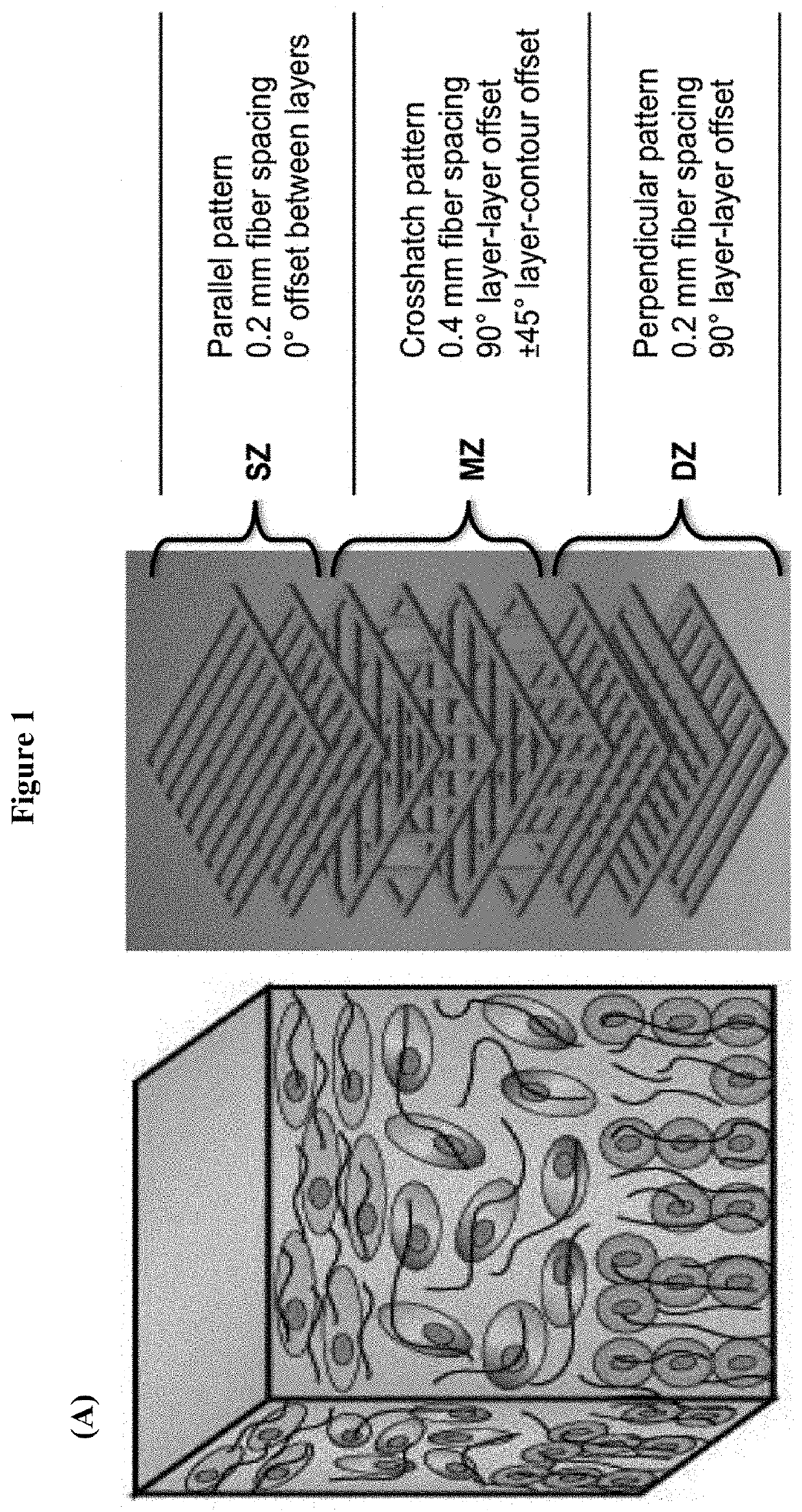
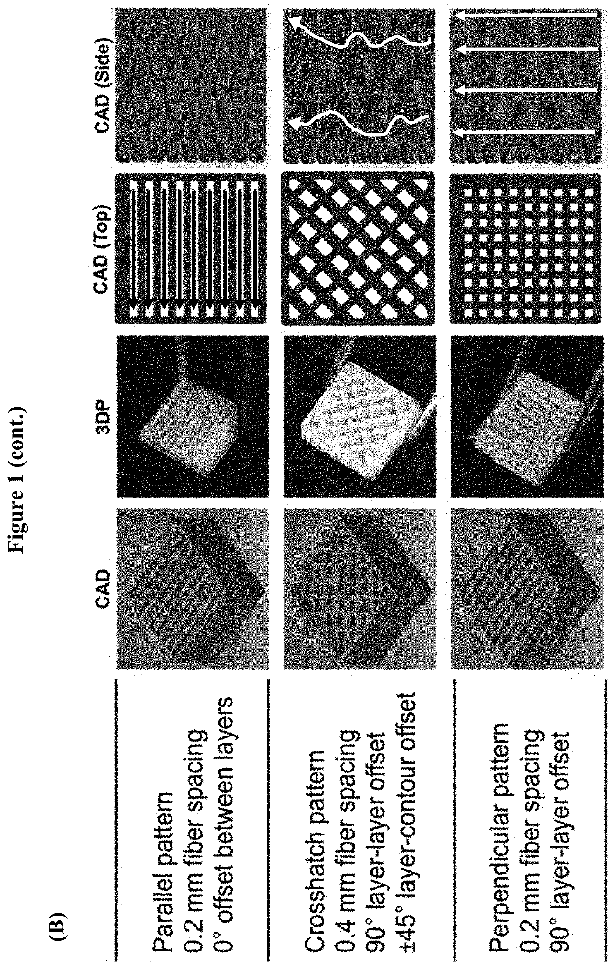
[0070]Scaffold Fabrication

[0071]An exemplary scaffold implant device suitable for cartilage implant was fabricated in a layer-by-layer fashion via extrusion-based 3D printing. A scaffold implant customized for a particular patient may be readily fabricated starting from a CAD model that can be either designed using commonly available software, or directly generated from imaging data routinely collected in the clinics (e.g. MRI, CAT scan, etc.). The scaffold was extruded in three different zonal layer patterns (FIG. 1, Panel A) designed to guide the growth and differentiation of resident stem cells according to the three different zones observed in native cartilage. The three layer patterns include: i) a parallel pattern, wherein tissue ingrowth occurs in a horizontal and parallel fashion as observed in the superficial zone (SZ) of articular cartilage; ii) a cross-hatch pattern, wherein the random presence of fibers and pores results in random alignment of cells and extracellular mat...

example 2

[0079]An animal study was conducted in order to evaluate the performance of microfracture surgery when combined with the disclosed scaffold implant. As demonstrated herein, the addition of an acellular PLCL scaffold device following microfracture provided effective guidance to the stem cells released from the bone marrow and resulted in substantially enhanced cartilage regeneration. The results were even more significant when using a PLCL scaffold functionalized with aggrecan, and resulted in an even higher cell / ECM retention from the bone marrow via microfracture, thicker cartilage formation, and deposition of ECM (collagen type I, glycosaminoglycans) similar to that of native cartilage.

[0080]3D-Printed Aggrecan-Functionalized Scaffold

[0081]A 3D-printed scaffold functionalized with aggrecan demonstrated a substantial improvement in the quality of regenerated cartilage tissue during microfracture as compared to conventional techniques. As the main proteoglycan component of native ar...

example 3

[0128]3D Printed Interface Scaffolds for Cartilage Regeneration

[0129]A multilayer scaffold composed of poly(L-lactide-co-caprolactone) (PLCL) and amine end-capped poly(lactic-co-glycolic acid) (amine-PLGA) was successfully printed with high resolution. The disclosed approach creates a regenerative environment with a robust bone-cartilage interface within an osteochondral implant. It is believed that co-printing the architecture at the bone-cartilage interface improves the adhesion between the bone and cartilage layers by increasing the contact points between the print strands of the same mixture at the interface.

[0130]Preliminary experiments demonstrated the feasibility of interface printing via a 3D printing strategy (FIG. 15). A thermoplastic polymer mixture of PLCL and amine-PLGA (as described in Example 2 above) served as the chondral matrix, while polycaprolactone (PCL) served as the primary osteo matrix. Exemplary printing patterns of the bone and cartilage layers were explore...


PUM

PropertyMeasurementUnit
Thicknessaaaaaaaaaa
Bioactiveaaaaaaaaaa

Abstract

An implantable acellular polymeric scaffold device functionalized with aggrecan is provided. Also provided are methods of fabricating a polymeric scaffold device, including methods of fabricating the scaffold device via 3D printing. Methods of treating a cartilage defect in a subject in need thereof comprise application of the disclosed scaffold device in combination with microfracture procedures. A specialized lid for a centrifugation well plate is also provided.

Description

CROSS-REFERENCE TO RELATED APPLICATIONS[0001]This application is based on U.S. Provisional Patent Application Ser. No. 62 / 792,722, entitled “Acellular Bioactive Polymeric Scaffold to Enhance Microfracture,” filed Jan. 15, 2019, and U.S. Provisional Patent Application Ser. No. 62 / 828,303, entitled “System, Device and Method for Cell Separation During Centrifugation,” filed Apr. 2, 2019, which applications are incorporated herein by reference in their entireties and to which priority is claimed.STATEMENT REGARDING FEDERALLY SPONSORED RESEARCH OR DEVELOPMENT[0002]This invention was made with government support under Grant Nos. CBET 1264517 and CBET 1604742 awarded by the National Science Foundation (NSF), and under Grant No. CECT, P41 EB023833 awarded by the National Institute of Biomedical Imaging and Bioengineering, Center for Engineering Complex Tissues. The United States government has certain rights in this invention.FIELD OF THE INVENTION[0003]The present disclosure relates to an...

Claims


Application Information

Patent Timeline
IPC IPC(8): A61F2/30A61L27/18
CPCA61F2/30756A61F2002/30985A61L27/18A61F2002/30766A61L2300/252A61L2300/412B33Y80/00A61F2/3094C08L67/04
Inventor GUO, TINGFISHER, JOHN PATRICKBAKER, HANNAHLERMAN, MAX JONAHCHOE, ROBERT
Owner UNIV OF MARYLAND
Why Eureka
Try Eureka