Microfluidic label-free isolation and identification of cells using fluorescence lifetime imaging (FLIM)

a lifetime imaging and microfluidic technology, applied in biochemistry apparatus and processes, laboratory glassware, instruments, etc., can solve the problems of low purity of recovered leukemia cells, difficult acquisition, and invasive methods, and achieve the effect of better trapping

Pending Publication Date: 2020-07-30
RGT UNIV OF CALIFORNIA +1
View PDF4 Cites 0 Cited by
  • Summary
  • Abstract
  • Description
  • Claims
  • Application Information

AI Technical Summary

Benefits of technology

The patent describes a method for identifying and classifying cancer cells using a combination of hydrodynamic control and fluorescence lifetime imaging microscopy (FLIM). The method allows for the separation and trapping of single cancer cells based on their size and metabolic state. The FLIM technique measures the decay time characteristics of the cells' intrinsic fluorescence, which can be used to identify and classify cancer cells without the need for labeling. The patent also describes a new platform for isolating and characterizing single plant cells using the same method. The platform includes a device with asymmetric gaps that enhance the trapping of large or less-deformable cells. The method and platform have potential applications in biological laboratories and clinical settings.

Problems solved by technology

Leukemia, a group of blood cancers that are characterized by the uncontrolled production and result in high numbers of abnormal leukocytes from the bone marrow, is the most prevalent pediatric cancer, and the sixth most common cause of cancer deaths in the U.S. Currently, bone marrow aspiration, and bone marrow biopsy has been used commonly as the standardized procedures for the diagnosis of leukemia and other blood disorders, however, these methods are usually invasive, risky, and difficult to acquire repeatedly over an extended time period.
However, the blood-based detection such as complete blood count (CBC) and peripheral blood smear is gentle but cannot specify leukemia types; while selective biomarker immunostaining and cytogenetic analysis can determine the specific types of leukemia, but the whole procedures are complicated, time-consuming, and expensive.
However, in case of separation of leukemia cells from blood, the low purity of recovered leukemia cells remains a significant challenge because the size of leukemia cells may exhibit overlap with the size of leukocytes.
In these approaches, however, the strong deformations may damage certain cancer cells due to the high shear stress, and the separation of cells through microstructured constrictions is limited by clogging, which reduces selectivity of cancer cells.
In addition, recovery of biomarker-conjugated cells from the antibody-coated surface induces leukemia cell damage and requires an additional non-trivial step for culture and enumeration, Jackson and Li et al. separated the peripheral blood to search for circulating leukemic cells and lymphoblasts within the antibody-immobilized microfluidic channel, respectively, but still, need time-consuming labeling process and have typically yielded low sample purities (<1%), causing challenges in downstream analysis.
During the past decades, the routine screening method was dominated by the inherited marker gene system which is very labor-intensive.

Method used


Image

  • Microfluidic label-free isolation and identification of cells using fluorescence lifetime imaging (FLIM)
  • Microfluidic label-free isolation and identification of cells using fluorescence lifetime imaging (FLIM)
  • Microfluidic label-free isolation and identification of cells using fluorescence lifetime imaging (FLIM)

Examples


example 1

nce Lifetime Imaging Microscopy for the Rapid Screening and Identification of Single Leukemia Cells in Blood from the High-Density Microfluidic Trapping Array

[0148]The rapid identification and analysis of single leukemia cells from blood has become critical for examination of the molecular-level tumor signatures and for early detection of human leukemia disease. However, isolation and identification of leukemia cells individually from peripheral blood requires immunological labeling and is extremely challenging due to the size overlap between leukemia cells and the more abundant white blood cells (WBCs). Herein is described a novel leukemia cell identification platform that combines deterministic single-cell separation and isolation, passive hydrodynamic trapping, and identification of single leukemia cells through phasor approach and Fluorescence Lifetime Imaging Microscopy (FLIM), which measures changes between free / bound nicotinamide adenine dinucleotide (NADH) as an indirect mea...

example 2

asive Single-Cell Transcriptomic and Metabolic Analysis Microfluidic Array

[0167]Single-cell analysis provides comprehensive information and reveals intracellular heterogeneity of the cell population, which is of critical importance in medical and biological research. However, most of the developed single-cell biomolecular and biochemical assays require cell lysing and complicated purification / labeling processes. Here is presented an integrated platform capable of collecting single-cell transcriptomic and metabolic information in a non-invasive and label-free manner. Cells are trapped in a 1-μm-thick PDMS membrane-sealed single-cell array, and a modified AFM probe penetrates through the membrane to extract single-cell mRNAs by dielectrophoresis (DEP), coupled with fluorescence lifetime imaging microscopy (FLIM) to acquire metabolic patterns through single-cells' intrinsic autofluorescence.

Materials and Methods

[0168]The cell-trapping array, which was made by soft lithography casting o...

example 3

and Identification of Single Plant Cells

Unique and Novel Features

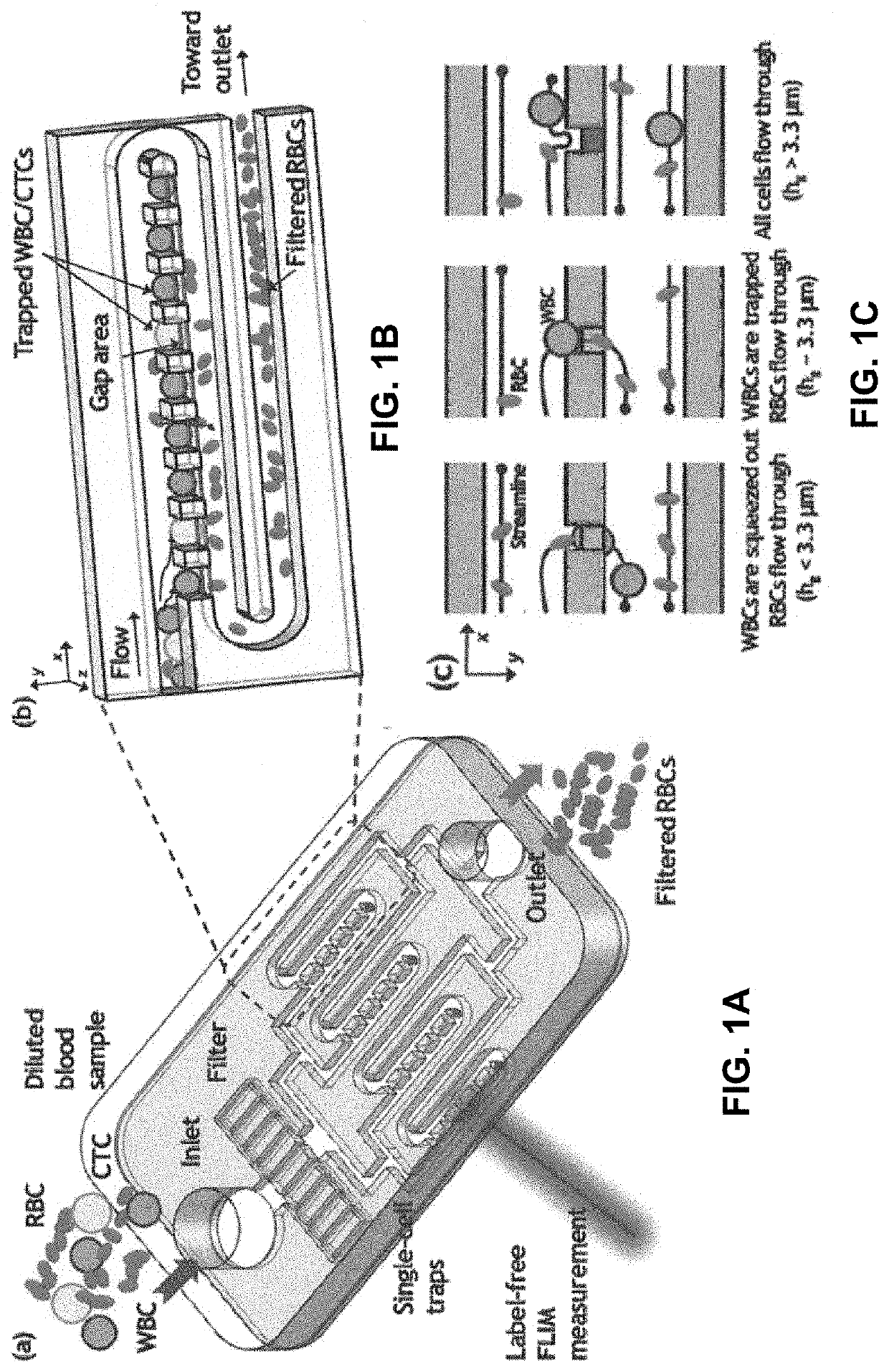
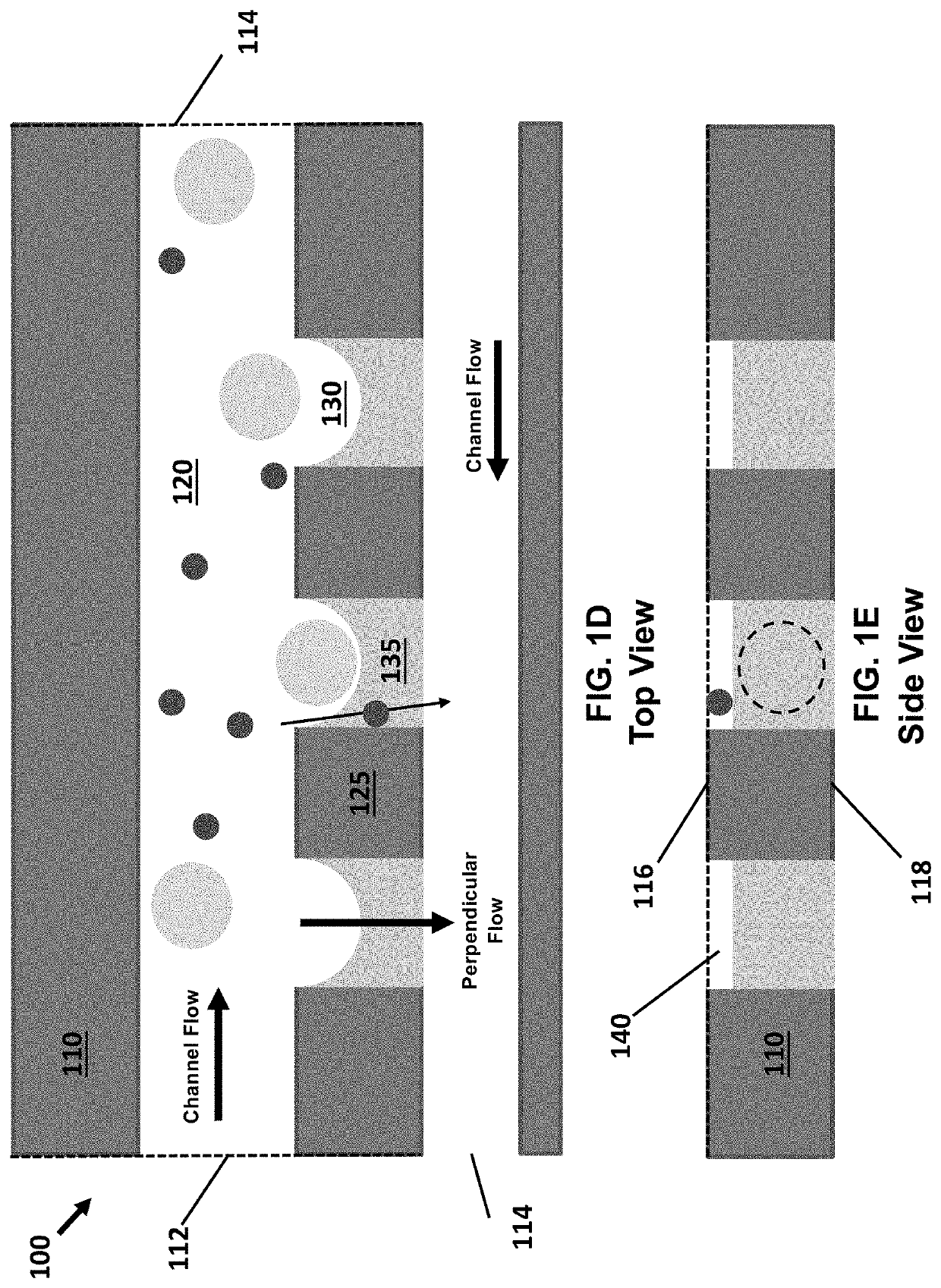
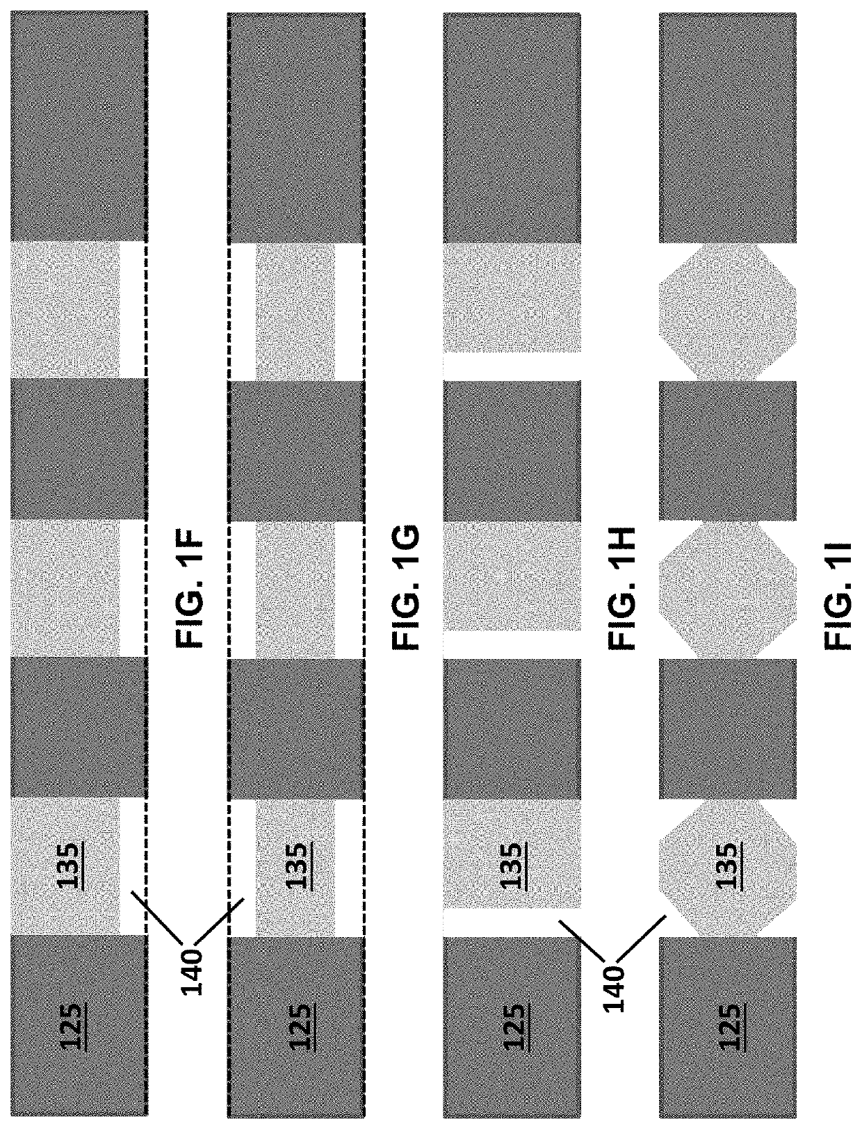
[0170]The high-density and high-efficiency cell traps were used to separate plant cells and debris in an array formed by a serpentine channel. This demonstrates the utility of deterministic single-cell trap for isolation and identification of single plant cells based on their average fluorescence lifetime in a label-free, gentle and scalable manner. The microfluidic single-cell trapping array consists of a serpentine channel with 20 traps along each row. The channel height is 120 μm, and the trap size is 75 μm, which is similar to the diameter of the microspores. For each single-cell trap, there is a 10 μm-height gap area to enable the perpendicular flow to push the cell into the trap. Gunk debris is blocked by the pillar array at the inlet, and small debris passes through the gap area and is filtered out.

Results and Discussion

Microfluidic Isolation of Single Plant Cells:

[0171]The combination of the phasor-FLIM imaging...


PUM

PropertyMeasurementUnit
Forceaaaaaaaaaa
Flow rateaaaaaaaaaa
Sizeaaaaaaaaaa

Abstract

Methods and devices for single cell analysis using fluorescence lifetime imaging microscopy (FLIM) are disclosed. The methods utilize microfluidic devices which use traps to immobilize cells for FLIM analysis. The analysed cells may be sorted before or after imaging and may be plant, animal, or bacterial cells. Analysis of the FLIM data may use a phasor plot and may be used to identify a metabolic pattern of the single cells.

Description

CROSS REFERENCE[0001]This application is a continuation-in-part and claims priority to PCT Application No. PCT / US2018 / 055722 filed Oct. 12, 2018, which claims priority to U.S. Provisional Patent Application No. 62 / 571,746, filed Oct. 12, 2017, the specifications of which are incorporated herein in their entirety by reference.GOVERNMENT SUPPORT[0002]This invention was made with government support under Grant No. IIP-1538813 awarded by the National Science Foundation. The government has certain rights in the invention.FIELD OF THE INVENTION[0003]The present invention relates to methods and devices for isolating and identifying cells using fluorescence lifetime imaging (FLIM). More specifically, the methods and devices are microfluidic.BACKGROUND OF THE INVENTION[0004]Single-cell analysis is a growing practice within the field of microfluidics. It is often beneficial to be able to analyze individual cells instead of collecting average data on a population of cells. Methods which allow ...

Claims


Application Information

Patent Timeline
IPC IPC(8): B01L3/00C12Q1/02C12N15/10
CPCB01L2300/0627B01L2300/0816B01L2300/0858C12Q1/02B01L3/502715B01L2300/0864B01L3/502761C12N15/1003B01L2200/0647B01L2300/0854G01N15/1429G01N15/0205G01N15/0255H05H3/02G01N2015/0288G01N2015/1006G01N2015/1493G01N15/1484G01N33/586G01N15/1433G01N15/149
Inventor LEE, ABRAHAM P.DIGMAN, MICHELLE A.LEE, DOHYUNLI, XUANMA, NINGYUN, YUE
Owner RGT UNIV OF CALIFORNIA
Why Eureka
Try Eureka