
Placental lipid bilayer for cell-free molecular interaction studies

a lipid bilayer and molecular interaction technology, applied in the field of heterogeneous membranes, can solve the problems of difficult study, increased perinatal and neonatal mortality, and difficult placental dysfunction, so as to minimize the risks of a developing fetus, improve the understanding of the placenta, and improve the effect of treatment plans

Pending Publication Date: 2020-11-26
BROWN UNIVERSITY
View PDF1 Cites 0 Cited by
  • Summary
  • Abstract
  • Description
  • Claims
  • Application Information

AI Technical Summary

Benefits of technology

The invention provides a way to mimic the lipid composition of the placenta using a lipid bilayer. This can be useful for studying interactions between pharmaceutical or environmental toxicants and the placenta at a molecular level. This can help in improving understanding of the placenta and minimizing risks to the developing fetus, better informing treatment plans, and screening new therapies. Ultimately, this system can help in understanding how medications may interact with the maternal-fetal interface, affecting prenatal and future maternal health. The invention also provides in vitro cell-free placenta models that can be used to quickly analyze the impact of molecule interactions on the placenta. These models enable rapid screening of potential detrimental effects of pharmaceuticals, environmental toxicants, and toxins on the prenatal and future maternal health.

Problems solved by technology

Placental dysfunction is associated with increased perinatal and neonatal mortality and lifelong health complications in the mother and child.
But the placenta remains one of the least understood organs in the human body.
Guttmacher et al., Placenta, 35(5), 303-304 (2014), partly because a lack of human placenta-representative in vitro and in vivo models make it difficult to study this dynamic organ.
Unfortunately, the techniques for assessing placental function are highly qualitative, such as histology and other descriptive methods.
The techniques have no capability for the rapid quantification of pharmaceutical interaction with the placenta, resulting in little quantified information on the environmental toxicant impact on placental structure and health.
Animal models cannot mimic the human placenta due to differences in the placental cell phenotype.
Primary trophoblasts (which form the embryo-derived portion of the placenta) can be used, but these cells have a finite proliferative ability, limiting experiment time.

Method used


Image

  • Placental lipid bilayer for cell-free molecular interaction studies
  • Placental lipid bilayer for cell-free molecular interaction studies
  • Placental lipid bilayer for cell-free molecular interaction studies

Examples


example 1

Synthetic Placenta Lipid Membranes for Nanomaterial Interaction Studies

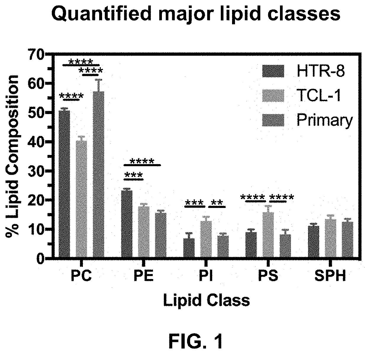
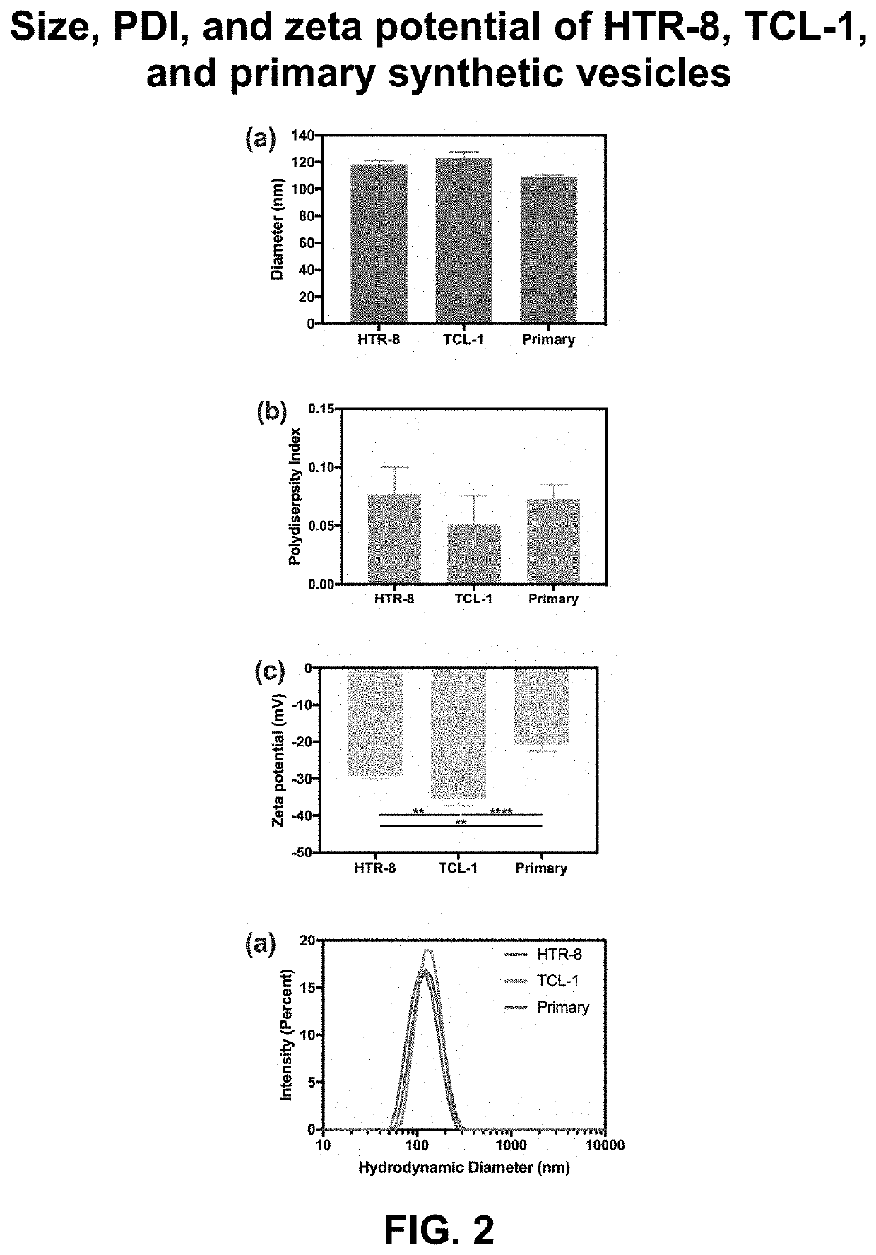
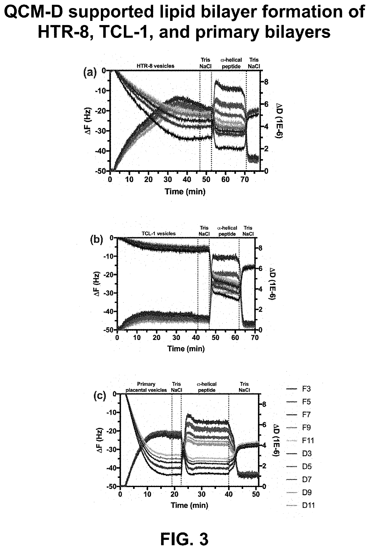
[0095]Rationale. The goal of this EXAMPLE 1 is to provide an in vitro system to monitor molecular interactions with a synthetic placental bilayer. A supported lipid bilayer (SLB) was developed from synthetic lipids mimicking the placenta cell membrane for adsorption and mass loss studies. Supported lipid bilayers are a useful tool to investigate adsorption or mass removal interactions occurring. To gain more information, the inventors developed suspended lipid bilayers across PVDF porous inserts. This development produced a parallel artificial membrane permeability assay (PAMPA) to investigate further DEHP, amphotericin B, and amBisome® interactions with HTR-8, TCL-1, and primary lipid models. The PAMPA studies can be used for permeation.

[0096]Analytical approach. First, the inventors used a reported composition to develop placental lipid bilayers. Previous studies have extracted lipids from placental tissue and ...

example 2

Trophoblast Cell Line-Derived Placental Lipid Vesicles and Lipid Bilayer

[0104]Rationale. The goal of this EXAMPLE 2 developed a lipid bilayer from trophoblast cell-extracted lipids. Trophoblast cells, specifically the BeWo villous trophoblast cell line, have been used to study maternal-fetal transport. Ali et al., Int. J. Pharm., 454(1), 149-157 (2013). The inventors are developing bilayers from the lipids extracted from the established BeWo villous trophoblast cell line. The inventors are comparing the bilayers with the synthetic composition from EXAMPLE 1.

[0105]Analytical approach. Lipid extraction from trophoblast cells. Quantification of lipids in BeWo trophoblast cells. The inventors first collected BeWo trophoblast cells, then solubilized and agitated the BeWo cell pellet. They next analyzed the lipid composition by gas chromatography. The lipid extracts are processed through the dry lipid film method and extrusion, as performed in EXAMPLE 1.

[0106]Cell-derived lipid vesicle an...

example 3

Tissue-Derived Placental Lipid Vesicles and Lipid Bilayer

[0111]Rationale. The goal of this EXAMPLE 3 is to extract the lipid composition from placental tissue, quantify this extract, and use it to develop placental lipid bilayers. This aim is also to allow comparison of the lipid composition with the placental cell lines, which has not previously been investigated.

[0112]Analytical approach. Lipid extraction from placenta tissue. Quantification of lipids in placenta tissue. The Women & Infants Hospital of Rhode Island kindly provided human placenta tissue samples to the inventors obtain for them to homogenize. The homogenized tissue is processed similarly to the processing of the BeWo cells, to extract the lipids using liquid-liquid extraction. The extracted lipids then went through the dry thin-film method and extrusion described in EXAMPLE 1 to obtain placental tissue-derived lipid vesicles. Both supported lipid bilayers and PAMPA are formed and characterized by the methods describ...


PUM

PropertyMeasurementUnit
hydrodynamic diameteraaaaaaaaaa
frequencyaaaaaaaaaa
sizeaaaaaaaaaa

Abstract

The invention provides a lipid bilayer mimicking the lipid composition of the placenta. The lipid composition provides an in vitro placenta model using the lipid composition of the placental cell membrane.

Description

REFERENCE TO RELATED APPLICATIONS[0001]This patent application claims priority under 35 U.S.C. § 119(e) to provisional patent applications U.S. Ser. No. 62 / 850,188, filed May 20, 2019; U.S. Ser. No. 62 / 905,756, filed Sep. 25, 2019; and U.S. Ser. No. 62 / 976,882, filed Feb. 14, 2020, all entitled “Placental Lipid Bilayer for Cell-Free Molecular Interaction Studies.”STATEMENT REGARDING FEDERALLY SPONSORED RESEARCH OR DEVELOPMENT[0002]This invention was made with government support under Grant No. 1644760 awarded by the National Science Foundation (NSF). The government has certain rights in the invention.FIELD OF THE INVENTION[0003]This invention generally relates to heterogeneous membranes, e.g., containing dispersed material or mixed matrix membranes with “carriers” containing embedded or bound biomolecules.BACKGROUND OF THE INVENTION[0004]The placenta controls fetal development during pregnancy, being a semi-permeable barrier that controls the exchange of nutrients and wastes. See Ha...

Claims


Application Information

Patent Timeline
Patent Type & Authority Applications(United States)
IPC IPC(8): C12N5/073G01N33/50
CPCG01N33/5014C12N5/0605G01N2405/04G01N33/92A61K9/1617A61K35/50A61K49/1839C07K14/4715C12N2506/025
Inventor SHUKLA, ANITABAILEY-HYTHOLT, CHRISTINATRIPATHI, ANUBHAV
Owner BROWN UNIVERSITY
Learn More