Instruments and methods for imaging collagen structure in vivo

a collagen structure and instrument technology, applied in the field of instruments and methods for imaging can solve the problems of inability to perform in vivo or in real time, inability to yield high contrast, high resolution images of collagen structure in vivo, etc., to improve the surgeon's estimation of tumor margins, reduce variability in total number, and streamline the overall procedure

Active Publication Date: 2021-02-25
UNIV OF MASSACHUSETTS +1
View PDF0 Cites 4 Cited by
  • Summary
  • Abstract
  • Description
  • Claims
  • Application Information

AI Technical Summary

Benefits of technology

[0009]In view of the above, a continuing need exists for improvements for in vivo imaging of the skin and skin related tumors. A non-invasive and simple-to-use technology to improve the surgeon's estimation of the tumor margins preoperatively reduces variability in the total number of surgical stages per case and in streamlining the overall procedure. A variety of imaging tools have been examined in the past to aid detection of cutaneous pathology, including reflectance confocal microscopy (RCM), optical coherence tomography (OCT), Raman spectroscopy, and second harmonic generation. While each device has certain advantages, shortfalls limit their use in the clinical setting for Mohs surgery. For instance, reflectance confocal microscopy provides high resolution imaging at superficial depths up to 200 μm limiting the application in imaging lesions extending into the dermis. Furthermore, technical barriers such as artifact formation due to patient motion and the slow pace of imaging can be limiting factors.

Problems solved by technology

These techniques require biopsy, which may cause discomfort, scarring and infection and cannot be performed either in vivo or in real time.
Confocal microscopy is an imaging method with cellular resolution but due to the shallow imaging depths, this approach does not yield high contrast, high resolution images of collagen structure in vivo.
Non-linear microscopy techniques such as two photon microscopy and second harmonic generation offer high-resolution morphological detail and deeper light penetration depth, but thus far it has not shown potential for in vivo visualization of dermal structure, due to high power densities required for imaging and the very small field of view.
Surgery is the most common treatment for these tumors, but pre-operative identification of surgical margins is challenging.
Mohs surgeons find identifying adequate surgical margins presurgically to be challenging because of the lack of information from simple visual inspection.
This is not only inefficient and erratic but prolongs the duration and extent of the Mohs procedure and adds substantial cost.
One challenge leading to the need for multiple surgical stages during MMS is the difficulty of clinically identifying the boundaries of the tumor prior to surgery.
These techniques require biopsy, which may cause scarring and infection and cannot be performed either in vivo or in real time.
Confocal microscopy is a mature imaging technique with submicron cellular resolution but has inadequate imaging depths, so collagen structure is not visualized in vivo.
But due to a requirement for higher power densities needed to achieve good quality images and a very small field of view, this imaging method is also not suitable for in vivo analysis of deeper dermal collagen structure.
While each device has certain advantages, shortfalls limit their use in the clinical setting for Mohs surgery.
Furthermore, technical barriers such as artifact formation due to patient motion and the slow pace of imaging can be limiting factors.
Optical coherence tomography provides deeper penetration but suffers from inaccuracy and unreliable differentiation between lesions such as basal cell carcinomas and actinic keratoses.
Raman spectroscopy is time consuming, requiring up to 60 minutes for diagnosis.

Method used


Image

  • Instruments and methods for imaging collagen structure in vivo
  • Instruments and methods for imaging collagen structure in vivo
  • Instruments and methods for imaging collagen structure in vivo

Examples


Embodiment Construction

[0056]The subject technology overcomes many of the prior art problems associated with in vivo examination of skin. The advantages, and other features of the instruments and methods disclosed herein, will become more readily apparent to those having ordinary skill in the art from the following detailed description of certain preferred embodiments taken in conjunction with the drawings which set forth representative embodiments of the present invention and wherein like reference numerals identify similar structural elements.

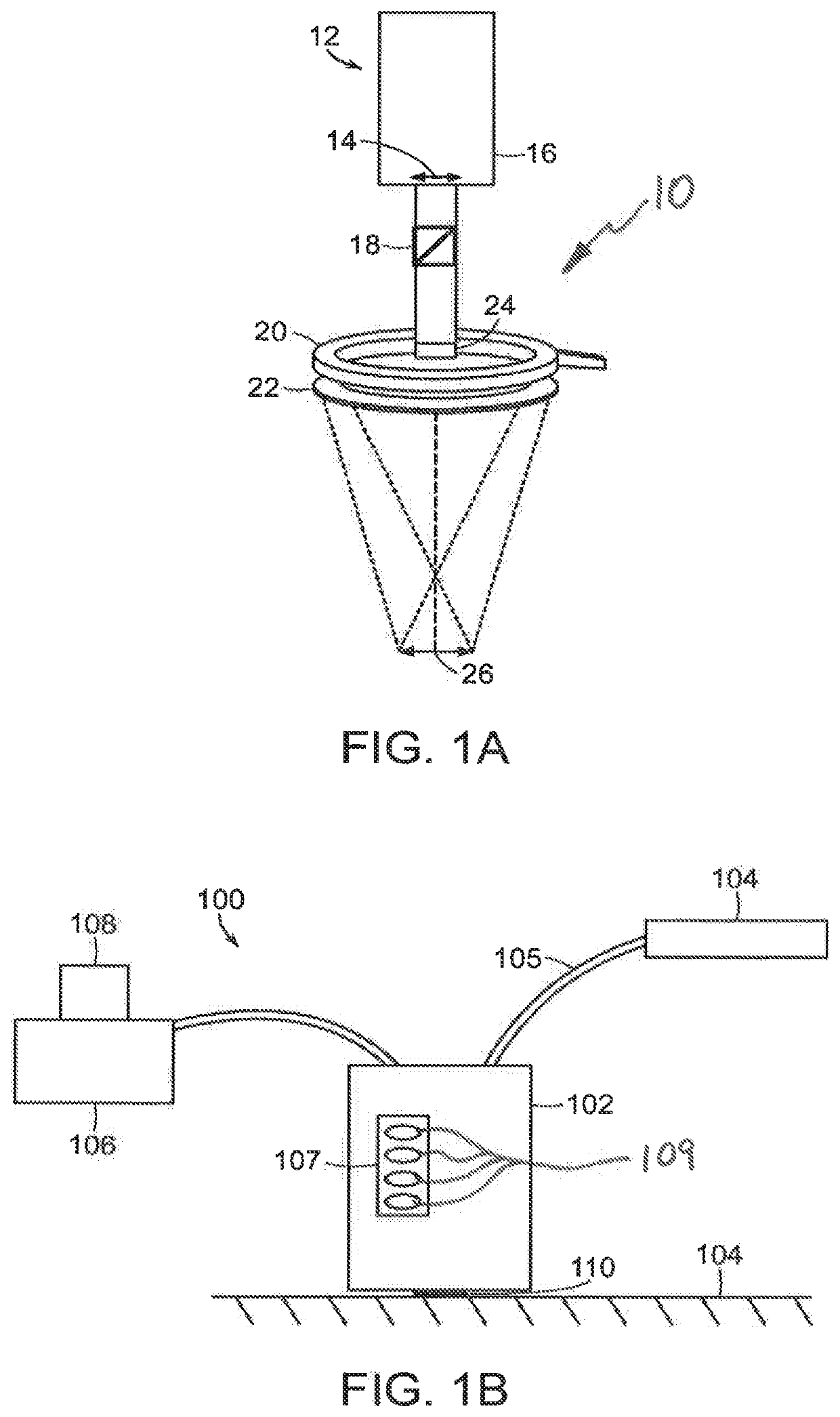
[0057]Referring now to FIG. 1A, a schematic view of the light delivery and detector elements 10 of a wide-field imaging instrument 100 (FIG. 1B) is shown. The instrument 100 can generate images of collagen structures of the skin with intact epidermis. FIG. 1B shows a schematic view of the instrument 100 including a handheld unit 102. As shown in FIG. 1A, the light delivery and detector elements 10 include a camera housing 12 in which a detector 16 such as CCD or CM...


PUM


Abstract

Instruments and methods for wide-field polarized imaging of the skin to determine an outer lesion margin objectively in vivo to provide guidance to a surgeon. Quantitative characterization of collagen structures in the skin can be used to determine the outer lesion margin or monitor skin treatment.

Description

FIELD OF THE INVENTION[0001]The subject disclosure relates to instruments and methods for using Optical Polarization Imaging (OPI) prior to surgery to detect the extent of subclinical tumor spread by monitoring disruption in collagen.BACKGROUND OF THE RELATED ART[0002]Skin is the first layer of protection for the human body against outside environment. Skin diseases and degeneration can be related to sun exposure, working environment and personal habits, which can be observed in the change of internal structure of skin. Collagen, which is the major component of the dermal structure, is an important factor related to dermal changes. Thus, it is beneficial to inspect collagen structure and quantitatively define the status of skin. Histopathological and immunohistochemical studies are commonly used for diagnosing diseases and evaluating dermal changes. These techniques require biopsy, which may cause discomfort, scarring and infection and cannot be performed either in vivo or in real t...

Claims


Application Information

Patent Timeline
Patent Type & Authority Applications(United States)
IPC IPC(8): A61B5/00G06T7/00G06T7/136G16H30/40A61B90/00
CPCA61B5/441A61B5/0064A61B5/0082G06T7/0016A61B2090/3904G16H30/40A61B90/39G06T2207/30096G06T2207/30088G06T7/136A61B5/6835A61B5/7207A61B5/742A61B5/0066A61B5/0068A61B5/0071A61B5/0077A61B5/443A61B5/444A61B5/4836G01N21/21A61B2576/02
Inventor YAROSLAVSKY, ANNA N.NEEL, VICTOR
Owner UNIV OF MASSACHUSETTS
Why Eureka
Try Eureka