
Methods of production of fibrous fibrinogen scaffolds and products thereof

Pending Publication Date: 2021-07-15
UNIV OF BREMEN
View PDF0 Cites 1 Cited by
  • Summary
  • Abstract
  • Description
  • Claims
  • Application Information

AI Technical Summary

Benefits of technology

The patent text describes a method for creating fibrous fibrinogen biomaterials, which can be used as a scaffold for tissue regeneration and wound healing applications. The method allows for controlled self-assembly of fibrinogen fibers, resulting in the formation of scaffolds with high yield, coverage, and dimensions. These scaffolds can be detached and used as free-standing protein patches or selectively immobilized on a substrate. The method is simple, reproducible, and can be tailored to create scaffolds for different areas of regenerative medicine. Additionally, the patent text suggests that the fibrous fibrinogen biomaterial can be used as a biological filter for filtration purposes in the biotechnological field.

Problems solved by technology

However, fibrinogen binds and reduces the activity of thrombin.
Accordingly, loss or reduction in antithrombin 1 activity due to mutations in fibrinogen genes or hypo-fibrinogen conditions can lead to excessive blood clotting and thrombosis.
However, fibrillogenesis of fibrinogen by previously reported methods have only led to fiber formation with a very low yield and a low surface coverage on substrates.
Moreover, methods to prepare fibrin hydrogels require the addition of the enzyme thrombin, which cleaves off the fibrinopeptides A and B from the original fibrinogen molecule, thus inducing a substantial change in the molecular structure.
Importantly, the use of the enzyme thrombin makes the preparation of fibrin hydrogels a very expensive method.
These can interfere with the native protein conformation and result in reduced bioactivity of fibrinogen.
Additionally, buffer-induced fibrillogenesis methods have so-far only achieved to produce fibrous scaffolds with a low fiber yield.
However, using these methods, fibrinogen fiber yield was very low and these fibers were only sparsely distributed on the underlying substrate and no dense fibrous scaffold suitable for tissue engineering could be obtained.
Due to low fiber density, no three-dimensional fibrous scaffolds were obtained.
Besides, hydrophobic substrates, which can induce fibrinogen fibrillogenesis, can lead to undesired side effects in cell culture systems and can result in (partial) protein denaturation upon adsorption.
However, the use of high electric fields in combination with organic solvents, such as 1,1,1,3,3,3-hexafluoro-2-propanol in electrospinning can negatively influence the bioactivity of electrospun fibrinogen scaffolds.
Both these factors make electrospinning a very expensive method.
This fiber collection adds another step into the electrospinning procedure, which makes the electrospinning of fibrous fibrinogen scaffolds more complex and more expensive.
However, so far none of the previous approaches has overcome the limitations and disadvantages stated above.

Method used


Image

  • Methods of production of fibrous fibrinogen scaffolds and products thereof
  • Methods of production of fibrous fibrinogen scaffolds and products thereof
  • Methods of production of fibrous fibrinogen scaffolds and products thereof

Examples


Embodiment Construction

Preparation of Substrates and Protein Solutions

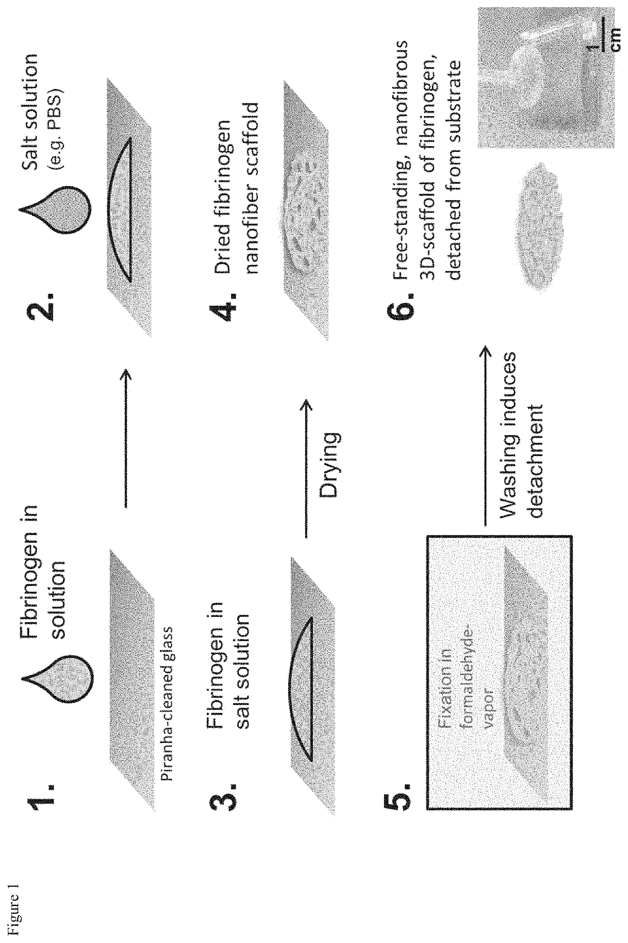
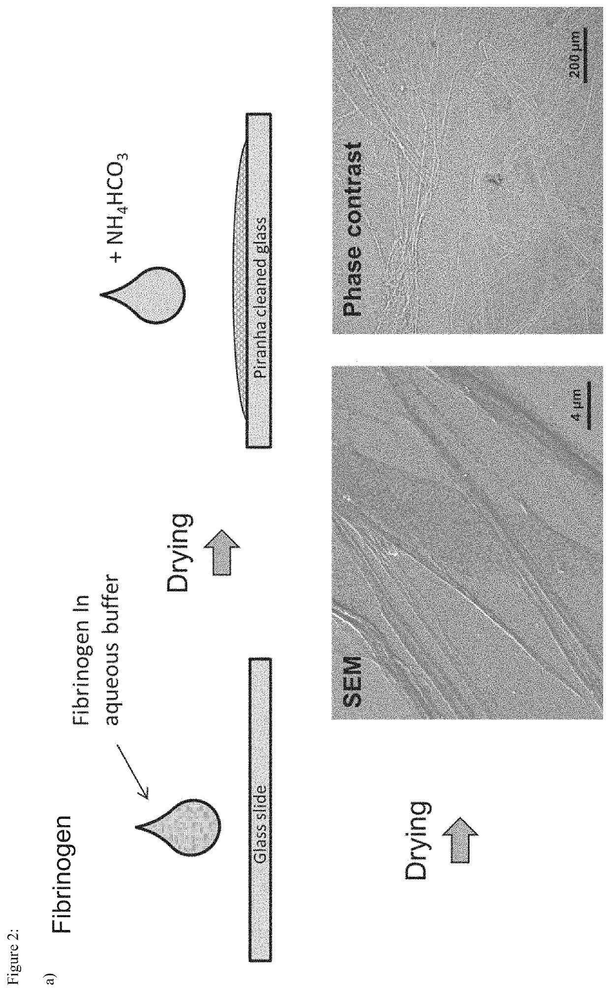
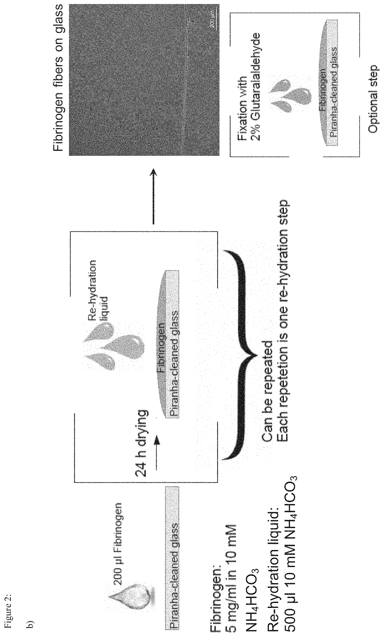
[0106]Round glass coverslips with a diameter of 15 mm (VWR, Darmstadt, Germany) and square glass slides (Gerhard Menzel GmbH, Braunschweig, Germany) were cleaned by 5 min immersion into H2SO5 (piranha solution). Piranha solution was freshly prepared by mixing 95% sulfuric acid (VWR) with 30% hydrogen peroxide solution (VWR) in a 3:1 ratio. After washing with deionized water from a TKA water purification system (Thermo Fisher Scientific, Schwerte, Germany) the activated glass coverslips were stored in deionized water and dried under nitrogen flow directly before further use. Moreover, the inventors studied the self assembly of fibrinogen on polystyrene petri dishes (Sarstedt, Numbrecht, Germany) and on glass, which was sputter-coated with a 25 nm gold layer in a Bal-Tec SCD 005 system (Leica Microsystems, Wetzlar, Germany). The inventors also analyzed fibrillogenesis of fibrinogen on glass slides, which were modified with (3-Aminopropyl)...


PUM

PropertyMeasurementUnit
Diameteraaaaaaaaaa
Diameteraaaaaaaaaa
Concentrationaaaaaaaaaa

Abstract

The present invention relates to methods for producing fibrous fibrinogen biomaterials that can be used as three-dimensional scaffolds. The methods of this invention enable the controlled detachment of fibrous fibrinogen scaffolds in vitro. The fibrous fibrinogen biomaterials generated by the methods of this invention can be detached in a solution. Alternatively, the fibrous fibrinogen scaffolds of this invention can be immobilized on a surface. The fibrous fibrinogen biomaterial can be used in medicine, such as in wound healing, regenerative medicine, dermal reconstruction, skin repair, bone vessel repair, blood vessel regeneration, tissue engineering, and implant coatings. The biomaterials can be generated “on-demand” and can be transferred to a site of injury, such as to a wound.

Description

[0001]The present invention relates to methods for producing fibrous fibrinogen biomaterials that can be used as three-dimensional scaffolds. The methods of this invention enable the controlled detachment of fibrous fibrinogen scaffolds in vitro. The fibrous fibrinogen biomaterials generated by the methods of this invention can be detached in a solution. Alternatively, the fibrous fibrinogen scaffolds of this invention can be immobilized on a surface. The fibrous fibrinogen biomaterial can be used in medicine, such as in wound healing, regenerative medicine, dermal reconstruction, skin repair, bone vessel repair, blood vessel regeneration, tissue engineering, and implant coatings. The biomaterials can be generated “on-demand” and can be transferred to a site of injury, such as to a wound.BACKGROUND OF THE INVENTION[0002]Fibrinogen, also referred to as ‘Factor I’, is a glycoprotein that circulates in the blood of vertebrates. During tissue and vascular injury, it is converted enzymat...

Claims


Application Information

Patent Timeline
IPC IPC(8): A61L27/22
CPCA61L27/225A61K38/363A61L24/106A61L26/0042C07K14/75
Inventor BRÜGGEMANN, DOROTHEASTAPELFELDT, KARSTENMEDNIKOVA, POLINA
Owner UNIV OF BREMEN
Learn More