Check patentability & draft patents in minutes with Patsnap Eureka AI!

Visual imaging device based on ps-oct for early demineralization and caries of dental hard tissues

a visual imaging and caries technology, applied in the field of medical equipment, can solve the problems of tooth loss, early caries that occurs in the pit and fissure area of the tooth or the adjacent area of the tooth that is difficult to detect with naked eyes, and the destruction of the dental hard tissues, etc., to achieve accurate and quantitative evaluation of the lesion range and depth, and reliable basis for diagnosis and treatment

Active Publication Date: 2021-11-04
XUANWU HOSPITAL OF CAPITAL UNIV OF MEDICAL SCI
View PDF6 Cites 0 Cited by
  • Summary
  • Abstract
  • Description
  • Claims
  • Application Information

AI Technical Summary

Benefits of technology

The present application aims to design a visual imaging system based on PS-OCT for quantitative evaluation of early dental caries and non-carious dental hard tissue demineralization in the oral cavity. This system can effectively monitor the dental hard tissues with early demineralization, accurately and quantitatively evaluating the lesion range and depth of early dental caries and providing a reliable basis for the diagnosis and treatment by clinicians. The new method detects early demineralization of dental hard tissue with high resolution.

Problems solved by technology

If caries are not effectively treated, it would cause the destruction of the dental hard tissues, further develop into pulpal and periapical diseases, even cause inflammations of the jaw, and eventually lead to tooth loss.
However, early carious demineralization that occurs in the pit and fissure area of the tooth or the adjacent area of the tooth is difficult to detect with naked eyes.
The undermining development of caries in these parts often leads to the fact that when the surface of tooth enamel is clinically found damaged, the deep tissues of the tooth have been damaged in a large range and dental hard tissue defects have been formed.
However, the internal demineralization of the tooth hard tissue formed in the early stage of the disease can hardly be confirmed by clinical examination such as visual examination and explorer detection because there are often no obvious signs on the surface of the tooth.
X-ray imaging technology can not only cause radioactive contamination, but also can hardly obtain clear X-ray images of the early demineralization of tooth enamel, thereby severely limiting the accurate diagnosis of early caries.

Method used


Image

  • Visual imaging device based on ps-oct for early demineralization and caries of dental hard tissues
  • Visual imaging device based on ps-oct for early demineralization and caries of dental hard tissues
  • Visual imaging device based on ps-oct for early demineralization and caries of dental hard tissues

Examples


Embodiment Construction

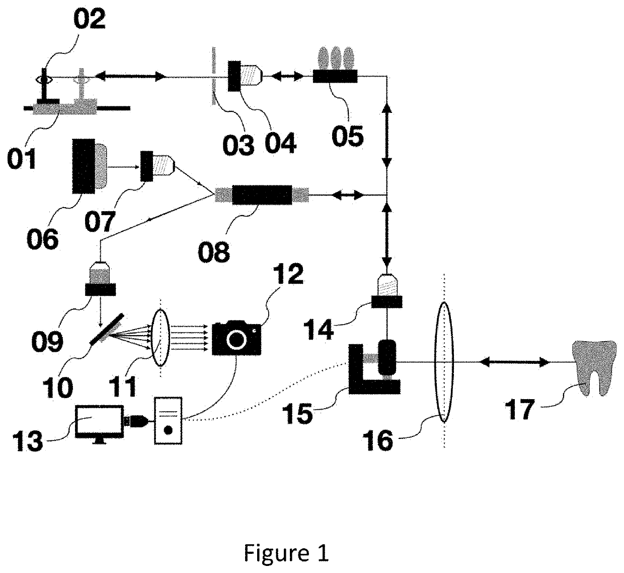
[0031]As shown in FIG. 1, a visual imaging device based on PS-OCT comprises a diode laser light source, a reference arm, a sample arm and a spectrometer. A polarized diode laser light from a laser light source 06 is coupled by passing through an optical fiber coupler 07, input into a coupler 08 and then input into the reference arm and the sample arm respectively. The light beam which enters the reference arm is polarized by passing through a polarizer 05 to obtain a polarized light. The polarized light is adjusted by passing through an optical fiber coupler 04 and an optical grating 03, then passed through an optical path length adjuster 01 and reflected by a reflecting mirror 02 to form a reference arm beam. The light beam which enters the sample arm passes through an optical fiber coupler 14 to reach a scanning galvanometer 15. The scanning galvanometer 15 includes a galvanometer in X-axis direction and a galvanometer in Y-axis direction. The vibration of the scanning galvanomete...


PUM


Abstract

Disclosed is a visual imaging device based on PS-OCT for early demineralization and caries of dental hard tissues, comprising a laser light source for emitting a laser light, a coupler for receiving and dividing the laser light emitted by the laser light source into a reference laser light and a detection laser light; wherein the reference laser light backtracking to the coupler and the detection laser light backtracking to the coupler are coupled and then passed through a transmission grating and a convex lens to input as an optical signal into a linear CCD detector for converting the optical signal into an electrical signal which is then input to a computer control system and collected by a built-in capture card; and the computer is configured to perform a three-dimensional reconstruction of an image and perform two-dimensional cross-section image analysis; and the computer control system is connected to the scanning galvanometer to control a vibration of the scanning galvanometer. The device of the present application can be used for performing PS-OCT imaging detection of the dental hard tissue surface in the oral cavity of a subject. The computer control system automatically performs two-dimensional cross-section imaging and three-dimensional reconstruction imaging of the image, thereby completing a three-dimensional quantitative evaluation. The present application provides a new method for clinically detecting early demineralization of dental hard tissue with high resolution.

Description

CROSS REFERENCE TO RELATED APPLICATION[0001]This application claims the benefit of Chinese Patent Application No. 202010357732.1, filed on Apr. 29, 2020, the entire disclosure of which is incorporated herein by reference in its entirety for all intended purposes.TECHNICAL FIELD[0002]The present application relates to the field of medical equipment, and in particular to a visual imaging device based on Polarization Sensitive-Optical Coherence Tomography (PS-OCT) for early demineralization and caries of dental hard tissues and a method of use thereof.BACKGROUND[0003]Optical Coherence Tomography (OCT) is an optical imaging technology that has developed rapidly in the past ten years. The technology involves use of the basic principles of weak coherence light interferometers for detecting signals of back reflections or several scatterings of incident weak coherent light at different depth levels of biological tissues, and obtaining two-dimensional or three-dimensional images of biologica...

Claims


Application Information

Patent Timeline
IPC IPC(8): A61B5/00A61B1/05
CPCA61B5/0088A61B5/0066A61B1/051A61B5/7475A61B5/742A61B5/4547G06T11/003G06T17/00G06T2210/41A61B1/247A61B1/00172A61B1/00009A61B1/0615A61B1/042A61B1/045A61B1/00117A61B1/00177A61B1/00042A61B1/00194
Inventor ZHAO, YINGBAI, YUHAODUAN, XIAOLEI
Owner XUANWU HOSPITAL OF CAPITAL UNIV OF MEDICAL SCI
Features
  • R&D
  • Intellectual Property
  • Life Sciences
  • Materials
  • Tech Scout
  • Unparalleled Data Quality
  • Higher Quality Content
  • 60% Fewer Hallucinations
Learn More