Ultrasound-mediated gene and drug delivery

a gene and gene technology, applied in the field of ultrasonic/sonic/infrasonic diagnostics, therapy, genetic material ingredients, etc., can solve the problems of direct injection to tissue-specific sites, the challenge of traversing the plasma membrane of target cells, and the challenge of systemic administration of genetic vectors, so as to reduce associated cell damage, increase transgene expression, and increase the effect of gene transfer efficiency

Pending Publication Date: 2021-12-02
SEATTLE CHILDRENS HOSPITAL
View PDF0 Cites 0 Cited by
  • Summary
  • Abstract
  • Description
  • Claims
  • Application Information

AI Technical Summary

Benefits of technology

[0014]While ultrasound-mediated gene delivery (UMGD) has been accomplished using high peak negative pressures (PNPs) of 2 MPa or above, it may not be a requirement for microbubble (MB) cavitation. Thus, lower-pressure conditions close to the MB inertial cavitation threshold were investigated, and additional efforts were directed towards increasing gene transfer efficiency and reducing associated cell damage. Longer pulse duration conditions yielded significant increase in transgene expression relative to sham with local maxima between 20 J and 100 J energy curves. A local maxima between 1 J and 10 J energy curves was observed in treated mice. Of these, several low pressure conditions showed a decrease in ALT and AST levels while maintaining better or comparable expression to the positive control, indicating a clear benefit to allow for effective transfection with minimized tissue damage versus the high-intensity control. The data presented here indicate that it is possible to eliminate the requirement of high PNPs by prolonging pulse durations for effective UMGD in vitro and in vivo, circumventing the peak power density limitations imposed by piezo-materials used in US transducers. Overall, these results demonstrate the advancement of UMGD technology for achieving efficient gene transfer and potential scalability to larger animal models and human application.
[0015]Thus, transcutaneous, ultrasound-mediated delivery methods for administering a therapeutic compound to a target tissue in a subject are provided. Examples of such methods involve positioning a positionable occluding device (e.g., a balloon catheter) in a blood vessel of the subject such that the resultant blockage is adjacent to the target tissue; engaging the occluding device to occlude outflow from a region adjacent to the target tissue; administering the therapeutic compound to the vessel of the subject such that it is substantially retained adjacent to the target tissue by the occluding device; determining the location of the therapeutic compound and / or a detectable adjunct compound (such as an ultrasound contrast agent, radioisotope, or the like) administered with the therapeutic compound using at least one of diagnostic ultrasound, radiography, or fluorography; administering therapeutic ultrasound energy (sonication) transcutaneously, such that the energy mediates delivery of the therapeutic compound across the vessel wall and into the adjacent target tissue. Specific methods described herein employ a therapeutic US device with at least some of the following features: small form factor, ergonomic to subject, high effective treatment volume, good penetration, operation at 1 MHz, and high peak pressures with low duty cycle. Specifically, such a tUS is used to insonicate microbubbles transcutaneously, for instance where such microbubbles have been localized in the subject and that localization confirmed using one more of diagnostic ultrasound, fluorography, or radiography.

Problems solved by technology

However, use of lipid or polymer encased pDNA may be hindered by difficulty in packaging, expelling genetic load, and avoiding cytoplasmic degradation.
In addition, direct injection to tissue-specific sites faces the challenge of traversing the plasma membrane of target cells.
Alternatively, systemic administration of genetic vectors is also challenged by the multiple barriers hindering entry of pDNA into cells.
Given the 2.7 MPa threshold necessary for effective gene transfer into the liver cells however, acoustic pressures beyond the capabilities of art-recognized piezo-material would be required due to the loss of acoustic energy passing through several tissue layers.

Method used


Image

  • Ultrasound-mediated gene and drug delivery
  • Ultrasound-mediated gene and drug delivery
  • Ultrasound-mediated gene and drug delivery

Examples


example 1

g Pulse Duration in Ultrasound-Mediated Gene Delivery Lowers the Acoustic Pressure Threshold for Efficient Gene Transfer to Cells and Small Animals

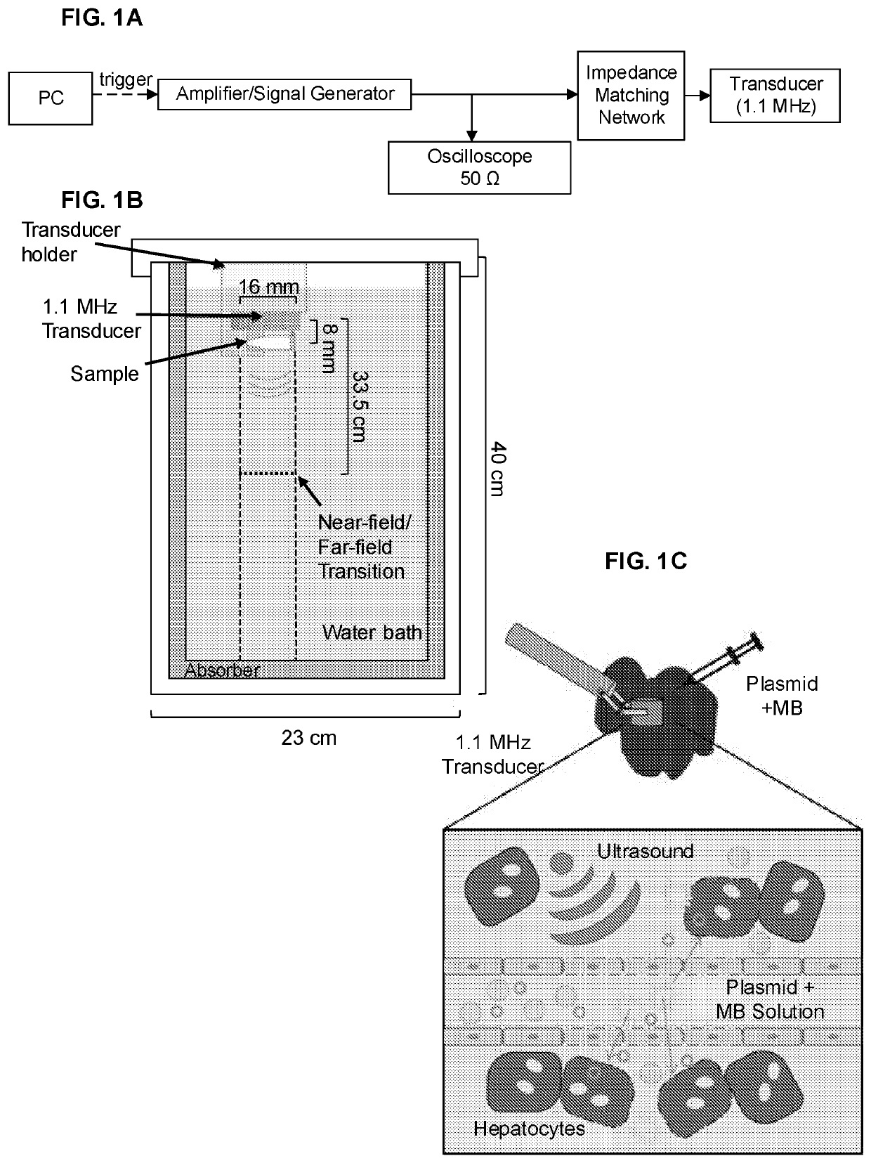
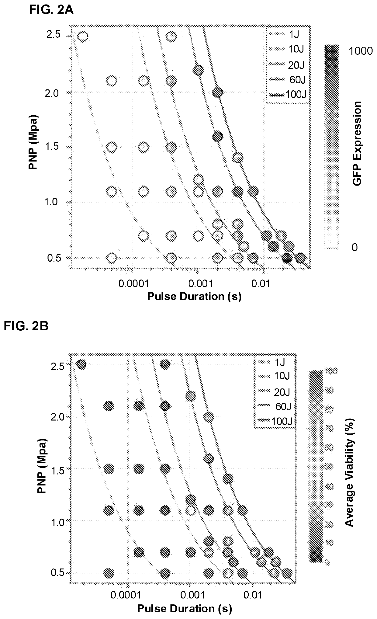
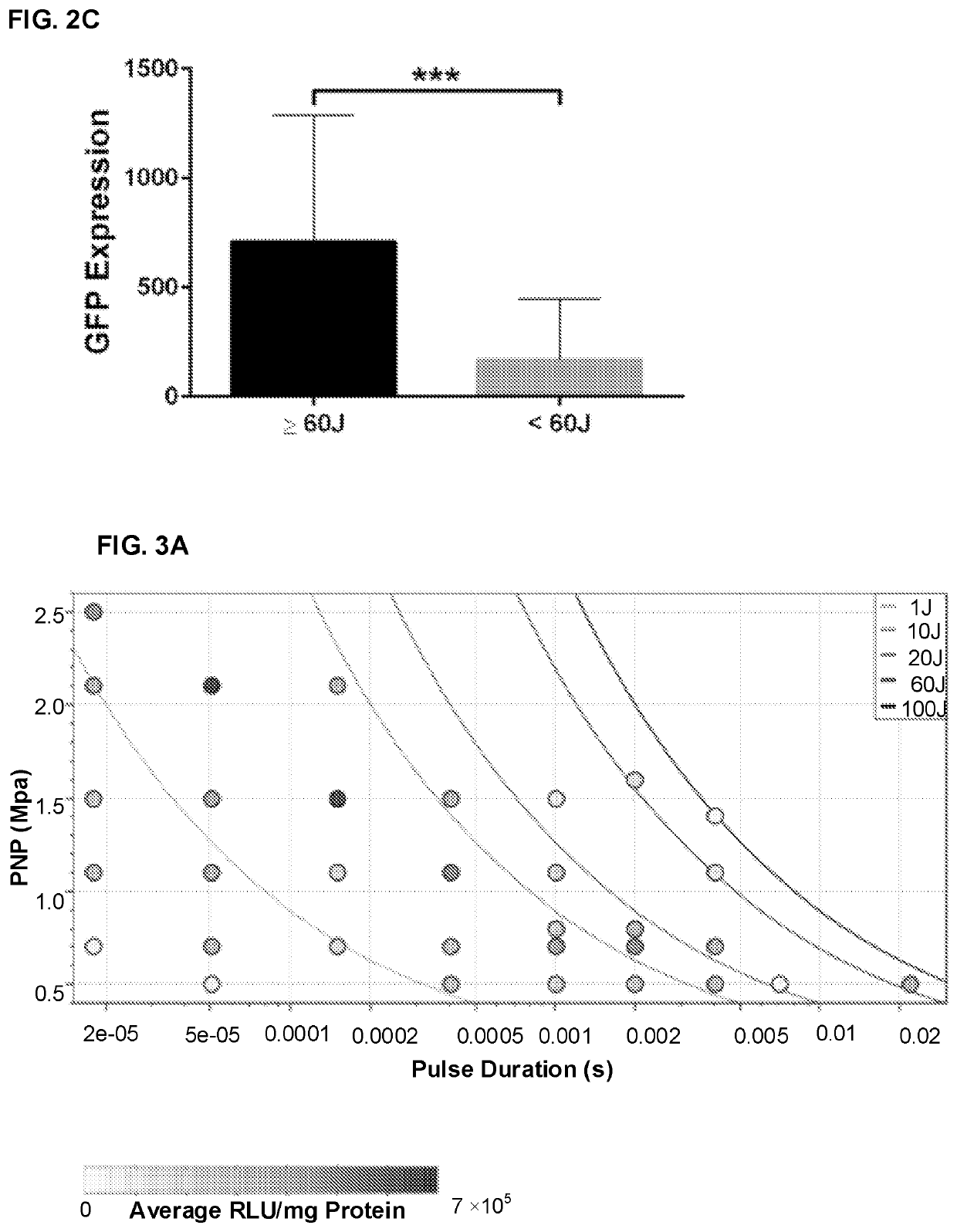
[0115]This Example describes development of clinically applicable procedures involving transcutaneous ultrasound-mediated gene deliver (UMGD). At least some of the material described in this example was published in Tran et al., J Controlled Release 279:345-354, 2018.

[0116]Introduction. Non-viral gene therapy confers appreciable benefits over viral methods including lower risk of immunopathogenicity, greater flexibility in vector construction, and better spatial and temporal control. Delivery of plasmid DNA (pDNA) is particularly attractive as manipulation of the host genome can be avoided and the vector can more easily be engineered for episomal persistence and long-term promoter activation. Ultrasound (US)-mediated gene delivery (UMGD) has long been recognized as a potential method to perform minimally invasive, non-viral gene transfer ...

example 2

Invasive Procedure for Ultrasound-Mediated Non-Viral Gene Delivery to Liver in a Porcine Model

[0160]In this example, changes in beam patterns such as focused, unfocused, or cylindrically focused beams in transducer designs are evaluated. Other changes including number and configuration of elements, or driving center frequency are also considered, as are various US parameter settings and treatment energy, which appear to play a role in UMGD bioeffects.

[0161]Significant gene transfer enhancement was realized using targeted, ultrasound (US)-mediated gene delivery (UMGD) of non-viral vectors in large animal models via an open surgery procedure; see Example 1. The goal to develop a minimally invasive treatment protocol that involves therapeutic US (tUS) across the skin for ease of clinical translation was handicapped because gene transfer efficiency was significantly reduced with transcutaneous UMGD due to US power attenuation across multiple tissue layers. Therefore, different US transd...

example 3

neous Ultrasound-Mediated Nonviral Gene Delivery to the Liver in a Porcine Model

[0178]Ultrasound (US)-mediated gene delivery (UMGD) of non-viral vectors was demonstrated in this study to be an effective method to transfer genes into the livers of large animals via a minimally invasive approach. A transhepatic venous non-viral gene delivery protocol was developed in combination with transcutaneous, therapeutic US (tUS) to facilitate significant gene transfer in pig livers. A balloon catheter was inserted into the pig hepatic veins of the target liver lobes via jugular vein access under fluoroscopic guidance. tUS exposure was continuously applied to the lobe with simultaneous infusion of pGL4 plasmid (encoding a luciferase reporter gene) and microbubbles. tUS was delivered via an unfocused, 2-element disc transducer (H105), or a novel, focused single-element transducer (H114). Supplying transcutaneous US using H114 and H105 with longer pulses and reduced acoustic pressures resulted in...


PUM

PropertyMeasurementUnit
energyaaaaaaaaaa
energyaaaaaaaaaa
frequencyaaaaaaaaaa

Abstract

Transcutaneous, ultrasound-mediated methods for administering compound(s) to subject tissue(s) are provided. Examples involve positioning an occluding device in a vessel such that the blockage is adjacent to target tissue; engaging the device to occlude outflow from a region adjacent to the tissue; administering compound(s) to the vessel such that it is substantially retained adjacent to the target tissue; determining the location of the compound and / or a detectable adjunct compound optionally administered with the compound, using diagnostic ultrasound, radiography, or fluorography; administering therapeutic ultrasound energy transcutaneously to mediate delivery of the compound across the vessel wall and into adjacent target tissue.

Description

CROSS-REFERENCE TO RELATED APPLICATION[0001]This application claims priority to U.S. Provisional Application No. 62 / 663,939 filed on Apr. 27, 2018, which is incorporated herein by reference in its entirety as if fully set forth herein.STATEMENT REGARDING FEDERALLY SPONSORED RESEARCH OR DEVELOPMENT[0002]This invention was made with government support under Grant No. HL128139-01 awarded by the NIH / NHLBI. The government has certain rights in the invention.FIELD OF THE DISCLOSURE[0003]The present disclosure provides systems and methods for administering a compound to a targeted tissue. More particularly, it relates to using ultrasound to deliver a compound (such as a nucleic acid molecule or a drug) to tissue(s) within a subject.BACKGROUND OF THE DISCLOSURE[0004]Non-viral gene therapy confers appreciable benefits over viral methods, including lower risk of immunopathogenicity, greater flexibility in vector construction, and better spatial and temporal control. Delivery of plasmid DNA (p...

Claims


Application Information

Patent Timeline
Patent Type & Authority Applications(United States)
IPC IPC(8): A61M37/00A61K48/00
CPCA61M37/0092A61N2007/0039A61K48/0075A61K9/0009A61B6/12A61B8/0841A61M2025/1052
Inventor MIAO, CAROL HSINGZHANG, FENG
Owner SEATTLE CHILDRENS HOSPITAL
Why Eureka
Try Eureka