Methods of monitoring mucosal healing

a mucosal and mucosal technology, applied in the field of mucosal monitoring, can solve the problems of intestinal perforation and hemorrhage, affecting the integrity of the barrier, and the cost of endoscopic monitoring, so as to achieve the effect of less absorbed and greater barrier integrity

Pending Publication Date: 2022-10-13
MEDIBEACON
View PDF0 Cites 0 Cited by
  • Summary
  • Abstract
  • Description
  • Claims
  • Application Information

AI Technical Summary

Benefits of technology

[0009]Other aspects of the present disclosure relate to methods for determining mucosal healing in a patient with a digestive disease. The method may include enterally administering a dosage of a composition comprising a fluorescent tracer that is not substantially absorbed by a healthy gut, measuring, via fluorescence, an amount of the administered dose that can be found outside the gut over a period of time, and determining a total percentage of the administered dose recovered. In some aspects, the total percentage of the administered dose recovered correlates inversely to the patient's mucosal healing, i.e., the less absorbed, the less excreted or measured via the skin, the greater the barrier integrity.

Problems solved by technology

However, patients with inflammatory bowel diseases (IBD) undergo many endoscopies, which can be a source of dissatisfaction, particularly because of the bowel prep.
Endoscopic monitoring is also expensive (including time off from work), and, on occasion, can cause intestinal perforation and hemorrhage.
While the DSAT is considered state of the art for assessing gut permeability, several limitations diminish its utility.
Most of these are technical limitations related to specimen timing, handling, and assay variability, each of which reduces the theoretical appeal of the DSAT.

Method used


Image

  • Methods of monitoring mucosal healing
  • Methods of monitoring mucosal healing
  • Methods of monitoring mucosal healing

Examples


example 1

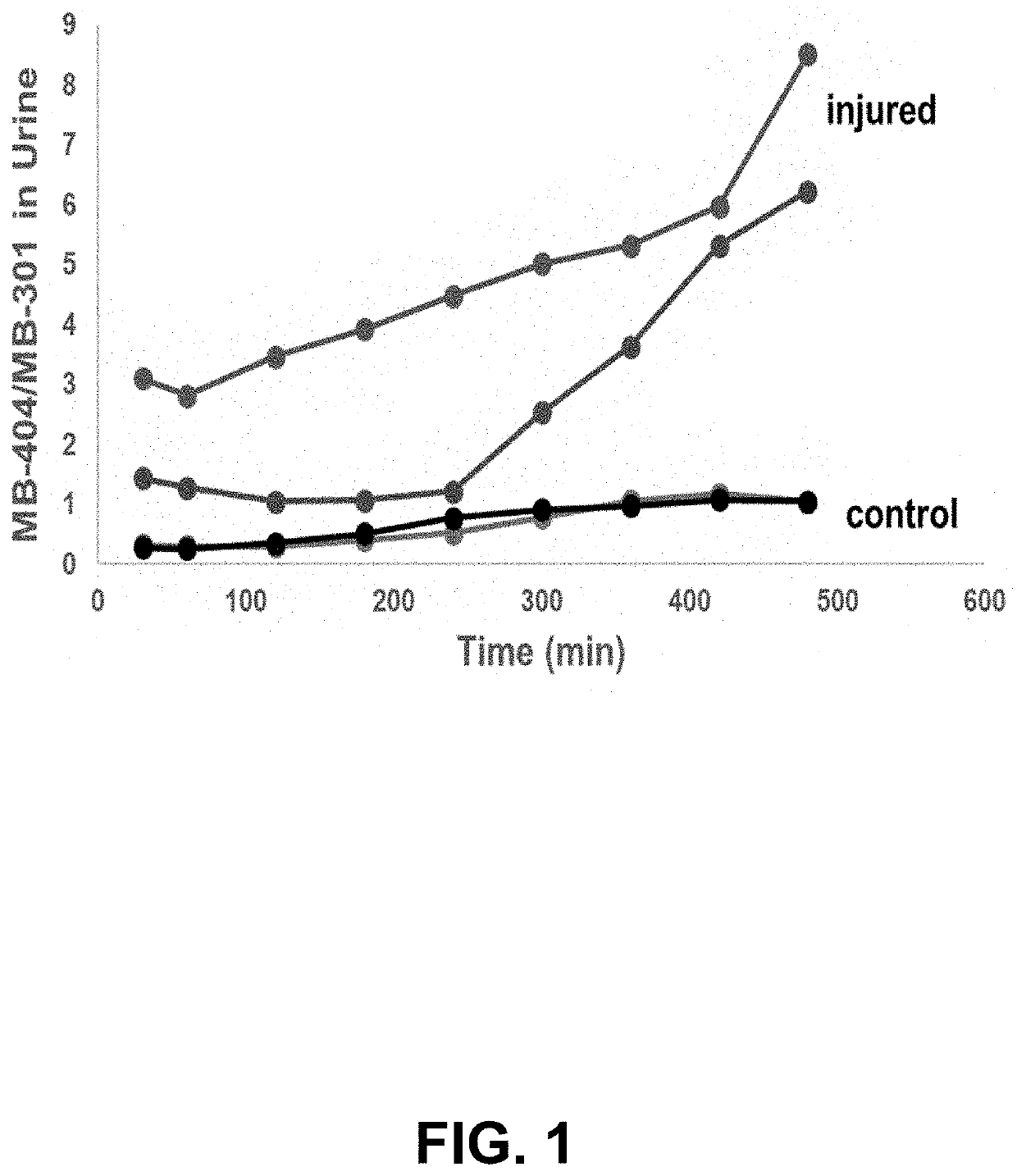
tio of Fluorophores Recovered in Urine of Rats with Small Bowel Injury

[0056]DSAT was mimicked using two fluorophores with similar molecular weights to mannitol and lactulose, MB-301 and MB-404 (see Table 2). Like lactulose, MB-404 is too large to be absorbed by a healthy gut, but can traverse intercellular spaces in a diseased gut exhibiting increased permeability.

TABLE 2Fluorescent Tracer AgentsTracer AgentFluorescent tracer agentsNameMB-301MB-102MB-404USANrelmapirazinMolecular198372492Weight (Da)Light405 / 540445 / 560500 / 620absorption / emissionspectra (nm)StructureScientific3,6-diamino-2,5-3,6-diamino-2,5-bis{N-[(1R)-1-N2,N5-bis(2,3-dihydroxypropyl)-namepyrazine-carboxy-2-3,6-bis[(S)-2,3-dicarboxylic acidhydroxyethyl]carbamoyl}pyrazinedihydroxypropylamino]pyrazine-2,5-dicarboxamide

[0057]Furthermore, like both sugars used in the DSAT, MB-301 and MB-404 are not metabolized and are excreted by glomerular filtration, with neither (or negligible) tubular secretion nor reabsorption. To test...

example 2

al Detection of High Molecular Weight Fluorophore in Rat Model of Bowel Injury

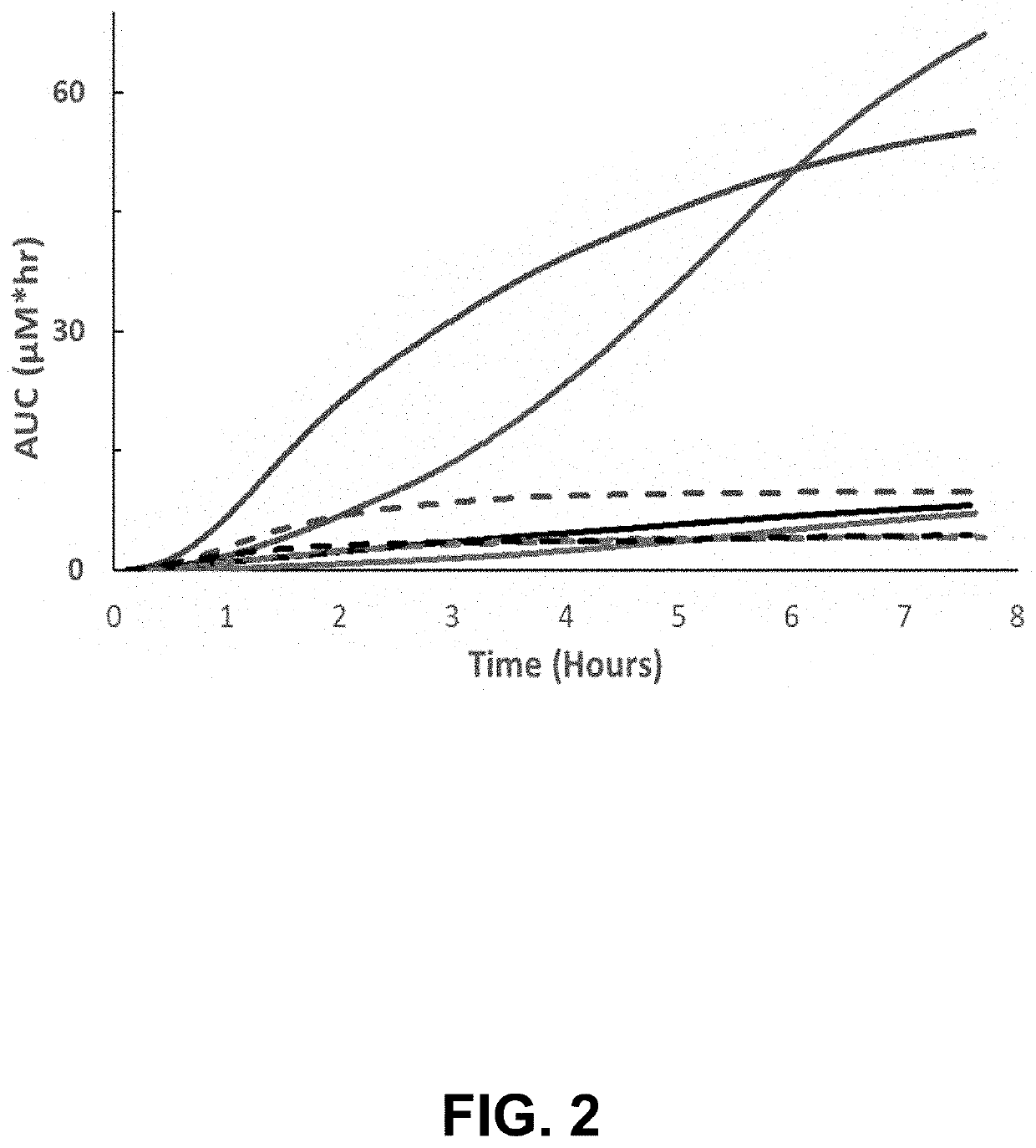
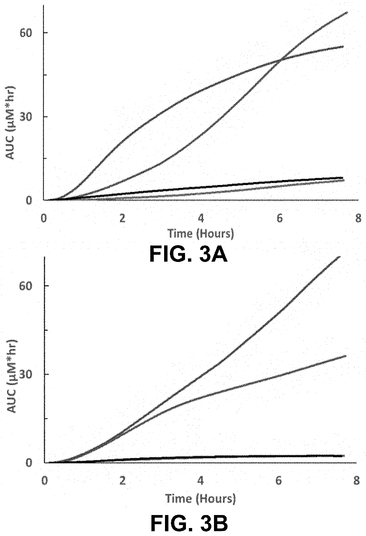
[0059]To probe the ability to assess gut function in a specimen-free and real-time manner, a dual-wavelength transdermal fluorescence detection system was utilized to monitor MB-301 and MB-404 in the rat model of bowel injury. As the excitation and emission spectra of these fluorophores do not overlap, their presence can be assayed simultaneously. Following anaesthetization, the fluorescence detection system was affixed with medical adhesive to a depilated area on the back of each rat. Transdermal fluorescence monitoring was then initiated to detect baseline fluorescence prior to delivery of MB-301 and MB-404 by oral gavage, and was monitored continuously. The concentration of MB-301 as determined by transdermal fluorescence measurements remained low in both injured and control animals (FIG. 2, dashed lines) as this fluorophore is comparatively rapidly absorbed by the small intestine and subsequently clear...

example 3

ation of Intestinal Permeability in Human Crohn's Disease

[0062]A single center, randomized, open label, crossover study was performed to evaluate a solution containing 18.6 mg / mL of a pyrazine derived fluorophore (MB-102) at 1.5 or 3.0 mg / kg versus DSAT (L, 1000 mg and R, 200 mg) administered enterally. The composition of MB-102 solution for use in this study is provided in Table 3.

TABLE 3Composition of MB-102 solution for oral ingestionQuantity per mL ofComponentFunctionFormulationMB-102Active Ingredient18.824mgSodium dihydrogen phosphateDiluent / Buffer2mgmonohydrate, BP, USPSodium chloride, Ph. EurDiluent / Buffer4.62mgWater for injection, Ph. EurDiluent / BufferTo 1014 mg1N Sodium hydroxideDiluent / BufferpH adjusted to7.0-7.4

[0063]To compare the dynamic ranges of the fluorophore and DSAT clearances, healthy adults and adults with evidence of small bowel Crohn's disease on magnetic resonance enterography were enrolled. Participants were randomized to receive either the fluorophore trace...


PUM

PropertyMeasurementUnit
emission wavelengthsaaaaaaaaaa
molecular weightaaaaaaaaaa

Abstract

The disclosure provides for methods for monitoring mucosal healing in a patient with a digestive disease, or for use in a pre-disease state, and includes intestinal as well as extra-intestinal disorders in which gut permeability is increased. The method may include establishing a baseline of the patient, treating the patient for the digestive disease or the pre-disease state, measuring gut permeability of the patient after treatment, and comparing a second total percentage of the administered dose recovered to the baseline total percentage of the administered dose recovered. Establishing the baseline may include enterally administering a first dosage of a composition comprising a fluorescent tracer, measuring a first amount of the administered dose that can be found outside the gut over a period of time, and determining a baseline total percentage of the administered dose recovered. Measuring gut permeability may include enterally administering a second dosage of the composition, measuring a second amount of the administered dose, and determining a second total percentage of the administered dose recovered.

Description

CROSS-REFERENCE TO RELATED APPLICATIONS[0001]This application claims priority to U.S. Provisional Application No. 63 / 169,568, filed Apr. 1, 2021, the contents of which are entirely incorporated by reference herein.FIELD[0002]The present disclosure relates generally to methods of monitoring intestinal mucosal healing in a patient.BACKGROUND[0003]In the healthy state, the intestinal barrier permits selective translocation (absorption) of water, electrolytes, and nutrients from the gut lumen into the systemic circulation, while preventing entry of undigested food, macromolecules, and microorganisms into the body. Increased gut permeability infers loss of barrier integrity, principally through disruption of tight junctions between epithelial cells. Gut permeability is increased in a variety of intestinal inflammatory conditions, including Crohn's Disease, graft versus host disease, and celiac disease.[0004]Endoscopy has been the primary method to evaluate mucosal healing. Endoscopy enab...

Claims


Application Information

Patent Timeline
Patent Type & Authority Applications(United States)
IPC IPC(8): G01N33/68A61B5/00A61K31/4965G01N33/493
CPCG01N33/6893A61B5/0071A61B5/4238A61K31/4965G01N33/493G01N2800/065A61B5/4848A61B5/4255G01N33/5091G01N2800/06
Inventor TARR, PHILLIP I.DORSHOW, RICHARD B.
Owner MEDIBEACON
Why Eureka
Try Eureka