
Method for identifying mismatch repair glycosylase reactive sites, compounds and uses thereof

a glycosylase and reactive site technology, applied in the field of mismatch repair glycosylase reactive site identification, can solve the problems of severe limitation, essentially restricted gel-electrophoresis-based methods, and mutations in those other genes that are likely not to be identified, so as to shorten the time required to screen tumor samples and improve cost-effectiveness

Inactive Publication Date: 2001-01-16
DANA FARBER CANCER INST INC
View PDF4 Cites 39 Cited by
  • Summary
  • Abstract
  • Description
  • Claims
  • Application Information

AI Technical Summary

Benefits of technology

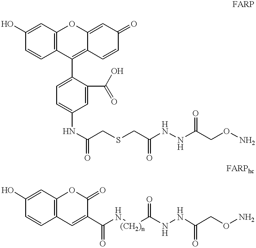
These reactive sites are identified by using a compound containing an aldehyde-binding moiety such as --O--NH2 (-hydroxylamine), or --NHNH2 (-hydrazine) or --NH2 (-amine) and also having a second moiety that reacts with a detectable entity (e.g. fluorescein, biotin, digoxigenin, which respectively react with antifluorescein antibody, avidin, and antidigoxigenin antibody. The antibodies may have chemiluminescence tags on them and thereby are detected). A unique feature of the present approach is that the aldehyde-binding moiety binds covalently to the enzyme-generated reactive sites. Combined with the specificity of the mismatch-repair enzymes, the use of covalently bound ligands to the position of mutations results in a sensitivity and specificity which is unparalleled by other methods for detection of mutations and polymorphisms.
5. With the sample immobilized on solid support, perform: hydroxylamine treatment / washing; MutY / TDG treatment(s) / washing; FARP / BARB / AED labeling / washing. Antibody labeling / washing; Chemiluminescence detection of mismatches. All these steps are very easy and convenient to perform with the DNA immobilized.

Problems solved by technology

A fundamental problem with currently existing mutation and polymorphism detection methods is that they only screen for mutations on a single gene at a time (i.e. the method is directed to looking at a `gene of interest`, that is suspected of having a mutation).
Given that the human genome has 50,000-100,000 genes, this is a severe limitation.
However, mutations in those other genes would likely not be identified.
Gel-electrophoresis-based methods are essentially restricted to examining mutations in a single gene at a time.
These methods, however, have had limited success [Nollau P and Wagener C., Clinical Chemistry 43: 1114-1128 (1997)] since they are complicated, typically requiring several enzymatic steps and they result in a large number of false positives, i.e. they frequently score mutations and polymorphisms in normal DNA.
For example, continuous cellular damages occur to nucleic acids during the cell life cycle; for example damage resulting from exposure to radiation, or to alkylating and oxidative agents, spontaneous hydrolysis and errors during replication.
Closely-spaced abasic sites generated within a few base pairs of each other by damaging agents may be a particularly significant set of lesions, as they may hinder repair [Chaudhry and Weinfeld, 1995a, 1997; Harrison et al., 1998], or they can be enzymatically converted to double strand breaks or other complex multiply-damaged sites [Dianov et al., 1991].
It has been postulated that such complex forms of DNA damage may be particularly difficult for cells to overcome [Ward 1985, 1988; Wallace, 1988; Goodhead, 1994; Chaudhry and Weinfeld, 1995a and b, and 1997; Rydberg, 1996; Hodgkins et al., 1996; Nikjoo et al., 1998; Harrison et al., 1998].
The binding efficiency of such systems has been relatively low.

Method used


Image

  • Method for identifying mismatch repair glycosylase reactive sites, compounds and uses thereof
  • Method for identifying mismatch repair glycosylase reactive sites, compounds and uses thereof
  • Method for identifying mismatch repair glycosylase reactive sites, compounds and uses thereof

Examples


example 2

BARP-Based Detection of Mismatches Formed via Self-Complementation of Single-Stranded M13 DNA

Samples of M13 single stranded DNA that contain approximately 1 MutY-recognizable mismatch per 2,500 bases were treated with MutY to generate aldehyde-containing reactive sites appropriate for reaction with BARP. Nominal gel electrophoretic studies as well as BARP-based chemiluminescent studies were then preformed. Control samples used were: Single stranded M13 without enzymatic treatment; Double stranded M13 DNA without any mismatches and no enzyme treatment; and double stranded M13 DNA without mismatches and enzyme. FIG. 8 (A and B) shows the results of both methods of detection. FIG. 8A (luminescence studies) show that only when mismatches are present (single stranded M13) and MutY is used is there a chemiluminescence signal. In agreement, gel electrophoresis (FIG. 8B) shows cuts in M13 are only generated under the same conditions. It can be seen that there is good agreement among the two...

example 3

Detection and Isolation of DNA Containing Base-Substitution Mutations: Detection of a Single A-To-C Transversion Engineered in a P53 Gene within a 7091-Long Plasmid

The ability of the present technology (A.L.B.U.M.S) to detect base mismatches (demonstrated in previous examples) is directly applicable to detection of base substitution mutations. For example, a standard procedure to generate mismatches at the positions of mutations in DNA, is to mix mutation-containing DNA with wild-type DNA. Upon heating and re-hybridization of the mixture, heteroduplexes with mismatches are generated at the positions of mutations (FIG. 1), which can then be detected with high sensitivity and specificity as demonstrated in example 1.

To isolate mutation-containing DNA from normal DNA, following BARP-labeling of the generated aldehydes at positions of mismatches (FIG. 1) the DNA is immobilized on neutravidin-coated microplates, followed by exhaustive washing to remove the homoduplex DNA. As a result, on...

example 4

Comparison of Small Versus Large Ligand Compounds in Binding to MutY- or TDG-Generated Reactive Sites in DNA: Synthesis and Advantage of AED Versus BARP and FARP. Chemiluminescence Signals by AED.

(a) To synthesize AED, O-(Carboxymethyl)hydroxylamine hydrochloride was conjugated to ethylenediamine (Aldrich) in distilled water using 1-Ethyl-3-[3-(dimethylamino)propyl] carbodiimide (EDAC) as the coupling reagent. An 100-fold excess of ethylenediamine over O-(Carboxymethyl)hydroxylamine hydrochloride was utilized during the reaction to allow preferential coupling of ethylenediamine to the carboxyl groups. The conditions for the catalysis of this reaction by EDAC is well known to those skilled in the art. TLC analysis and purification on silica gel with CHCl.sub.3 :CH.sub.3 OH:CH.sub.3 COOH in a 70:20:5 ratio indicated the product at an R.sub.f of 0.2-0.25. The certificate of analysis provided 1H NMR data consistent with the AED structure provided earlier.

(b) The ability of hydroxylamine...


PUM

PropertyMeasurementUnit
Molecular weightaaaaaaaaaa
Fluorescenceaaaaaaaaaa

Abstract

The present application discloses a method of identifying mutations in a target DNA sequence. The method involves:(a) hybridizing the target DNA sequence with a control DNA sequence wherein said control DNA sequence is the wild-type DNA sequence corresponding to the target DNA sequence to create a duplex;(b) treating the duplex to remove any spontaneous aldehydes;(c) reacting the duplex with a repair glycosylase to convert any mismatched sites in the duplex to reactive sites containing an aldehyde-containing abasic site;(d) reacting the duplex with a compound of the formula X-Z-Y, wherein X is a detectable moiety, Y is NHNH2, O-NH2 or NH2, and Z is a hydrocarbon, alkyhydroxy, alkylethoxy, alkylester, alkylether, alkylamide or alkylamine, wherein Z may be substituted or unsubstituted; and wherein Z may contain a cleavable group; for a sufficient time and under conditions to covalently bind to the reactive sites;(e) detecting the bound compound to identify sites of mismatches;(f) determining where the mismatch occurs; and(g) determining whether the mismatch is a mutation or polymorphisms.

Description

The present invention is directed to methods for identifying and labeling glycosylase-recognizable sites on nucleic acids, and to novel compounds that bind to such glycosylase-recognizable sites on nucleic acids. In a preferred embodiment the method can be used to identify mutations and / or polymorphisms on a nucleic acid segment, or in an arbitrary mixture of nucleic acid segments or genes.The detection of mutations has been an area of great interest in recent years. For example, mutations in certain genes have been associated with a variety of disorders--ranging from blood disorders to cancers. Genetic tests are thus becoming an increasingly important facet of medical care. Consequently, there has been an emphasis on the ability to rapidly and efficiently detect mutations and polymorphisms.Many electrophoretic techniques have been developed to rapidly screen DNAs for sequence differences by which such mutations can be detected. Denaturing Gradient Get Electrophoresis (DGGE) [Myers,...

Claims


Application Information

Patent Timeline
IPC IPC(8): C12Q1/68C12Q1/6827C12Q1/6837
CPCC12Q1/6827C12Q1/6837
Inventor MAKRIGIORGOS, GERASSIMOS M.
Owner DANA FARBER CANCER INST INC
Learn More