
Materials and methods relating to the attachment and display of substances on cell surfaces

Inactive Publication Date: 2001-02-20
INTREXON ACTOBIOTICS NV
View PDF3 Cites 37 Cited by
  • Summary
  • Abstract
  • Description
  • Claims
  • Application Information

AI Technical Summary

Benefits of technology

The high level of heterologous protein expression which can be achieved using the T7 polymerase system has been found to provide high levels of expression of the Protein A-fusion polypeptide on the surface of the bacteria. For many of the applications envisaged for the invention, the economics of the associated processes will be materially favoured by achieving the highest possible surface density of fusion molecules on the bacteria. However, for some applications the use of constitutive, or less active, promoters may give useful results, especially where lower levels of expression are required.

Problems solved by technology

However, in Gram-negative bacteria it has proved difficult to find an adaptable method of displaying polypeptides on the surface of the cells so that they are securely attached to the surface at high density.
The possibility of displaying foreign proteins by making C-terminal fusions to integral membrane proteins is complicated by the fact that the C-terminal regions of many integral membrane proteins appears to be necessary for targeting and correct assembly of the protein in the outer membrane.
The major problem with this approach has been that large amino acid insertions / substitutions disrupt the proper folding and / or localisation of the fusion protein in such a way that when used to immobilise the bacterial cells, the fusion proteins are extracted from the outer membrane (Steidler, PhD Thesis, University of Gent, Belgium).
Although significant progress has recently been made in the development of methods from the surface display of proteins in Gram-negative bacteria, the major drawback to the use of integral membrane proteins for such purposes is that the fusion proteins are not firmly attached to the cell surface.
Furthermore, the high level expression of membrane proteins is known to be detrimental to the cell, a factor which may limit the possible applications for such systems in cases where high density surface expression is required.
By comparison with E. coli, as relatively few heterologous proteins have been expressed at high level in L. lactis, before the present work it was impossible to predict with certainty whether the expression of any particular protein would be possible or whether such expression would be toxic to the cell.
Such toxicity is likely to occur when a secreted or membrane associated protein is expressed at high level, as protein export may adversely affect the integrity of the cell membrane or the function of the general secretory pathway.
As is shown below, it is not possible to remove the protein by boiling the cells in the presence of an ionic detergent which would normally release non-covalently bound proteins.
This binding could not have been readily predicted from the possession by protein A of the LPXTG motif and C-terminal hydrophobic domains common to many surface proteins of Gram-positive bacteria, since these motifs are just necessary, but not sufficient conditions for the occurrence of covalent cell wall anchoring.
However, the need for an abundant supply of an immobilised form of the ligand can render affinity chromatography uneconomic.
They may also inactivate anti-microbial substances such as antibiotics or toxins emanating from other organisms; and they may degrade toxic or recalcitrant substances left in the environment by geological or industrial processes.
Although methods for producing bifunctional heterodimers are known, there are at present no generally applicable methods available for separating them from the unavoidably co-produced homodimeric (hence monofunctional) molecules.

Method used


Image

  • Materials and methods relating to the attachment and display of substances on cell surfaces

Examples


example 2

Display of Tetanus Toxin Fragment C (TTFC) Antigen on the Surface of L. lactis

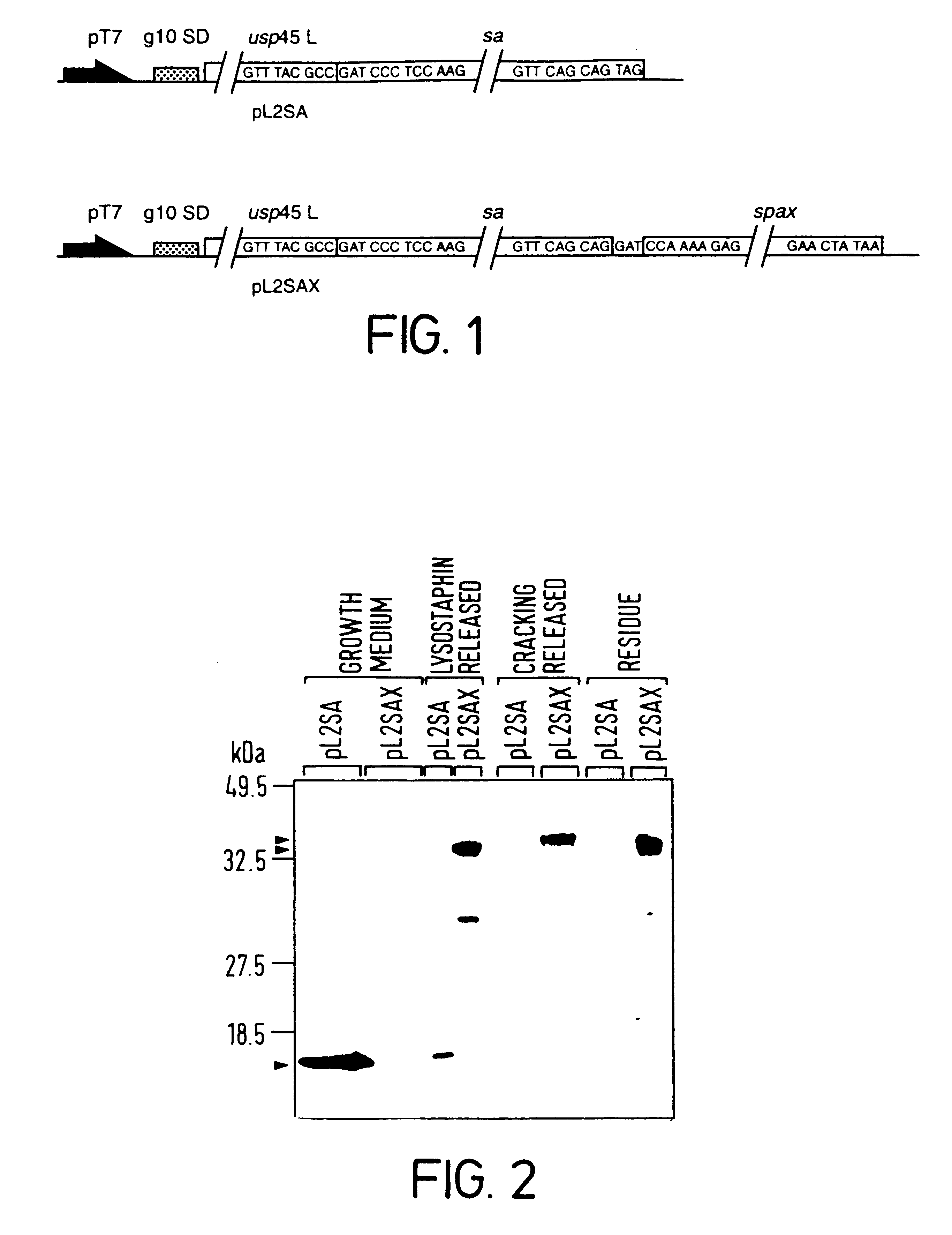
This additional example provides evidence that protein antigens can be firmly attached to the surface of L. lactis by constructing chimeric genes encoding a fusion protein which contains nucleic acid encoding (1) an N-terminal secretion signal (2) the protein antigen to be surface expressed and (3) the cell wall sorting and anchoring domain of S. aureus protein A.

Stage 1: Cloning and genetic manipulation of TTFC and the S. aureus protein A anchoring domain.

(1a) Construction of pTREX and pTREX1

Synthetic oligonucleotides encoding a putative RNA stabilising sequence, a translation initiation region and a multiple cloning site for target genes were annealed by boiling 20 .mu.g of each oligonucleotide in 200 ul of 1.times.TBE, 150 mM NaCl and allowing to cool to room temperature. Annealed oligonucleotides were extended using Tfl DNA polymerase in 1.times.Tfl buffer containing 250 .mu.M deoxynucleotide triphosph...


PUM

PropertyMeasurementUnit
Volumeaaaaaaaaaa
Compositionaaaaaaaaaa
Surfaceaaaaaaaaaa

Abstract

Methods for obtaining surface expression of a desired protein or polypeptide in Gram-positive host organisms are provided. In addition, vectors useful in such methods as well as Gram-positive host organisms transformed with such vectors are disclosed.

Description

The present invention relates to materials and methods for the exportation, cell wall attachment and display of substances, in particular proteins and polypeptides, on the cell surface of Gram-positive bacteria such as Lactococcus lactis. Methods are disclosed providing for the surface expression of cell wall anchoring of proteins, including antigens, specific binding proteins and enzymes.BACKGROUND TO THE INVENTIONThe display of polypeptides on the surface of bacteria has been a subject of investigation for several years. This interest has arisen because recombinant DNA technology makes it possible to use bacterial cells as factories for the inexpensive production of a wide range of different proteins. Work has centred on the display of polypeptides on the surface of Gram-negative bacteria, see for example the review provided in U.S. Pat. No. 5,348,867. However, in Gram-negative bacteria it has proved difficult to find an adaptable method of displaying polypeptides on the surface o...

Claims


Application Information

Patent Timeline
IPC IPC(8): C07K14/195C07K14/36C07K14/31C07K14/33C12N15/62C12N11/16C12N15/74C12N11/00A61K38/00A61K39/00G01N33/569A61K39/08C12N1/21C12N11/02C12N15/00C12N15/09C12N15/31C12Q1/02C12R1/445
CPCC07K14/31C07K14/33C07K14/36C12N11/16C12N15/625C12N15/746A61K38/00A61K39/00C07K2319/00C07K2319/02C07K2319/035C07K2319/22C07K2319/55C12N15/74A61K39/08C12N15/11
Inventor STEIDLER, LOTHARREMAUT, ERIKWELLS, JEREMY MARK
Owner INTREXON ACTOBIOTICS NV
Learn More