

Peptides and peptidomimetics with structural similarity to human P53 that activate P53 function
a technology of p53 and peptides, applied in the field of peptides and peptidomimetics with structural similarity to human p53 that activate p53 function, can solve the problems of affecting the function of the antibody pab421, and the futility of administering it to a patient for this purpos
- Summary
- Abstract
- Description
- Claims
- Application Information
AI Technical Summary
Benefits of technology
Problems solved by technology
Method used
Image

Examples
example 1
Synthesis of Peptides
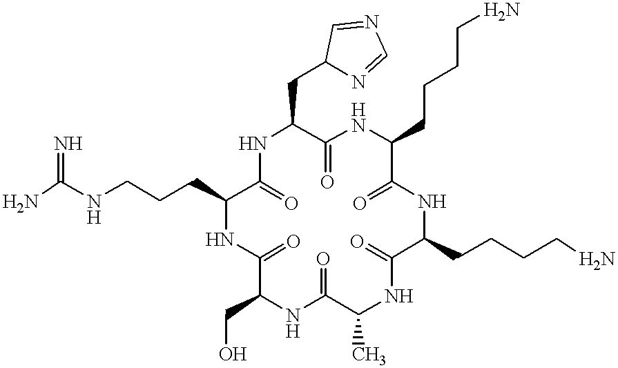
Peptides corresponding to the negative regulatory region 1 (NRR1) spanning residues 361-383 of human p53 were synthesized. Peptides were assembled on a Milligen 9050 automated synthesizer using the standard Fmoc-protocol [Fields and Noble (1990), Int. J. Pept. Protein Res., 35: 161-214]. After cleavage, the peptides were purified by reverse phase HPLC using a C18 column and a linear gradient of acetonitrile in 0.1% aqueous trifluoroacetic acid. The efficiency and accuracy of synthesis was monitored by amino acid composition analysis and / or mass spectroscopy. The present invention is not limited by the specific chemistry or synthesizer used to prepare the peptides of this invention; and such factors are not critical for the activities of the peptides of this invention.
The following peptides were synthesized:
All peptides were acetylated at their N-terminus and contained an amide chemical group attached to their C-terminus.
Amino acid composition analysis and mass s...
example 2
DNA Binding Assay
The ability of the peptides of this invention to activate DNA binding of human p53 was assayed using a standard DNA binding assay for p53 [Halazonetis et al. (1993), cited above; Halazonetis and Kandil (1993), cited above]. This assay utilizes in vitro translated human wild-type p53 or tumor-derived p53 mutants and specific oligonucleotides containing p53 binding sites. These reagents and methods for preparing them are described below.
Human p53 was produced by in vitro translation using plasmids containing the full-length p53 coding sequence. Standard cloning procedures [Ausubel et al. (1994), "Current Protocols in Molecular Biology," Greene Publishing Associates and John Wiley & Sons, New York; Innis et al. (1990), PCR Protocols: A Guide to Methods and Applications, Academic Press, San Diego] were used to prepare these plasmids.
Reagents
Plasmid pGEMhump53wt encodes full-length human wild-type p53. This plasmid was prepared by PCR [Innis et al. (1990), cited above] u...
example 3
Mapping of Negative Regulatory Regions Within Wild-type p53
The p53 DNA binding assay involves incubating in vitro translated p53 with radioactively-labeled DNAs containing p53 binding sites as described in Example 2 above. For these studies, incubation was performed with oligonucleotides BC.V4A. Wild-type p53 binds to this oligonucleotide. However, its DNA binding activity is subject to downregulation by the negative regulatory region 1 (NRR1), which maps to the C-terminus of p53. Thus deletion of residues 364-393 of human p53 activates DNA binding [Hupp et al. (1992), cited above; Halazonetis and Kandil (1993), cited above]. We reproduced this result using oligonucleotide BC.V4A and plasmid pGEMhump53D364-393. However, DNA binding of a deletion mutant lacking residues 353-360 was not activated (see Table 1).
To map more finely the NRR1 and to identify other NRR that might be present in the C-terminus of p53, we examined the DNA binding activities of the p53 deletion mutants describe...
PUM
| Property | Measurement | Unit |
|---|---|---|
| molecular weight | aaaaa | aaaaa |
| molecular weight | aaaaa | aaaaa |
| molecular weight | aaaaa | aaaaa |
Abstract
Description
Claims
Application Information