Kits & systems for ultrasonic imaging

a technology of ultrasonic imaging and ultrasonic imaging, applied in the field of ultrasonic imaging kits & systems, can solve the problems of inability to provide contrast enhancement lasting even a few seconds, let alone minutes, and the use of extremely expensive equipmen

Inactive Publication Date: 2001-08-28
TARON +1
View PDF110 Cites 21 Cited by
  • Summary
  • Abstract
  • Description
  • Claims
  • Application Information

AI Technical Summary

Benefits of technology

This spray dried composition, comprising gas-filled hollow microspheres, is an important product of the present invention. This product provides significant advantages over lyophilized liposome containing precursors. It is believed that the spherical structure of the present microbubble precursor material serves to quickly and uniformly form the relatively insoluble, relatively incompressible non-Newtonian viscoelastic surfactant film that is characteristic of preferred microbubbles of the present invention.

Problems solved by technology

While numerous conventional imaging technologies are available, e.g., magnetic resonance imaging (MRI), computerized tomography (CT), and positron emission tomography (PET), each of these techniques use extremely expensive equipment.
Moreover, ultrasound imaging does not use ionizing radiation.
However, such bubbles tend to shrink rapidly due to the diffusion of the trapped gas into the surrounding liquid.
Most microbubble compositions have failed to provide contrast enhancement that lasts even a few seconds, let alone minutes.
This greatly limits their usefulness.
However, solid phase shells that encapsulate gases have generally proven too fragile or too permeable to the gas to have satisfactory in vivo life.
Furthermore, thick shells (e.g., albumin, sugar, or other viscous materials) reduce the compressibility of the bubbles, thereby reducing their echogenicity during the short time they can exist.
Solid particles or liquid emulsion droplets that evolve gas or boil when injected (as in Quay, PCT / U.S. Pat. No. 94 / 00,422) pose the danger of supersaturating the blood with the gas or vapor.
This will lead to a small number of large embolizing bubbles forming at the few available nucleation sites rather than the intended large number of small bubbles.
In addition, bubbles created in vivo in this way will be "naked", and consequently will have the complement activation problem described above.
Moreover, liposome preparation involves sophisticated and time consuming manufacturing.
Although reduced water solubility and diffusivity can affect the rate at which the gas leaves the bubble, numerous problems remain with the Quay bubbles.
This is a disadvantage in that sophisticated bubble preparation systems must be provided at the site of use.
Moreover, The Quay gas selection criteria are incorrect in that they fail to consider certain major causes of bubble shrinkage, namely, the effects of bubble surface tension, surfactants and gas osmotic effects, and these errors result in the inclusion of certain unsuitable gases and the exclusion of certain optimally suitable gases.
Typical ultrasound contrast enhancement agents exhibit contrast enhancement potential for only about one pass through the arterial system, or a few seconds to about a minute, and thus do not survive past the aorta in a patient following intravenous injection.
While bubbles have been shown to be the most efficient ultrasound scatterers for use in intravenous ultrasound contrast agents, their main practical drawback is the extremely short lifetime of the small (typically less than 5 microns diameter) bubbles required to pass through capillaries in suspension.
This short lifetime is caused by the increased gas pressure inside the bubble, which results from the surface tension forces acting on the bubble.
The increased internal gas pressure forces the gas inside the bubble to dissolve, resulting in bubble collapse as the gas is forced into solution.
That is, they apparently evade the reticuloendothelial system more effectively than liposomes, and are thus not cleared from circulation as quickly.
The water at the surface of the droplet evaporates very rapidly, causing a buildup of the compounds which were dissolved in the atomized solution.
However, when a mixture of a modifier gases (e.g., air or carbon dioxide) are used with a gas osmotic agent (e.g., C.sub.6 F.sub.14) a slight reduction in volume of the smaller bubbles, due to one of the modifier gases leaving the bubble, will concentrate the osmotic gas and increases its osmotic pressure, thus, retarding further shrinkage.
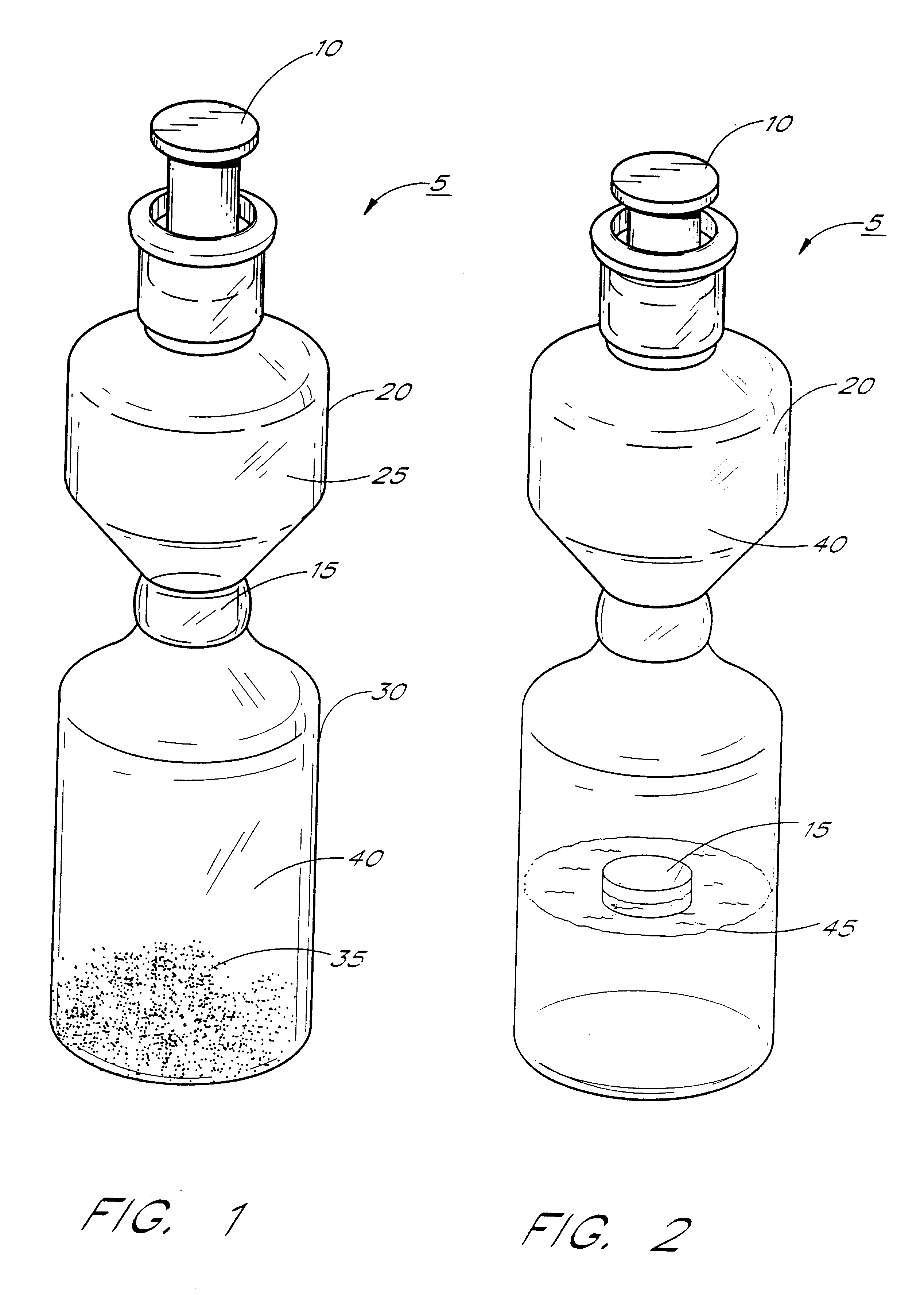
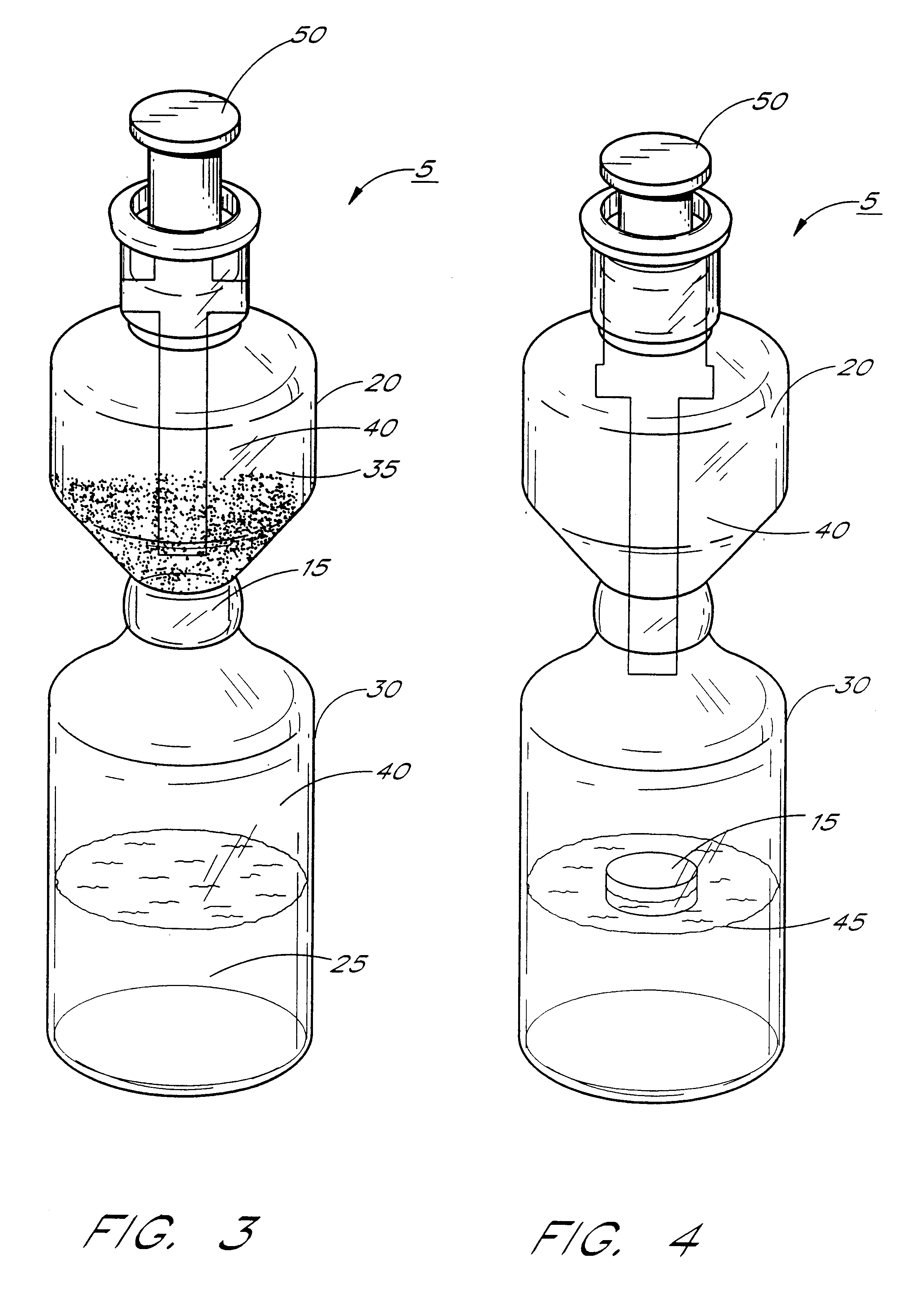
(In contrast, conventional reconstitution of a solute in a single chamber vial with a needle and syringe without a vent can result in the production of considerable intrachamber pressure which could collapse the microbubbles.)

Method used


Image

  • Kits & systems for ultrasonic imaging
  • Kits & systems for ultrasonic imaging

Examples


example ii

Comparison of phospholipid vs. sucrose ester gas emulsions

One liter of each of the following four solutions was prepared with water for injection:

Solution 1:

3.9% w / v m-HES hydroxyethylstarch (Ajinimoto, Tokyo, Japan)

3.25% w / v Sodium chloride (Mallinckrodt, St. Louis, Mo.)

2.83% Sodium phosphate, dibasic (Mallinckrodt, St. Louis, Mo.)

0.42% w / v Sodium phosphate, monobasic (Mallinckrodt, St:. Louis, Mo.)

Solution 2:

2.11% w / v Poloxamer 188 (BASF, Parsipany, N.J.)

0.32% w / v Ryoto Sucrose Stearate S-1670 (Mitsubishi-Kasei Food Corp., Tokyo, Japan)

0.16% w / v Ryoto Sucrose Stearate S-570 (Mitsubishi-Kasei Food Corp., Tokyo, Japan)

Solution 3:

3.6% w / v m-HES hydroxyethylstarch (Ajinimoto, Tokyo, Japan)

3.0% w / v Sodium chloride (Mallinckrodt, St. Louis, Mo.)

2.66 Sodium phosphate, dibasic (Mallinckrodt, St. Louis, Mo.)

0.39% w / v Sodium phosphate, monobasic (Mallinckrodt, St. Louis, Mo.)

Solution 4:

0.15% w / v Poloxamer 188 (BASF, Parsipany, N.J.)

0.45% w / v Hydrogenated Egg phosphotidylcholine EPC-3 (Lipoi...

example iii

Comparison of water insoluble phospholipid vs. water insoluble phospholivid / water soluble surfactant (Poloxamer 188) microbubbles

One liter of each of the following emulsions was prepared for spray drying as described in Example II:

Formulation A: Water Insoluble Phospholipid Formulation

3.6% w / v m-HES hydroxyethylstarch (Ajinimoto, Tokyo, Japan)

3.0% w / v Sodium chloride (Mallinckrodt, St. Louis, Mo.)

2.6% w / v Sodium phosphate, dibasic (Mallinckrodt, St. Louis, Mo.)

0.39% w / v Sodium phosphate, monobasic (Mallinckrodt, St. Louis, Mo.)

0.45% w / v Hydrogenated Egg Phospholipids E PC 3, (Lipoid, Ludwigshafen, Germany)

3.0% v / v 1,1,2-Trichlorotrifluoroethane (Freon 113; EM Science, Gibbstown, N.J.)

Formulation B: Water Insoluble Phospholipid / Water Soluble (Poloxamer 188) Formulation

3.6% w / v m-HES hydroxyethylstarch (Ajinimoto, Tokyo, Japan)

3.0% w / v Sodium chloride (Mallinckrodt, St. Louis, Mo.)

2.6% w / v Sodium phosphate, dibasic (Mallinckrodt, St. Louis, Mo.)

0.39% w / v Sodium phosphate, monobasic (M...

example iv

Comparison of water insoluble phospholipid vs. water insoluble phospholipid / water soluble (Polysorbate 20) microbubbles

One liter of each of the following emulsions was prepared for spray drying as described in Example II:

Formulation A: Water Insoluble Phospholipid Formulation

3.6% w / v m-HES hydroxyethylstarch (Ajinimoto, Tokyo, Japan)

3.0% w / v Sodium chloride (Mallinckrodt, St. Louis, Mo.)

2.6% w / v Sodium phosphate, dibasic (Mallinckrodt, St. Louis, Mo.)

0.39% w / v Sodium phosphate, monobasic (Mallinckrodt, St. Louis, Mo.)

0.45% w / v Hydrogenated Egg Phospholipids E PC 3 (Lipoid, Ludwigshafen, Germany)

3.0% v / v 1,1,2-Trichlorotrifluoroethane (Freon 113; EM Science, Gibbstown, N.J.)

Formulation B: Water Insoluble Phospholipid / Water Soluble (Polysorbate 20) Formulation

3.6% w / v m-HES hydroxyethylstarch (Ajinimoto, Tokyo, Japan)

3.0% w / v Sodium chloride (Mallinckrodt, St. Louis, Mo.)

2.6% w / v Sodium phosphate, dibasic (Mallinckrodt, St. Louis, Mo.)

0.39% w / v Sodium phosphate, monobasic (Mallinckrod...


PUM


Abstract

A gas emulsion forming composition comprising a dry, hollow, particulate, approximately microspherical material permeated with a gas or gas mixture, which upon dissolution in aqueous liquid forms a gas emulsion comprising a plurality of bubbles surrounded by a layer of at least a first and a second surfactant, wherein the first surfactant consists essentially of a phospholipid or mixture of phospholipids having at least one acyl chain which comprises at least 10 carbon atoms, and comprising at least about 5% w / w of total surfactant, and wherein the second surfactant may or may not be a phospholipid and is more water soluble than the first surfactant; kits for preparing such microbubbles; and methods for using such microbubbles as contrast agents.

Description

1. Field of the InventionThe present invention includes a method for preparing stable, long-lived gas emulsions for ultrasound contrast enhancement and other uses, and to compositions of the gas emulsions so prepared.2. Background of the ArtUltrasound technology provides an important and more economical alternative to imaging techniques which use ionizing radiation. While numerous conventional imaging technologies are available, e.g., magnetic resonance imaging (MRI), computerized tomography (CT), and positron emission tomography (PET), each of these techniques use extremely expensive equipment. Moreover, CT and PET utilize ionizing radiation. Unlike these techniques, ultrasound imaging equipment is relatively inexpensive. Moreover, ultrasound imaging does not use ionizing radiation.Ultrasound imaging makes use of differences in tissue density and composition that affect the reflection of sound waves by those tissues. Images are especially sharp where there are distinct variations i...

Claims


Application Information

Patent Timeline
Patent Type & Authority Patents(United States)
IPC IPC(8): A61K49/22A61K9/127A61B8/00A61K49/00
CPCA61K49/227A61K49/223A61K49/00
Inventor TREVINO, LEO A.SCHUTT, ERNEST GEORGEKLEIN, DAVID H.TARARA, THOMAS E.WEERS, JEFFRY G.KABALNOV, ALEXEY
Owner TARON
Why Eureka
Try Eureka