Optical disk-based assay devices and methods

an optical disk and assay technology, applied in the field of chemical assays and diagnostics analytic instruments, can solve the problems of inability to design, inability to meet the needs of centralized laboratories and large hospitals, and high cost of analyzers

Inactive Publication Date: 2002-01-29
VINDUR TECH
View PDF58 Cites 171 Cited by
  • Summary
  • Abstract
  • Description
  • Claims
  • Application Information

AI Technical Summary

Benefits of technology

The cleavable spacers of the present invention, the backbone of which consists, in preferred embodiments, of PEG, are themselves an example of this principle: the reduction in nonspecific binding, with concomitant increase in specificity, occasioned by removing the recognition moieties from the device substrate to a PEG spacer, is a significant advantage of the present invention, and further argues for adapting existing nucleic acid detection and immunoassays to the cleavable spacers of the present invention.
, and further argues for adapting existing nucleic acid detection and immunoassays to the cleavable spacers of the present invention.
To reduce nonspecific binding of sample components, the assay device detection surface, and / or other surfaces of the assay device that contact sample, may also be coated with soluble proteins that do not have any specific interaction with other proteins or large biomolecules. Examples of these are albumin, ovalbumin, prionex, avidin, streptavidin, gelatin, casein, neutral IgG, .alpha.1-acid glycoprotein, and hemocyanin. Thus, albumin is a very good coating material for all assays, but especially for the immunoassays.
For nucleic acid assay devices, the surfaces can be made negatively charged by carboxylate, sulfonate or phosphate groups, to reduce nonspecific binding. Phosphorylated soluble proteins, such as casein and its fragments, can be immobilized to provide a negatively-charged surface. To effect the immobilization, the proteins can first be thiolated, for example, by 3-(2-pyridyldithio)propionic acid N-hydroxysuccinimide ester (SPDP) and then attached either on gold or on a plastic surface via thiol group. Alternatively, proteins can simply be adsorbed on surfaces due to hydrophobic interaction. Adsorption is best done at the isoelectric point (for human IgG, pH=7.8) or slightly higher pH of the protein. In order to mask charges during adsorption, the salt concentration should be at least 100 mM NaCl. Increased temperature and mixing favors adsorption. If the protein being adsorbed is to function not only to reduce nonspecific binding, but also for other purposes, such is the case when primary or auxiliary recognition molecules are adsorbed, too high a temperature is of course detrimental, as it may lead to denaturation. For similar reasons, high detergent concentration should be avoided, because they solubilize proteins. However, for the same reason, detergents are favored during the assay, because they diminish nonspecific binding. For this reason the covalent binding of proteins is preferred so that detergents can be used in the actual assay.
Coating the assay device surface, or portions thereof, with proteins offers the additional advantage of presenting, via the protein's many functional groups, further opportunities for coupling molecules to the surface of the device. Thus, proteins often have several reactive aliphatic amino groups that are amenable to cross-linking. Similarly, carboxylic or thiol groups can be further derivatized. The carbohydrates presented by glycoproteins can be oxidized and the aldehydo groups coupled with amino groups in the presence of reducing agent. Several other coupling chemistries are well known in the art. Avidin-biotin or streptavidin-biotin interaction is very well known and routinely used in immuno- and other assays.
In yet another approach, adsorption or coupling of specific antibodies onto the assay device signal detection surface allows specific localization of other molecules onto these sites by using antigen conjugates.

Problems solved by technology

However, these analyzers are expensive and only centralized laboratories and large hospitals can afford them.
Such centralization necessitates sample transport, and often precludes urgent or emergent analysis of time-critical samples.
The limit of such effort is the design of clinical tests suitable for use at the patient bedside or in the patient's home without dedicated detectors.
Otherwise, all these methods are only qualitative.
One of the newest is a thermometer-type assay device (Ertinghausen G., U.S. Pat. No. 5,087,556) that is not yet commercially available.
One disadvantage, however, of each of these formats is that only one, or a very limited number, of assays can conveniently be performed simultaneously.
Each cassette has a relatively complicated structure.
The indentations on the disk cause destructive interference within the reflected beam, which corresponds to a bit having a "zero" value.
While the smaller wavelength is backward compatible with standard pressed CDS, it is incompatible with current versions of the dye-based CD-R.
Despite the spatial addressability and high information density of optical media, these media have not previously been thought useful for detection of analytes.
Existing applications of waveguides to detection of analytes show poor spatial resolution.

Method used


Image

  • Optical disk-based assay devices and methods
  • Optical disk-based assay devices and methods
  • Optical disk-based assay devices and methods

Examples


example 1

6.1 Example 1

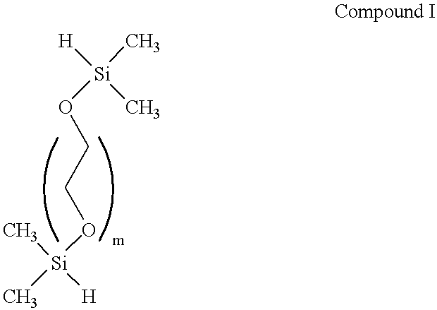
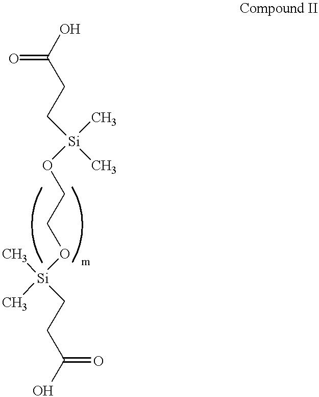
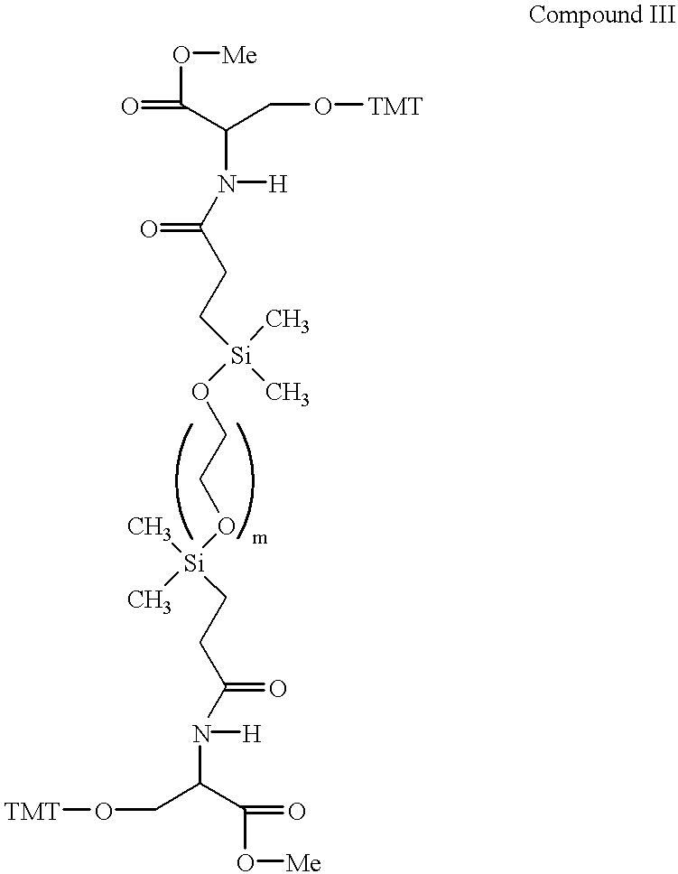
Synthesis of a Spacer with Cleavable Siloxane Site

A representative cleavable spacer, shown schematically in FIG. 5, is synthesized as follows.

In brief, the synthesis is begun by constructing the central portion of the spacer molecule first. Both ends of the poly(ethyleneglycol) are then silanized, e.g. with chlorodimethylsilane to afford a compound of the formula of Compound I.

The silane groups then are derivatized with an alkenoic acid, straight or branched chain (e.g., CH.dbd.CH(CH.sub.2).sub.n COOH, n=1-11, although the number of carbon atoms is immaterial, such as vinyl acetic acid, acrylic acid and the like) having a terminal double bond, such as vinyl acetic acid to form a compound having the structural formula of Compound II, and reacted further to provide a protected hydroxyl group on each side of the silane to provide for later attachment of oligonucleotides as illustrated by the compound having the structural formula of Compound III. Various common reactants c...

example 2

6.2 Example 2

Synthesis of a Cleavable Magnesium Dicarboxylate Spacer Recognizing Human IgG

Onto a gold-coated polycarbonate disk is added by ink-jet printer 2 .mu.l of 10 .mu.M biotindisulfide water solution in 64 circular spots having a diameter of 5 mm. Onto these same spots is added by ink-jet printer 2 .mu.l of a mixture of 1 .mu.M streptavidin and 1 .mu.M albumin.

Goat anti-human IgG (Bioprocessing, Inc., Scarborough, Me; Covalent Immunology, Monroe, N.H.) is reduced by thioethanolamine to produce univalent halves, each of which consists of one heavy chain and one light chain (HL). Thioethanolamine is removed by dialysis and maleimido-polyethyleneglycol-biotin (MAL-PEG-BIO; MW 3, 400, Shearwater Polymers, Inc., Alabama) is added. A small amount of thioethanolamine is added to render maleimido groups unreactive. The mixture is dialyzed against 10 mM phosphate buffer (pH 7) in a dialysis tube (molecular weight cut-off 30,000).

To this antibody derivative (Ab-PEG-BIO) is added a ten ...

example 3

6.3 Example 3

Detection of HIV-1 in a Nucleic Acid Assay

HIV-1 proviral DNA from clinical samples is amplified as follows, essentially as described in U.S. Pat. No. 5,599,662, incorporated herein by reference.

Peripheral blood monocytes are isolated by standard Ficoll-Hypaque density gradient methods. Following isolation of the cells, the DNA is extracted as described in Butcher and Spadoro, Clin. Immunol. Newsletter 12:73-76 (1992), incorporated herein by reference.

Polymerase chain reaction is performed in a 100 .mu.l reaction volume, of which 50 .mu.l is contributed by the sample. The reaction contains the following reagents at the following initial concentrations:

10 mM Tris-HCl (pH 8.4)

50 mM KCl

200 .mu.M each DATP, dCTP, dGTP, and dUTP

25 pmoles of primer 1, of sequence shown below

25 pmoles of primer 2, of sequence shown below

3.0 mM MgCl.sub.2

10% glycerol

2.0 units of Taq DNA polymerase (Perkin-Elmer)

2.0 units UNG (Perkin-Elmer)

Primer 1: 5'-TGA GAC ACC AGG AAT TAG ATA TCA GTA CAA TGT-...


PUM

PropertyMeasurementUnit
constant linear velocityaaaaaaaaaa
diameteraaaaaaaaaa
thicknessaaaaaaaaaa

Abstract

Optical disk-based assay devices and methods are described, in which analyte-specific signal elements are disposed on an optical disk substrate. In preferred embodiments, the analyte-specific signal elements are disposed readably with the disk's tracking features. Also described are cleavable signal elements particularly suitable for use in the assay device and methods. Binding of the chosen analyte simultaneously to a first and a second analyte-specific side member of the cleavable signal element tethers the signal-responsive moiety to the signal element's substrate-attaching end, despite subsequent cleavage at the cleavage site that lies intermediate the first and second side members. The signal responsive moiety reflects, absorbs, or refracts incident laser light. Described are nucleic acid hybridization assays, nucleic acid sequencing, immunoassays, cell counting assays, and chemical detection. Adaptation of the assay device substrate to function as an optical waveguide permits assay geometries suitable for continuous monitoring applications.

Description

1. FIELD OF THE INVENTIONThe present invention relates to the field of analytical instrumentation for chemical assays and diagnostics, and to the detection of small quantities of analytes in samples. More specifically, the invention relates to an assay device comprising an optical disk having analyte-specific signal elements disposed readably thereon2. BACKGROUND OF THE INVENTION2.1 Small Scale Clinical AssaysUntil recently, most clinical diagnostic assays for the detection of small quantities of analytes in fluids have been conducted as individual tests; that is, as single tests conducted upon single samples to detect individual analytes. More recently, efficiency and economy have been obtained by designing apparatus for multi-sample preparation and automated reagent addition, and by designing apparatus for rapid analysis of large numbers of test samples, either in parallel or in rapid serial procession. Often, such automated reagent preparation devices and automated multiplex anal...

Claims


Application Information

Patent Timeline
Patent Type & Authority Patents(United States)
IPC IPC(8): B01J19/00G01N33/543G01N33/551G01N33/553G01N35/00C12Q1/68C40B40/06C40B70/00G01N33/531G01N33/569G01N33/576
CPCB01J19/0046G01N35/00069C12Q1/6825C12Q1/6834C40B80/00G01N33/531G01N33/54353G01N33/54366G01N33/54373G01N33/553G01N33/56988G01N33/5767B01J2219/0054B01J2219/00585B01J2219/00596B01J2219/00605B01J2219/0061B01J2219/00612B01J2219/00619B01J2219/00621B01J2219/00626B01J2219/0063B01J2219/00635B01J2219/00637B01J2219/00648B01J2219/00659B01J2219/00702B01J2219/00722C40B40/06C40B70/00B01J2219/00536
Inventor VIRTANEN, JORMA
Owner VINDUR TECH
Why Eureka
Try Eureka