Mass spectrometric methods for biomolecular screening

a mass spectrometry and biomolecular technology, applied in the field of mass spectrometry methods for biomolecular screening, can solve the problems of large effects on the biological activity of compounds, large time-consuming, expensive and time-consuming, and therefore slow process of synthesis and evaluation of single compounds bearing incremental structural changes, and achieve rapid and simultaneous screening, rapid and simple operation, and enhanced efficiency

Inactive Publication Date: 2002-08-06
IONIS PHARMA INC
View PDF4 Cites 120 Cited by
  • Summary
  • Abstract
  • Description
  • Claims
  • Application Information

AI Technical Summary

Benefits of technology

In a preferred embodiment, the present invention also provides method for simultaneously screening multiple biomolecular targets against combinatorial libraries and mixtures or collections of compounds. This is a significant advantage of the present invention over current state-of-the-art techniques in the screening of compounds for such binding. There is believed to be no prior technique that allows the simultaneous and rapid screening of multiple targets, while providing structural detail on the target and binding ligand at the same time. In addition to providing methods for the rapid and simultaneous screening of multiple biomolecular targets, the present invention also provides methods for determining the structure and nature of binding of both the target and binding ligand.
over current state-of-the-art techniques in the screening of compounds for such binding. There is believed to be no prior technique that allows the simultaneous and rapid screening of multiple targets, while providing structural detail on the target and binding ligand at the same time. In addition to providing methods for the rapid and simultaneous screening of multiple biomolecular targets, the present invention also provides methods for determining the structure and nature of binding of both the target and binding ligand.
As discussed above, mass spectrometry methods of the present invention provide a direct means for screening and identifying those components of combinatorial mixtures that bind to a target biomolecule in solution. In order to enhance efficiency, it is preferable to multiplex the screening process by simultaneously screening multiple targets for binding activity against a combinatorial library or mixture of compounds. This strategy is normally limited by the distribution of charge states and the undesirable mass / charge overlap that will be generated from all possible noncovalent biomolecule-ligand complexes that could be formed during such a screening assay. This problem of overlapping peaks in the mass spectra is further exacerbated if the biomolecular targets being screened are of similar sequence, composition, or molecular weight. In such instances it would not be possible to ascertain in a rapid and simple operation the composition of biomolecule-ligand complexes because of the extensive mass redundancy present in the pool of biomolecules being studied and possible in the combinatorial library being screened.
The method of the present invention alleviates the problem of biomolecular target mass redundancy through the use of special mass modifying molecular weight tags. These mass modifying tags are typically uncharged or positively charged groups such as, but not limited to, alkyl and tetraalkylammonium groups, and polymers such as, but not limited to, polyethylene glycols (PEG), polypropylene, polystyrene, cellulose, sephadex, dextrans, cyclodextrins, peptides, and polyacrylamides. These mass modifying tags may be selected based on their molecular weight contribution and their ionic nature. These mass modifying tags may be attached to the biopolymeric targets at one or more sites including, but not limited to, the 2'--O--, 3'-terminus, 5'-terminus or along the sugar-phosphate backbone of nucleic acid targets. Addition of mass modifying tags to the 5'terminus of synthetic oligonucleotides can be realized either using conventional phosphoramidite chemistry, other conventional chemistry or by biochemical or enzymatic means. Such mass modification of a nucleic acid may be carried out using conventional, manual or automated techniques. Alternatively, addition of mass modifying tags may be performed at the 3'-terminus by the use of appropriately modified polymer or CPG supports for solid-phase synthesis of nucleic acids. Mass modification at the 3'terminus may also be done by biochemical or enzymatic means. It is also possible to attach mass modifying tags to the internucleotide linkages of a nucleic acid. This may be performed via the use of appropriately modified phosphoramidites, or other nucleoside building blocks during nucleic acid synthesis or via post-synthetic modification of the internucleotide linkage. Further, attachment of mass modifying tags to nucleic acid targets may also be accomplished via the use of bifunctional linkers at any functional site on the nucleic acid. Similarly, when working with other classes of biomolecular targets these mass modifying tags may likewise be incorporated at one or more positions on the biomolecule. As will be apparent, inclusion in either target or ligand of isotopic mass labels may also be useful.
Thus, similar nucleic acid and other biological targets may be differentially tagged for rapid mass spectrometric screening by the methods of this invention. When noncovalent complexes are observed from this multiplexed screening of multiple nucleic acid targets with mixtures of small molecular weight combinatorial libraries, the constituent ligand and biomolecule are readily identified using conventional mass analyzers such as quadrupole, ion trap, ICR, magnetic sector, or TOF and followed by MS / MS. This is because the mass modifying tags make the m / z (mass to charge ratio) of the signal arising from each target biomolecule-ligand complex ion of similar charge, distinct in the mass spectrum, and which results in cleanly separated ion peaks. Mass redundancy and peak overlap are both avoided by the use of mass modifying tags.
The present invention is also highly useful in combination with other techniques for the identification of ligands which interact with molecular interaction sites on RNA and other nucleic acids. Molecular interaction sites attend RNA and are believe to be highly important in the functioning of such RNA. The nucleotide sequences of molecular interaction sites are highly conserved, even among taxonomically diverse species. Moreover, such molecular interaction sites have specific structures which provide opportunities for ligand binding. Ascertaining which ligands bind to such sites as well as determining the relative affinities and specificities for the binding of each ligand provides lead compounds for drug discovery, therapeutics, agricultural chemistry, industrial chemistry and otherwise.

Problems solved by technology

However, significant hurdles need to be overcome when trying to identify or develop high affinity ligands for a particular biological target.
These include the difficulty surrounding the task of elucidating the structure of targets and targets to which other molecules may be bound or associated, the large numbers of compounds that need to be screened in order to generate new leads or to optimize existing leads, the need to dissect structural similarities and dissimilarities between these large numbers of compounds, correlating structural features to activity and binding affinity, and the fact that small structural changes can lead to large effects on biological activities of compounds.
Traditionally, drug discovery and optimization have involved the expensive and time-consuming, and therefore slow, process of synthesis and evaluation of single compounds bearing incremental structural changes.
However, solid phase screening assays can be rendered difficult by non-specific interactions.
Despite these advances, the process of drug discovery and optimization entails a sequence of difficult steps.
This process can also be an expensive one because of the costs involved at each stage and the need to screen large numbers of individual compounds.
Moreover, the structural features of target receptors can be elusive.
Another drawback of present sequencing techniques is their inability to reveal anything more than the primary structure, or sequence, of the target.
While X-ray crystallography is a very powerful technique that can allow for the determination of some secondary and tertiary structure of biopolymeric targets (Erikson and Fesik, Ann. Rep. in Med. Chem., 1992, 27, 271-289), this technique can be an expensive procedure and very difficult to accomplish.
Crystallization of biopolymers is extremely challenging, difficult to perform at adequate resolution, and is often considered to be as much an art as a science.
Further confounding the utility of X-ray crystal structures in the drug discovery process is the inability of crystallography to reveal insights into the solution-phase, and therefore the biologically relevant, structures of the targets of interest.
The surface-plasmon resonance technique is more straightforward to use, but is also quite costly.
The tedious nature, high cost, multi-step character, and low sensitivity of many of the above-mentioned screening technologies are shortcomings of the currently available tools.
Further, available techniques do not always afford the most relevant structural information--the structure of a target in solution, for example.
Also, the need for customized reagents and experiments for specific tasks is a challenge for the practice of current drug discovery and screening technologies.
Current methods also fail to provide a convenient solution to the need for deconvolution and identification of active members of libraries without having to perform tedious re-syntheses and re-analyses of discrete members of pools or mixtures.

Method used


Image

  • Mass spectrometric methods for biomolecular screening
  • Mass spectrometric methods for biomolecular screening
  • Mass spectrometric methods for biomolecular screening

Examples


example 1

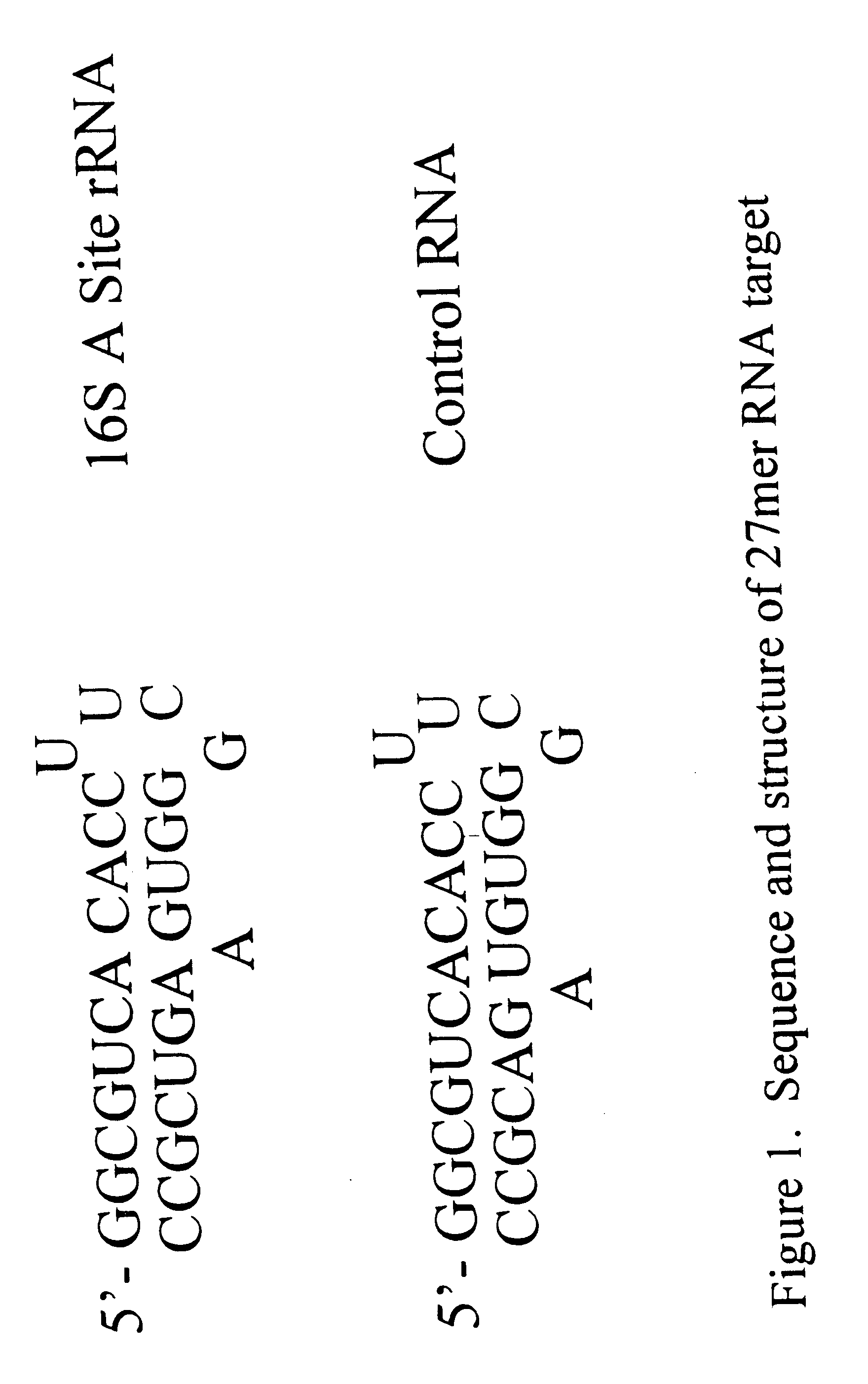
Determining the Structure of a 27-mer RNA Corresponding to the 16S rRNA A Site

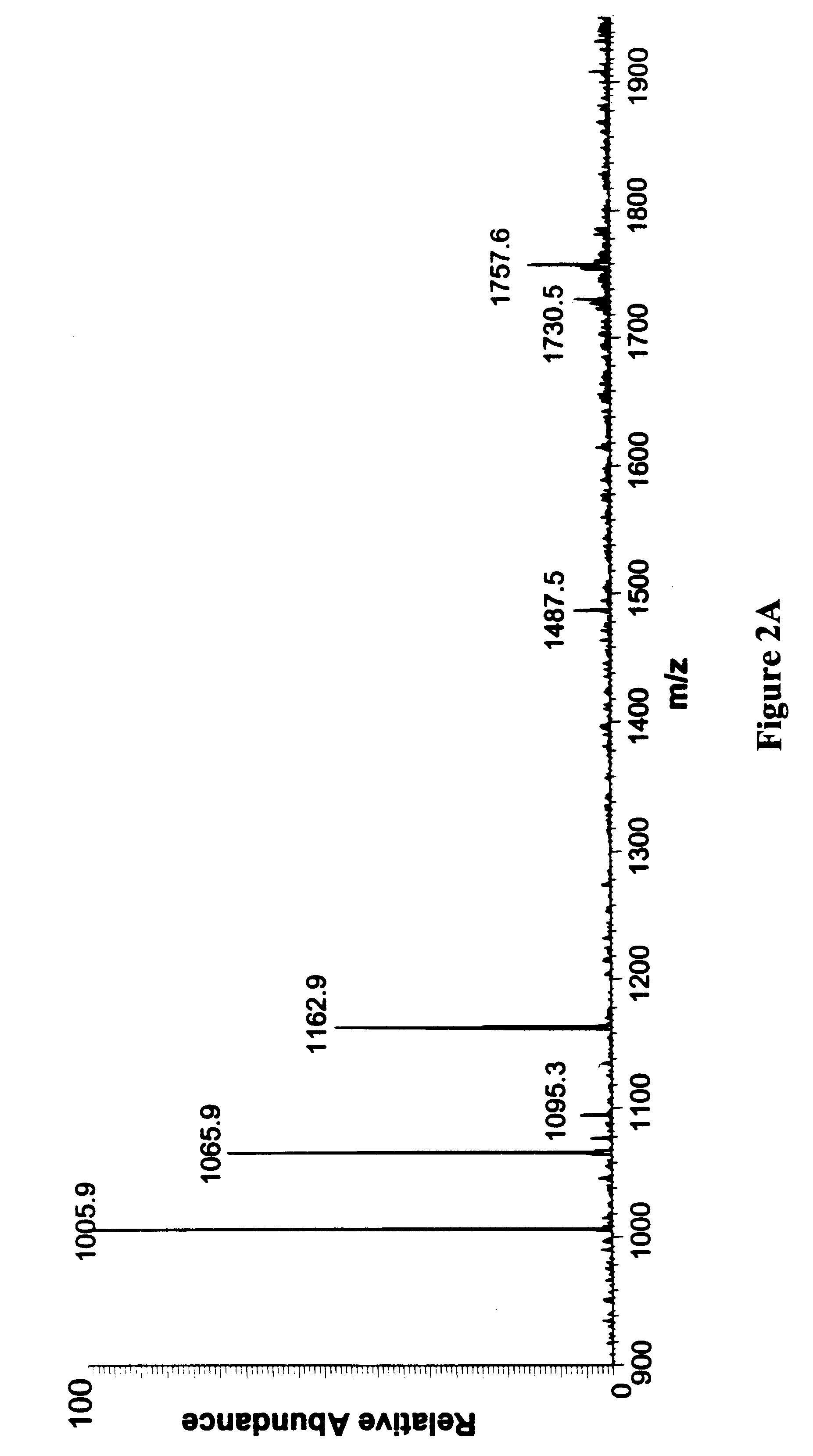
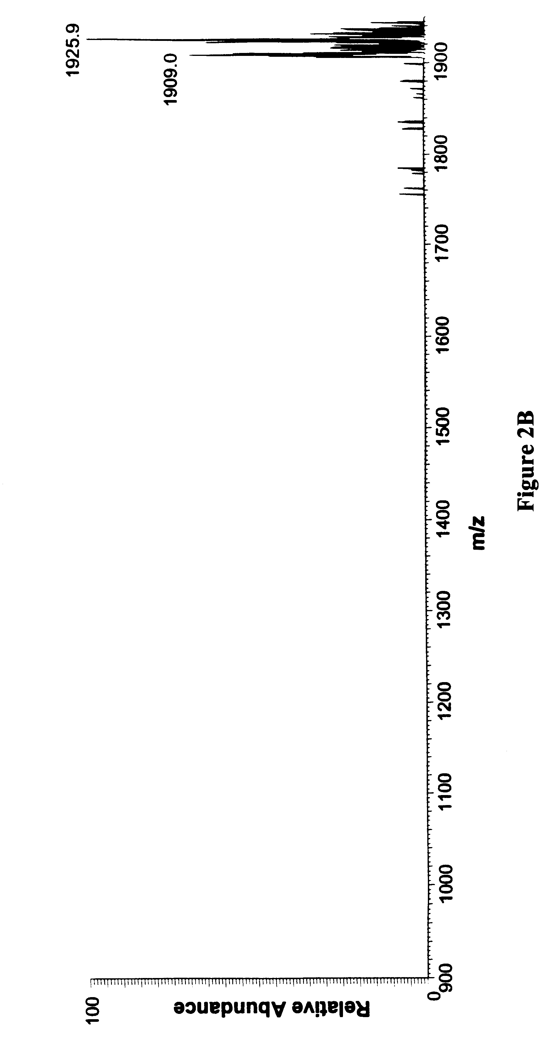
In order to study the structure of the 27-mer RNA corresponding to the 16S rRNA A site, of sequence 5'-GGC-GUC-ACA-CCU-UCG-GGU-GAA-GUC-GCC-3' [SEQ. ID NO. 1] a chimeric RNA / DNA molecule that incorporates three deoxyadenosine (dA) residues at positions 7, 20 and 21 was prepared using standard nucleic acid synthesis protocols on an automated synthesizer. This chimeric nucleic acid of sequence 5'-GGC-GUC-dACA-CCU-UCG-GGU-GdAdA-GUC-GCC-3' [SEQ. ID NO. 2] was injected as a solution in water into an electrospray mass spectrometer. Electrospray ionization of the chimeric afforded a set of multiply charged ions from which the ion corresponding to the (M-5H).sup.5- form of the nucleic acid was further studied by subjecting it to collisionally induced dissociation (CID). The ion was found to be cleaved by the CID to afford three fragments of m / z 1006.1, 1162.8 and 1066.2. These fragments correspond to the w.sub.7.su...

example 2

Determining the Binding Site for Paromomycin on a 27-mer RNA Corresponding to the 16S rRNA A Site

In order to study the binding of paromomycin to the RNA of example 1, the chimeric RNA / DNA molecule of example 1 was synthesized using standard automated nucleic acid synthesis protocols on an automated synthesizer. A sample of this nucleic acid was then subjected to ESI followed by CID in a mass spectrometer to afford the fragmentation pattern indicating a lack of structure at the sites of dA incorporation, as described in Example 1. This indicated the accessibility of these dA sites in the structure of the chimeric nucleic acid.

Next, another sample of the chimeric nucleic acid was treated with a solution of paromomycin and the resulting mixture analyzed by ESI followed by CID using a mass spectrometer. The electrospray ionization was found to produce a set of multiply charged ions that was different from that observed for the nucleic acid alone. This was also indicative of binding of t...

example 3

Determining the Identity of Members of a Combinatorial Library that Bind to a Biomolecular Target

1 .mu.L (0.6 O.D.) of a solution of a 27-mer RNA containing 3 dA residues (from Example 1) was diluted into 500 uL of 1:1 isopropanol:water and adjusted to provide a solution that was 150 mM in ammonium acetate, Ph 7.4 and wherein the RNA concentration was 10 .mu.M. To this solution was added an aliquot of a solution of paromomycin acetate to a concentration of 150 nM. This mixture was then subjected to ESI-MS and the ionization of the nucleic acid and its complex monitored in the mass spectrum. A peak corresponding to the (M-5H).sup.5- ion of the paromomycin-27mer complex is observed at an m / z value of 1907.6. As expected, excess 27-mer is also observed in the mass spectrum as its (M-5H).sup.5- peak at about 1784. The mass spectrum confirms the formation of only a 1:1 complex at 1907.6 (as would be expected from the addition of the masses of the 27-mer and paromomycin) and the absence o...


PUM


Abstract

The present invention provides methods for the determination of the structure of biomolecular targets, as well as the site and nature of the interaction between ligands and biomolecular targets. The present invention also provides methods for the determination of the relative affinity of a ligand for the biomolecular target it interacts with. Also provided are methods for screening ligand or combinatorial libraries of compounds against one or more than one biological target molecules. The methods of the invention also allow determination of the relative binding affinity of combinatorial and other compounds for a biomolecular target. The present invention further provides methods for the use of mass modifying tags for screening multiple biomolecular targets. In a preferred embodiment, ligands which have great specificity and affinity for molecular interaction sites on biomolecules, especially RNA can be identified. In preferred embodiments, such identification can be made simultaneously with libraries of ligands.

Description

The present invention is directed to methods for the use of mass spectrometry for the determination of the structure of a biomolecule especially a nucleic acid target, the site(s) of interaction between ligands and the target, the relative binding affinity of ligands for the target and other useful information. The present invention also provides methods for the use of mass spectrometry for screening chemical mixtures or libraries, especially combinatorial libraries, for individual compounds that bind to a selected target and can be used in pharmaceuticals, veterinary drugs, agricultural chemicals industrial chemicals and otherwise. The present invention is further directed to methods for screening multiple targets simultaneously against, e.g. a combinatorial library of compounds.A further aspect of the invention provides methods for determining the interaction between one or a plurality of molecular species, especially "small" molecules and a molecular interaction site on a nucleic...

Claims


Application Information

Patent Timeline
Patent Type & Authority Patents(United States)
IPC IPC(8): C12Q1/68G01N27/62C12N15/09C40B40/06G01N27/64G01N33/15G01N33/50G01N33/566G01N33/68
CPCC12Q1/6806C12Q1/6811C12Q1/6816B01J2219/00702B01J2219/00722Y10T436/24C40B40/06C12Q2565/627C12Q2525/101C12Q2565/133C12Q2525/121C12Q2565/607H01J49/004
Inventor CROOKE, STANLEY T.GRIFFEY, RICHARDHOFSTADLER, STEVE
Owner IONIS PHARMA INC
Why Eureka
Try Eureka