Tissue engineered tendons and ligaments

a technology of tendons and ligaments, applied in the field of tissue engineered tendons and ligaments, can solve the problems of disordered structure which imparts this ability to the connective tissue, tendon defects, and proves difficult problems for orthopedic surgery and hand surgery, and achieves the effect of moderate tensile strength

Inactive Publication Date: 2005-01-11
MASSACHUSETTS INST OF TECH +1
View PDF134 Cites 111 Cited by
  • Summary
  • Abstract
  • Description
  • Claims
  • Application Information

AI Technical Summary

Benefits of technology

As shown by the examples, transplanted tenocytes attached to biodegradable synthetic polymer scaffolds to generate new tendon in mice. Tenocytes were isolated from freshly slaughtered newborn calf tendon and were seeded onto a non-woven mesh of polyglycolic acid, arranged as either a random array of fibers, or as fibers in parallel. The cell-polymer constructs were implanted into the mice subcutaneously. Specimens were harvested after six to ten weeks and examined. On gross examination, all specimens closely resembled tendons from which the cells had initially been isolated. Histologic evaluation demonstrated that collagen bundles appeared to be organizing in a parallel fashion at the lateral aspects and appeared very similar to the collagen bundles seen in normal tendon. Centrally, the collagen fibrils appeared to be randomly oriented. Specimens that were created from implantation of parallel polymer fibers appeared to have a greater degree of parallel collagen fibril orientation at an earlier time period. The neo-tendon constructs demonstrated moderate tensile strength when stretched.
In the preferred embodiment, the bioactive factors are growth factors. Examples of growth factors include heparin binding growth factor (hbgf), transforming growth factor alpha or beta (TGFβ), alpha fibroblastic growth factor (FGF), epidermal growth factor (TGF), vascular endothelium growth factor (VEGF), some of which are also angiogenic factors. In some embodiments it may be desirable to incorporate factors such as nerve growth factor (NGF) or muscle morphogenic factor (MMP). Steroidal antiinflammatories can be used to decrease inflammation to the implanted matrix, thereby decreasing the amount of fibroblast tissue growing into the matrix.
The results demonstrate that tenocytes will adhere to synthetic biodegradable polymers'survive and multiply in vitro, and that tenocyte-polymer construct implantation in vivo results in formation of tendon with characteristics similar to normal mature tendon. The collagen fibrils undergo organization over time. Histological evaluation of the neo-tendon construct at six weeks shows linear organization, like normal tendon, peripherally and random array of collagen fibrils centrally. With parallel arranged polymer fibers, linear orientation of collagen centrally was achieved at an earlier time. At ten weeks, the collagen fibers were arranged in parallel linear fashion throughout the showed proper anatomic cellular organization regardless of polymer fiber orientation. Thus, parallel structural fibers facilitates early organization of collagen, but the ultimate architectural organization is related to cellular communication and interaction and not of polymer orientation.

Problems solved by technology

When damaged, the orderly structure which imparts this ability to the connective tissue is disrupted and usually does not heal to yield a fully functional tissue.
Tendon defects, regardless of their origin, often prove to be difficult problems for orthopedic surgery and hand surgery.
Autogenous tendon usage is limited by availability and donor site morbidity.
Homo- or heterografts, though, suffer from a scarcity of supply, increased susceptibility to infection, and concerns about possible transmission of infectious agents.
The original structure of the tendons is not maintained, however, and the repaired tendon lacks strength and flexibility.
Artificial prostheses, although not infectious, are also susceptible to infection, extrusion, and uncertain long-term immunologic interactions with the host.
Moreover, prostheses cannot adapt to environmental stresses as do living tendon tissue and have a high incidence of adhesive breakdown at their interface with the host.

Method used


Image

  • Tissue engineered tendons and ligaments

Examples


example 1

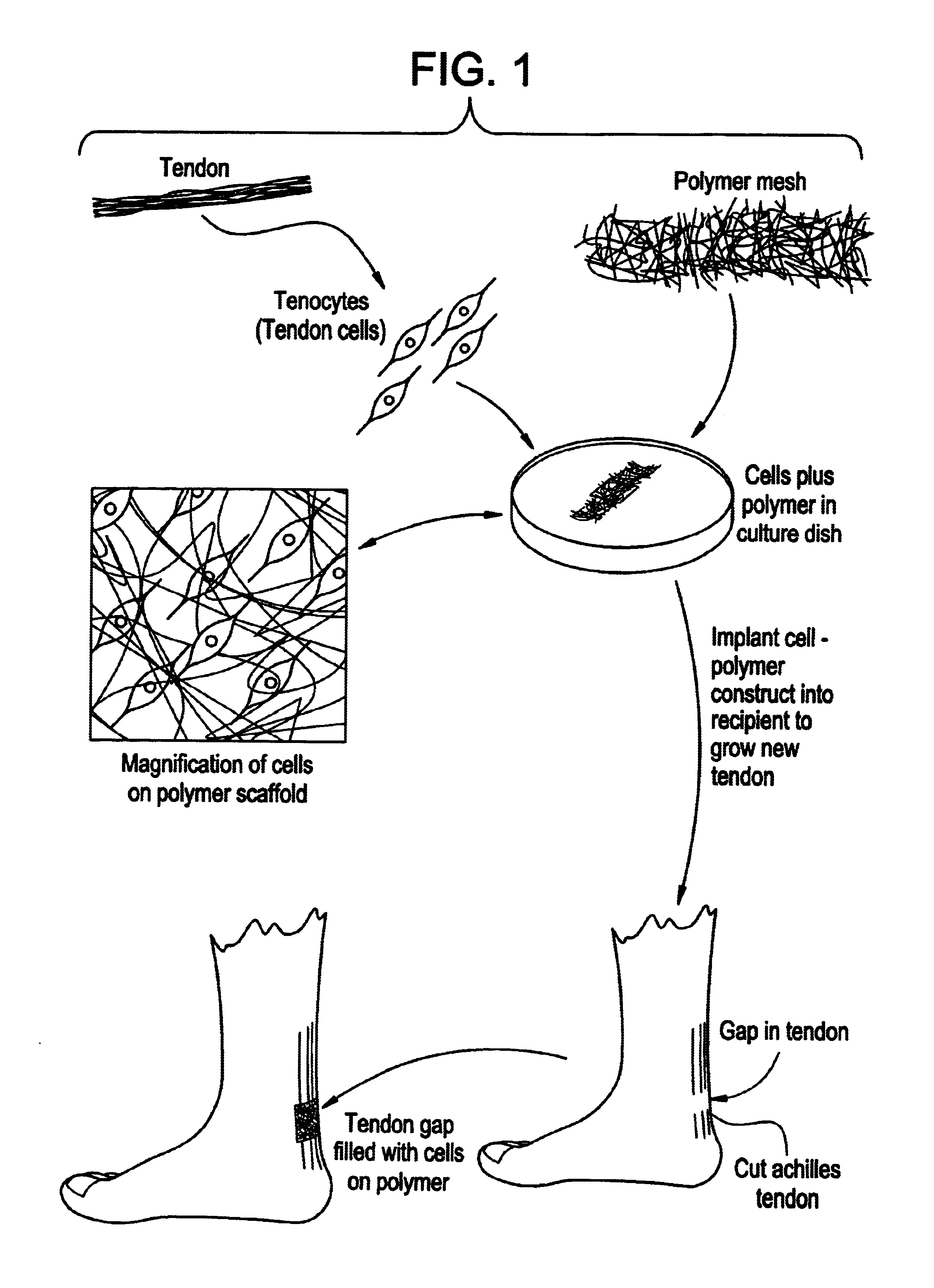
Construction of Neo-tendon

Method and Materials

Sheets approximately 100 microns thick composed of an embossed non-woven mesh of polyglycolic acid with interfiber interstitial spacing averaging 75 to 100 microns in diameter, arranged as either a random array of fibers, or as fibers in parallel (Dexon, Davis and Geck), were cut into pieces approximately 0.4 cm×4 cm and set aside. Tendon was obtained from the shoulder of newborn calves within six hours of sacrifice. The tendons were diced into pieces approximately 0.5 cm×0.5 cm and placed in a sterile 50 ml conical tube. The tendon pieces were washed twice with Dulbecco's phosphate buffered saline (PBS) (Gibco, Grand Island, N.Y.). A sterile 0.396 Collagenase solution was prepared by mixing 75 mg of type collagenase (Worthington, Freehold, N.J.) with 25 ml of Hamm's F-12 medium (Gibco, Grand Island, N.Y.). The tendon fragments were incubated in the collagenase solution for 12-16 hours at 37° C. on a shaker. After digestion, the solution...


PUM

PropertyMeasurementUnit
diameteraaaaaaaaaa
diameteraaaaaaaaaa
thickaaaaaaaaaa

Abstract

Connective tissue, including neo-tendons and ligaments, has been constructed using biodegradable synthetic scaffolds seeded with tenocytes. The scaffolds are preferably formed from biodegradable fibers formed of a polymer such as polyglycolic acid-polylactic acid copolymers, and seeded with cells isolated from autologous tendon or ligament by means of enzymatic digestion or direct seeding into tissue culture dishes from explants. The cell polymer constructs are then surgically transplanted to replace missing segments of functioning tendon or ligament.

Description

BACKGROUND OF THE INVENTIONThe present invention is generally in the area of forming new tissues by implantation of appropriate cells on a polymer matrix, and is specifically directed towards construction of new tendons and ligaments.Tissues connecting bones and muscles are collectively referred to herein as “connective tissue”. Tendons are tissues which attach muscles to bones; aponeuroses are sheet-like tendons connecting one muscle with another or with bones; ligaments hold bones together at joints. Tendons and ligaments are elongated, cylindric structures formed of dense connective tissue, adapted for tension in one direction, with fibers having an orderly, parallel arrangement. The most common variety of dense regularly arranged connective tissue has a predominance of collagenous (white) fibers arranged in bundles. Fibroblasts are placed in rows between the bundles. The tissue is silvery white, tough, yet somewhat pliable. The collagen bundles of the tendons aggregate into larg...

Claims


Application Information

Patent Timeline
Patent Type & Authority Patents(United States)
IPC IPC(8): A61L27/58A61L27/38A61L27/00A61L27/18A61F2/08
CPCA61L27/18A61L27/3604A61L27/3662A61L27/3804A61L27/3817A61L27/58A61L27/3843A61L27/386C08L67/04A61F2/08A61L2430/10
Inventor VACANTI, CHARLES A.CAO, YI LINLANGER, ROBERT S.VACANTI, JOSEPH P.PAIGE, KEITHUPTON, JOSEPH
Owner MASSACHUSETTS INST OF TECH
Why Eureka
Try Eureka