

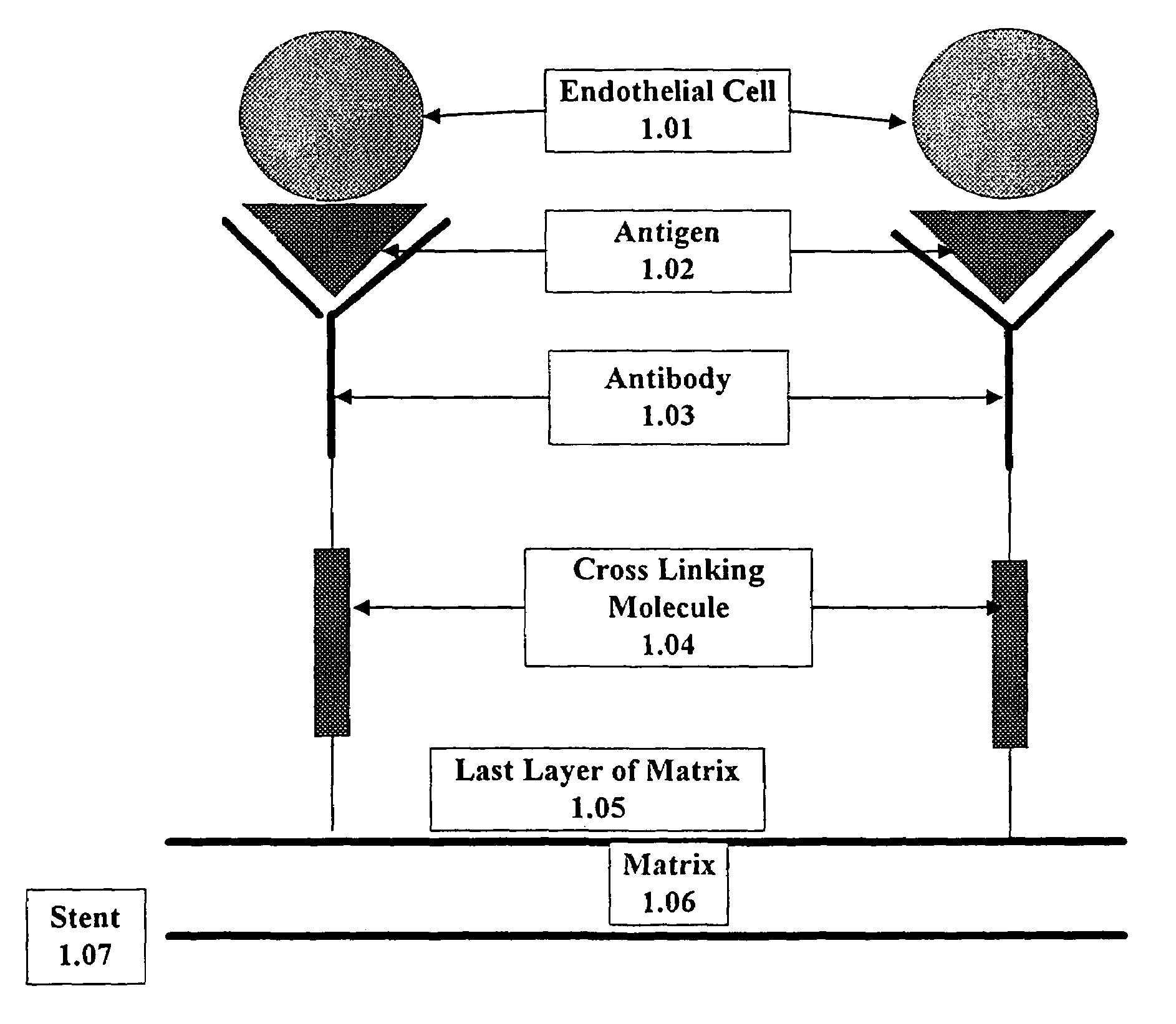
Medical device with coating that promotes endothelial cell adherence
a technology of endothelial cells and medical devices, which is applied in the direction of prosthesis, drug compositions, peptides, etc., can solve the problems of permanent opening of the affected coronary artery, large underestimate of the actual incidence of the disease in the population, and ischemic damage to the tissues supplied by the artery, so as to prevent restnosis and other thromboembolic complications.
- Summary
- Abstract
- Description
- Claims
- Application Information
AI Technical Summary
Benefits of technology
Problems solved by technology
Method used
Image

Examples
experimental examples
[0068]This invention is illustrated in the experimental details section which follows. These sections set forth below the understanding of the invention, but are not intended to, and should not be construed to, limit in any way the invention as set forth in the claims which follow thereafter.
example 1
Adherence of Human Endothelial Cells to Cd34 Fab-Coated Stents
Materials and Methods
1. Cells
[0069]HUVEC will be prepared from human umbilical cords by the method of Jaffe (Jaffe, E. A. In “Biology of Endothelial Cells”, E. A. Jaffe, ed., Martinus-Nijhoff, The Hague (1984), incorporated herein by reference) and cultured in Medium 199 supplemented with 20% fetal calf serum (FCS), L-glutamine, antibiotics, 130 ug / ml heparin and 1.2 mg / ml endothelial cell growth supplement (Sigma-Aldrich, St. Louis, Mo.).
[0070]Progenitor endothelial cells will be isolated from human peripheral blood by the method of Asahara et al. (Isolation of Putative progenitor endothelial cells for angiogenesis. Science 275:964–967). Monoclonal anti-CD34 antibodies will be coupled to magnetic beads and incubated with the leukocyte fraction of human whole blood. After incubation, bound cells will be eluted and cultured in M-199 containing 20% fetal bovine serum and bovine brain extract. (Clonetics, San Diego, Calif.)....
example 2
Proliferation of Human Endothelial Cells on CD34 Fab-Coated Stents
Endothelial Cell Proliferation Assay
[0074]R stents coated with fibrin that incorporates anti-CD34 Fab fragments will be incubated with HUVEC or progenitor endothelial cells for between 4 and 72 hours in M199 containing 0.5% BSA. After incubation of the stents with the HUVEC or progenitor endothelial cells, the stents will be washed 5 times with M199 containing 0.5% BSA and then incubated with [3H]-thymidine. [3H]-thymidine incorporation will be assessed in washed and harvested HUVEC or progenitor endothelial cells (cells will be harvested with trypsin). Proliferation of HUVEC or progenitor endothelial cells on fibrin-coated stents will be compared with endothelial cell proliferation in standard microtiter dishes. Proliferation of HUVEC or progenitor endothelial cells on fibrin-coated stents will be equal to or greater than proliferation of endothelial cells in microtiter dishes
PUM
| Property | Measurement | Unit |
|---|---|---|
| thickness | aaaaa | aaaaa |
| concentration | aaaaa | aaaaa |
| concentration | aaaaa | aaaaa |
Abstract
Description
Claims
Application Information