Methods for treating an artery or vein in a human subject

a technology of biological conduits and arteries, which is applied in the direction of medical preparations, pharmaceutical active ingredients, peptide/protein ingredients, etc., can solve the problems of obstructing tissue removal by dilation methods, no treatment that can sustain patency, and frequent obstruction of biological conduits

Inactive Publication Date: 2006-06-20
PROTARA THERAPEUTICS INC
View PDF19 Cites 24 Cited by
  • Summary
  • Abstract
  • Description
  • Claims
  • Application Information

AI Technical Summary

Benefits of technology

[0024]Treatment methods of the invention provide significant advantages over prior treatment methodologies. For example, enzymatic degradation of one or more key components of the extracellular matrix gently removes the tissue obstructing the lumen. Additionally, collagenolysis or other thera

Problems solved by technology

Obstructions to biological conduits frequently result from trauma to the conduit which can result from transplant, graft or other surgical procedures wherein the extracellular matrix of the obstructing tissue largely comprises collagen.
However, this dilation method does not remove the obstructing tissue.
There is currently no treatment that can sustain patency over the long term.
Not surprisingly, when the body builds new tissue collagen provides the extracellular structural framework such that the deposition of hard collagen in the lesion can result in duct obstruction.
Benign biliary stricture results in obstruction of the flow of bile from the liver and can result in jaundice and hepatic dysfunction.
If untreated, biliary obstruction can result in hepatic failure and death.
These long and difficult surgeries have significant morbidity and mortality due to bleeding, infection, biliary leak, and recurrent biliary obstruction at the anastomosis.
However, balloon dilatio

Method used


Image

  • Methods for treating an artery or vein in a human subject
  • Methods for treating an artery or vein in a human subject
  • Methods for treating an artery or vein in a human subject

Examples


example 1

Tissue Digestion Analysis

[0059]The protocol of the following example is a detailed description of a “standard in vitro tissue digestion assay” as referred to herein.

[0060]The rate of tissue digestion, which is composed mostly of collagen, by a mixture of collagenase and elastase, proteolytic enzymes with activity respectively against collagen and elastin, was determined. Trypsin inhibitor was added to negate the effect of any residual trypsin activity. Briefly, fresh pig tendon was excised, trimmed, washed, blotted dry and weighed. Individual tendon pieces were suspended in 3.58 mg / ml HEPES buffer at neutral pH and various concentrations of enzymes were added. Iodinated radiographic contrast was added in various concentrations to some of the enzyme solutions. The tissue digestion was carried out in a water bath at 37° C. At various time points, the tendon pieces were removed from the enzyme solution, washed, blotted dry and weighed. Each time point was derived from the average of th...

example 2

Determining Dose Dependent In Vitro Activity of a Therapeutic Agent Including Collagenase, Elastase, and a Trypsin Inhibitor

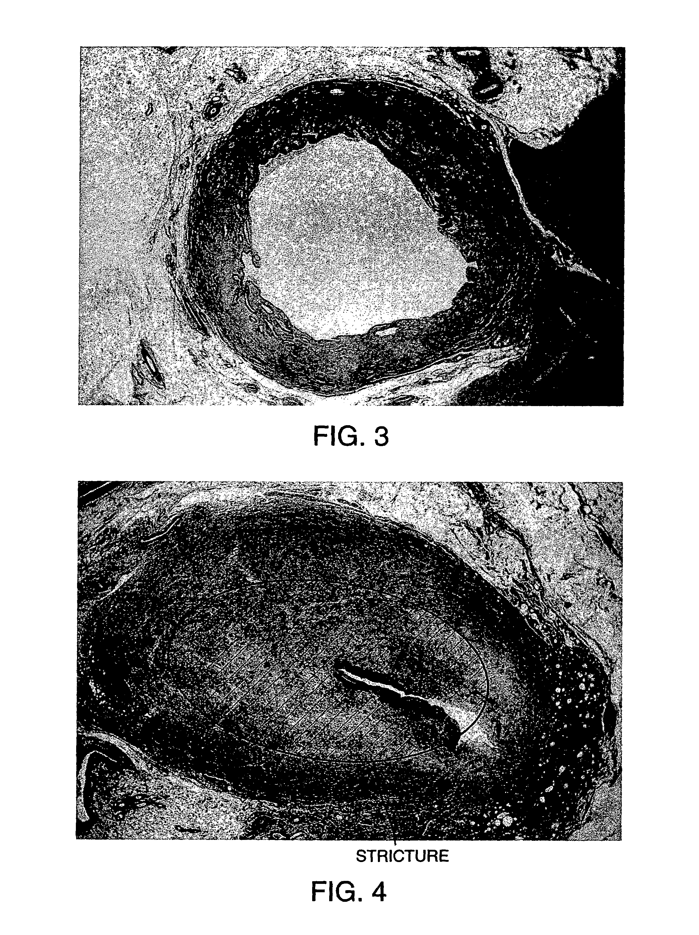
[0062]The effect of enzyme concentration on tissue digestion rates was studied (FIG. 3). The “1×” tissue sample was treated with collagenase 156 Mandel units / ml+elastase 0.125 mg / ml+trypsin inhibitor 0.38 mg / mg, The “2×” sample was treated with collagenase 312 Mandel units / ml+elastase 0.25 mg / ml+trypsin inhibitor 0.76 mg / ml. The “5×” sample was treated with collagenase 780 Mandel units / ml+elastase 0.625 mg / ml+trypsin inhibitor 1.9 mg / ml. All digestion volumes were 0.5 ml. Increasing the concentration of enzymes in vitro increased the rate of tissue digestion (FIG. 3). Buffer alone had no effect on the tissue. An effective in vivo dose was found to be 10,000 ABC units.

example 3

Determining the Effect of Iodinated Radiographic Contrast Material on Tissue Digestion Rates to Facilitate Monitoring Enzyme Delivery Prior to Injection of a Therapeutic Agent Comprising a Contrast Material into a Patient

[0063]The “35% Omnipaque” tissue sample was treated with collagenase 156 Mandel units / ml+elastase 0.125 mg / ml+0.38 trypsin inhibitor with 35% Onmipaque 350 contrast (volume:volume). The “5% Omnipaque” sample was treated with collagenase 312 Mandel units / ml+elastase 0.25 mg / ml+0.76 trypsin inhibitor with 5% Omnipaque 350 (volume:volume). The “1% Onmipaque” sample was treated with collagenase 312 Mandel units / ml+elastase 0.25 mg / ml+0.76 trypsin inhibitor with 1% Omnipaque 350. All digestion volumes were 0.5 ml. These results demonstrate that Omnipaque 350 iodinated contrast material inhibits enzyme activity at radiographically visible (35%) concentrations, but not at lower (1–5%) concentrations (FIG. 4). Similar results were observed with Hypaque 60 contrast.


PUM

PropertyMeasurementUnit
Diameteraaaaaaaaaa
Volumeaaaaaaaaaa

Abstract

The invention provides methods for treating an obstructed biological conduit that include administering to the conduit an agent that can degrade extracellular matrix of obstructing tissue. Particular methods include delivery of an enzyme or a mixture of several enzymes to the area or region of obstruction wherein the enzyme(s) have the capability to degrade extracellular matrix components within the obstruction thereby restoring the normal flow of transported fluid through the conduit. The invention also includes preventively dilating a section of conduit to minimize the risk of obstruction formation.

Description

[0001]This application claims the benefit of U.S. Provisional Application Ser. No. 60 / 155,938 filed Sep. 24, 1999, which is incorporated by reference herein in its entirety.STATEMENT REGARDING GOVERNMENT RIGHTS[0002]This invention was made with government support under grant no. HL07712-07 awarded by the National Institutes of Health. The government has certain rights in the invention.BACKGROUND OF THE INVENTION[0003]1. Field of the Invention[0004]The present invention relates to methods of opening obstructed biological conduits. Preferred methods of the invention include methods and systems for opening obstructed biological conduits using local delivery of a therapeutic agent, particularly a protease, to lyse the extracellular matrix of the obstructing tissue.[0005]2. Background[0006]Obstructions to biological conduits frequently result from trauma to the conduit which can result from transplant, graft or other surgical procedures wherein the extracellular matrix of the obstructing...

Claims


Application Information

Patent Timeline
IPC IPC(8): A61K38/48A61K38/55
CPCA61K38/486A61K38/4886A61K38/55
Inventor FRANANO, NICHOLAS F.
Owner PROTARA THERAPEUTICS INC
Why Eureka
Try Eureka