
Ultrasound imaging and treatment

a technology applied in the field of ultrasonic imaging and treatment, can solve the problems of inability to carry out portable examination, inability to carry out mri, and unfavorable patient care, and achieve the effect of cost saving

Inactive Publication Date: 2006-07-18
IMARX THERAPEUTICS
View PDF444 Cites 84 Cited by
  • Summary
  • Abstract
  • Description
  • Claims
  • Application Information

AI Technical Summary

Benefits of technology

"The present invention provides methods and apparatus for preparing temperature activated gaseous precursor-filled liposomes that can be used as contrast agents for ultrasonic imaging or drug delivery agents. The methods are simple and cost-effective. The liposomes have several advantages such as biocompatibility, ease of membrane penetration, and long storage life. They also exhibit intense echogenicity, stability to pressure, and can be targeted to tissues such as tumors. The liposomes can be used for ultrasound, magnetic imaging, and x-ray contrast agents. They can also encapsulate lipophilic drugs and can be modified easily."

Problems solved by technology

X-rays, however, are known to be somewhat dangerous, since the radiation employed in X-rays is ionizing, and the various deleterious effects of ionizing radiation are cumulative.
This technique, however, has various drawbacks such as expense and the fact that it cannot be conducted as a portable examination.
In addition, MRI is not available at many medical centers.
Nuclear medicine techniques, however, suffer from poor spatial resolution and expose the animal or patient to the deleterious effects of radiation.
Furthermore, the handling and disposal of radionuclides is problematic.
However, despite these various technological improvements, ultrasound is still an imperfect tool in a number of respects, particularly with regard to the imaging and detection of disease in the liver and spleen, kidneys, heart and vasculature, including measuring blood flow.
The methods and materials in the prior art for introduction of genetic materials, for example, to living cells is limited and ineffective.
These methods have all been relatively ineffective in vivo and only of limited use for cell culture transfection.
None of these methods potentiate local release, delivery and integration of genetic material to the target cell.
Great strides have been made in characterizing genetic diseases and in understanding protein transcription but relatively little progress has been made in delivering genetic material to cells for treatment of human and animal disease.
A principal difficulty has been to deliver the genetic material from the extracellular space to the intracellular space or even to effectively localize genetic material at the surface of selected cell membranes.
Whole virus has been used but the amount of genetic material that can be placed inside of the viral capsule is limited and there is concern about possible dangerous interactions that might be caused by live virus.
Despite extensive work on viral vectors, it has been difficult to develop a successfully targeted viral mediated vector for delivery of genetic material in vivo.
Conventional, liquid-containing liposomes have been used to deliver genetic material to cells in cell culture but have mainly been ineffective in vivo for cellular delivery of genetic material.
For example, cationic liposome transfection techniques have not worked effectively in vivo.

Method used


Image

  • Ultrasound imaging and treatment
  • Ultrasound imaging and treatment
  • Ultrasound imaging and treatment

Examples


example 1

Preparation of Gas-Filled Lipid Spheres from Perfluorobutane

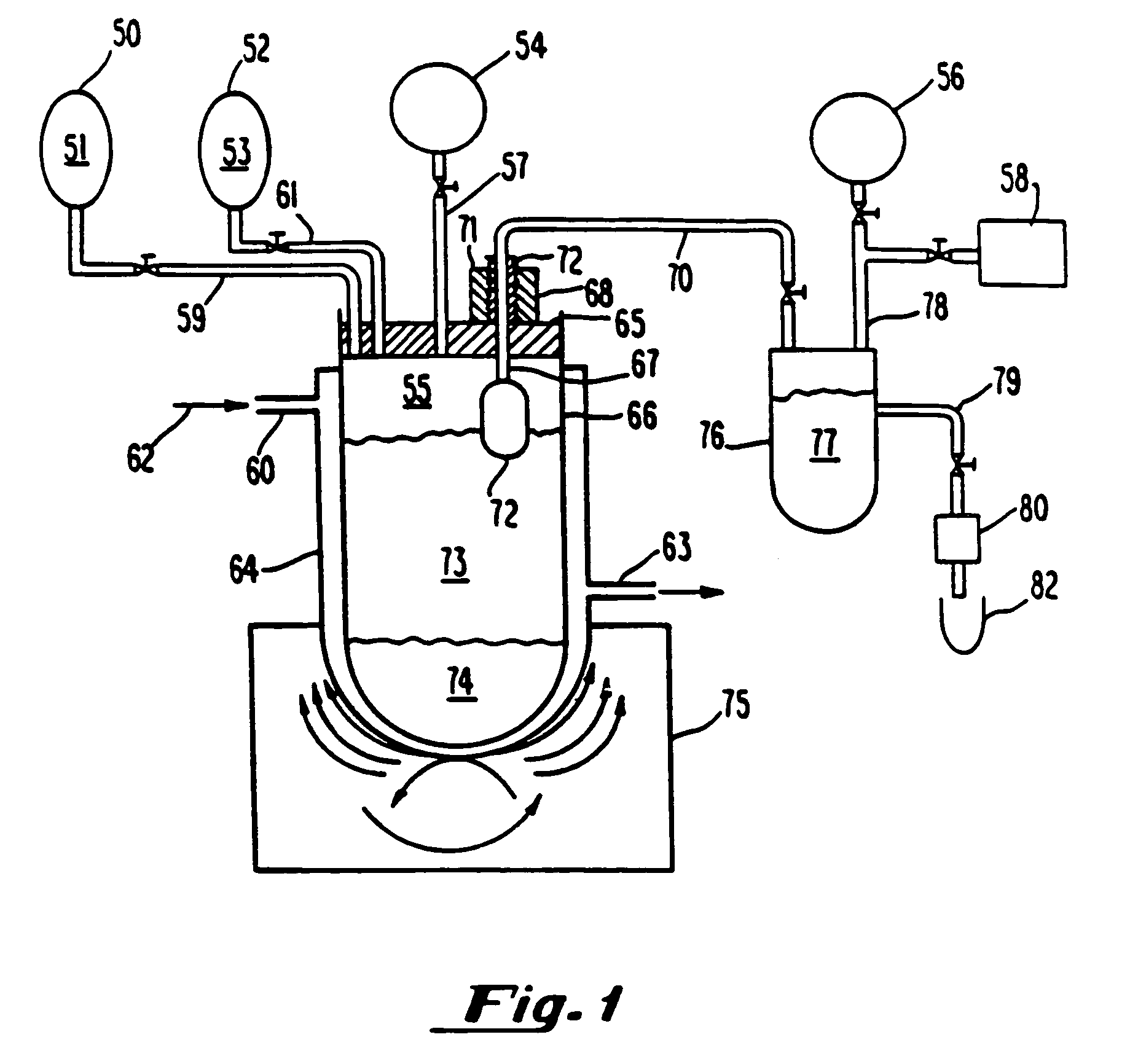
[0214]Gaseous precursor-containing liposomes were prepared using perfluorobutane (Pfaltz and Bauer, Waterbury, Conn.) as follows: A 5 mL solution of lipid, 5 mg per ml, lipid=87 mole percent DPPC, 8 mole percent DPPE-PEG 5,000, 5 mole percent dipalmitoylphosphatidic acid (all lipids from Avanti Polar Lipids, Alabaster, Ala.), in 8:1:1 normal saline:glycerol:propylene glycol, was placed in a glass bottle with a rubber stopper (volume of bottle=15.8 ml). Air was evacuated from the bottle using a vacuum pump, Model Welch 2-Stage DirecTorr Pump (VWR Scientific, Cerritos, Calif.) by connecting the hose to the bottle through a 18 gauge needle which perforated the rubber stopper. After removing the gas via vacuum, perfluorobutane was placed in the stoppered bottle via another 18 gauge needle connected to tubing attached to the canister of perfluorobutane. This process was repeated 5 time such that any traces of air were removed fr...

example 2

Preparation of Gaseous Precursors Via Microfluidization

[0218]Gaseous precursor-filled lipid bilayers were prepared as in Example 1 except, after addition of the gaseous precursor, the contents were microfluidized through six passes on a Microfluidics microfluidizer (Microfluidics Inc., Newton, Mass.). The stroke pressure ranged between 10,000 and 20,000 psi. Continuing with the preparation as per Example 1, produced gas-filled lipid bilayers with gaseous precursor encapsulated.

example 3

Formulation of Gas-filled Lipid Bilayers Using Phosphatidic Acid and Dipalmitoyphosphatidylcholine

[0219]Gas-filled lipid bilayers were prepared as set forth in Example 6 except for the fact that DPPC was used in combination with 5 mole % phosphatidic acid (Avanti Polar Lipids, Alabaster, Ala.). Formulation of gas-filled lipid bilayers resulted in an increase in solubility as exemplified by a decrease in the amount of lipid particulate in the lower aqueous vehicle layer. Resultant sizing appeared to decrease the overall mean size vs. DPPC alone to less than 40 um.


PUM


Abstract

Methods of and apparatus for preparing temperature activated gaseous precursor-filled liposomes are described. Gaseous precursor-filled liposomes prepared by these methods are particularly usefull for example, in ultrasonic imaging applications and in therapeutic drug delivery systems.Gas, gaseous precursors and perfluorocarbons are presented as novel potentiators for ultrasonic hyper-thermia. The gas, gaseous precursors and perfluorocarbons which may be administered into the vasculature, interstitially or into any body cavity are designed to accumulate in cancerous and diseased tissues. When therapeutic ultrasonic energy is applied to the diseased region heating is increased because of the greater effectiveness of sound energy absorption caused by these agents.

Description

CROSS-REFERENCE TO RELATED APPLICATIONS[0001]This application is a continuation of applicant's copending U.S. application Ser. No. 10 / 113,577, filed Apr. 2, 2002, which in turn is a continuation of U.S. application Ser. No. 08 / 796,798, filed Feb. 6, 1997 (now abandoned), which is a continuation of U.S. application Ser. No. 08 / 487,230, filed Jun. 6, 1995 (now U.S. Pat. No. 5,853,752), which is a divisional of application U.S. Ser. No. 08 / 159,687, filed Nov. 30, 1993 (now U.S. Pat. No. 5,585,112), which is a continuation-in-part of copending applications U.S. Ser. No. 08 / 160,232 (now U.S. Pat. No. 5,542,935) and Ser. No. 08 / 159,674 (now abandoned), filed concurrently on Nov. 30, 1993, which are continuations-in-part of copending application U.S. Ser. No. 08 / 076,239, filed Jun. 11, 1993 (now U.S. Pat. No. 5,469,854), which is a continuation-in-part of copending applications U.S. Ser. No. 07 / 717,084 (now U.S. Pat. No. 5,228,446) and U.S. Ser. No. 07 / 716,899 (now abandoned), both of whic...

Claims


Application Information

Patent Timeline
Patent Type & Authority Patents(United States)
IPC IPC(8): A61B8/00A61B17/00A61B17/22A61K8/14A61K9/127A61K41/00A61K47/48A61K49/18A61K49/22A61M5/31A61N7/02A61Q19/00
CPCA61K9/127A61K9/1277A61K9/1278A61K41/0028A61K41/0052A61K47/48815A61K47/48869A61K49/1815A61K49/22A61K49/223A61K49/227A61N7/02A61B2017/00716A61N2007/025A61M5/3145A61M2202/0476A61B2017/22088A61K47/6911A61K47/6925
Inventor UNGER, EVAN C.
Owner IMARX THERAPEUTICS
Learn More