Protein microarrays on mirrored surfaces for performing proteomic analyses

a technology of protein microarrays and mirrored surfaces, applied in the field of cell product analysis and materials, can solve the problems of not providing sufficient blocking of non-specific protein binding, and achieve the effects of improving understanding of disease processes, facilitating drug discovery, and improving signal quality

Inactive Publication Date: 2006-12-12
CHIRON CORP
View PDF35 Cites 20 Cited by
  • Summary
  • Abstract
  • Description
  • Claims
  • Application Information

AI Technical Summary

Benefits of technology

[0010]To achieve the foregoing, the present invention provides protein-displaying microarrays (also referred to herein as “protein microarrays”), their manufacture, use, and application. In particular, the invention provides arrays that produce enhanced signals due to the use of a mirrored array substrate surface in an assay in which fluorescently labeled binding partners interact with an array element bound to the substrate surface (e.g., Cy3 or Cy5 other fluorescent probes), referred to herein as “fluorescence array scanning...

Problems solved by technology

In such extreme cases, conventional blocking agents such as casein, bovine serum albumin a...

Method used


Image

  • Protein microarrays on mirrored surfaces for performing proteomic analyses
  • Protein microarrays on mirrored surfaces for performing proteomic analyses
  • Protein microarrays on mirrored surfaces for performing proteomic analyses

Examples


example 1

Method Used to Make Silanized Slides

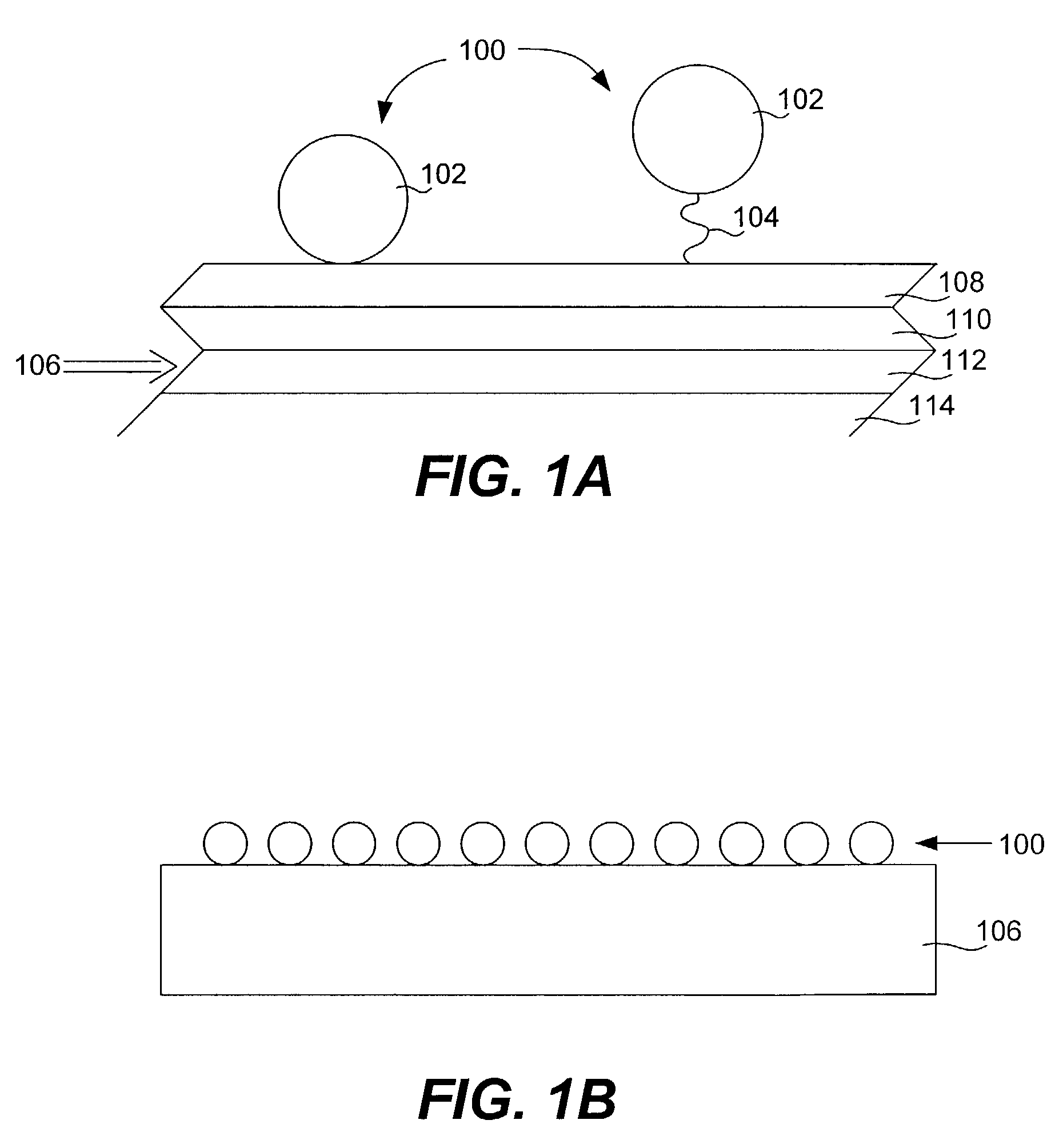
[0136]Slide preparation: Glass microscope slides were pre-cleaned by sonicating in a soap bath for 10 minutes, followed by high pressure de-ionized water wash. The slides were further cleaned 5 min in Nochromix / H2SO4, rinsed with high pressure de-ionized water wash. Finally, they were immersed for 5 minutes in isopropyl alcohol and dried with a stream of N2. Pre-cleaned slides are also commercially available from, for example, Erie Scientific (Ultraclean) or Bioslide (Superclean).[0137]E-beam: A CHA SEC-600-RAP e-beam was fitted with special wafer-sized inserts in the planetaries to accommodate the rectangular shape of the glass microscope slides. The e-beam crucibles were loaded with aluminum metal and fresh Si02. The chamber was then pumped down to at least 2×10⁻⁷ torr. After pumpdown, the planetaries containing the slides were rotated (to allow even coating) while the high voltage (about 10 V) was applied. The electron beam vaporized the materi...

example 2

Use of a Homobifunctional Adapter

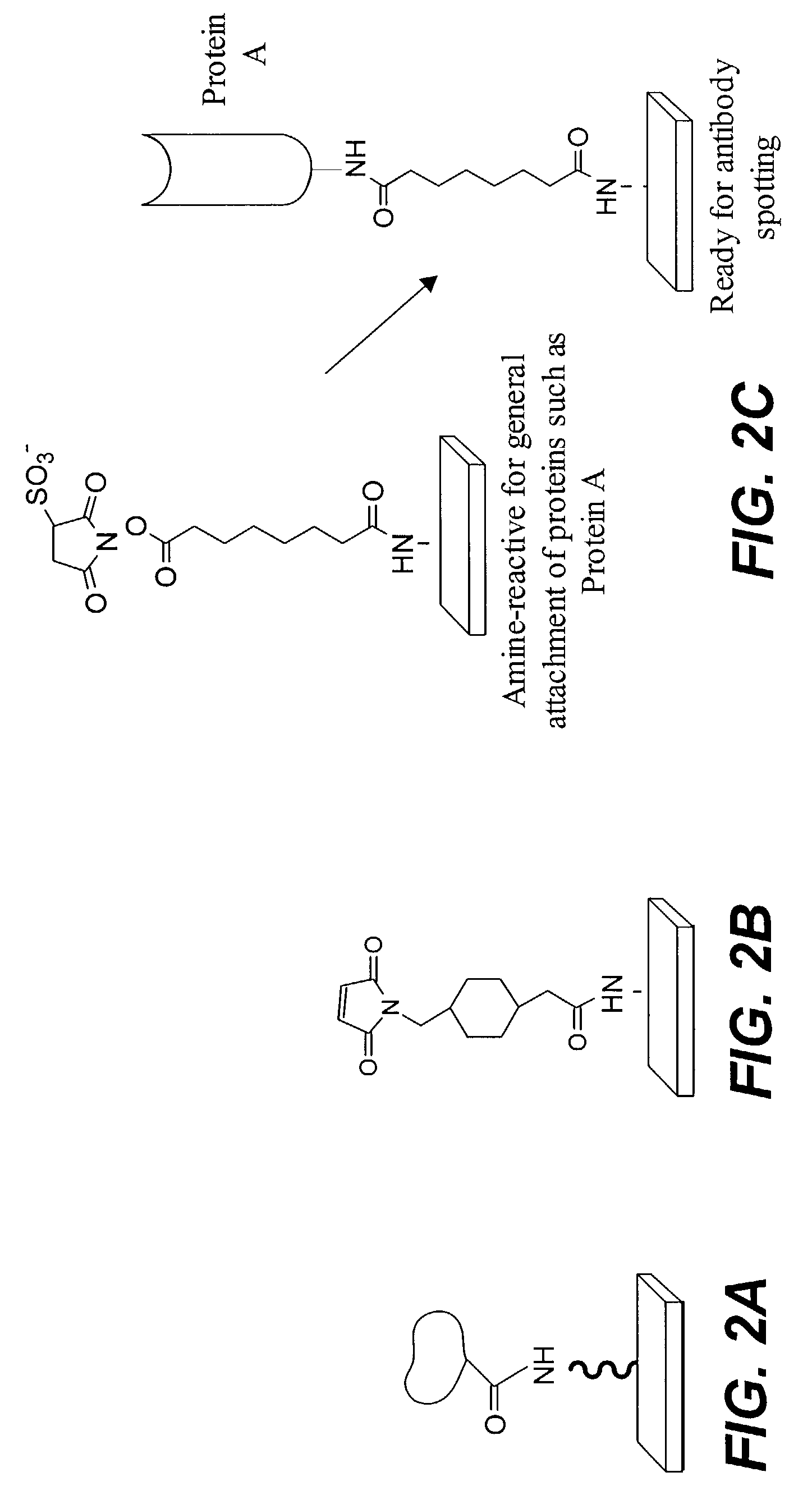
[0139]Amine functionalized substrate surfaces were derivatized with an activated ester by reacting amine groups displayed on the substrate surface with the homobifunctional crosslinker bis-NHS ester. The NHS ester at one terminus of the adapter reacted with the amines on the substrate to produce an NHS ester functionalized slide as follows: 50 mg of adaptor was dissolved in 30 mL DMF, then 170 mL of 1×PBS or other suitable buffer was added. This was applied to the slides and incubated 1–2 hours with mixing. The slides were then rinsed in deionized water. The solution should be fresh to ensure that the NHS ester has not hydrolyzed.) Proteins containing exposed amine groups were spotted onto such a slide to produce covalently bound protein microarrays.

example 3

Use of a Heterobifunctional Maleimide Adapter

[0140]Amine functionalized substrate surfaces were derivatized with maleimide groups by reacting amine groups displayed on the substrate surface with heterobifunctional crosslinkers. SMCC (having an N-hydroxysuccinimide (NHS) ester at one terminus, and a maleimide group at the other) was applied using the same protocol described above in Example 2. The NHS ester reacted with the amines on the substrate to produce a maleimide functionalized slide. Proteins containing exposed thiol groups were spotted onto such a slide to produce covalently bound protein microarrays.


PUM

PropertyMeasurementUnit
Thicknessaaaaaaaaaa
Metallic bondaaaaaaaaaa
Reflectionaaaaaaaaaa

Abstract

Provided are protein microarrays, their manufacture, use, and application. Protein microarrays in accordance with the present invention are useful in a variety preoteomic analyses. Various protein arrays in accordance with the present invention may immobilize large arrays of proteins that may be useful for studying protein-protein interactions to improve understanding of disease processes, facilitating drug discovery, or for identifying potential antigens for vaccine development. The protein array elements of the invention are native or modified proteins (e.g., antibodies or fusion proteins). The protein array elements may be attached directly to a organic functionalized mirrored substrate by a binding reaction between functional groups on the substrate (e.g., amine) and protein (e.g., activated carboxylic acid). Techniques for chemical blocking of the arrays are also provided. The invention contemplates spotting of array elements onto solid planar substrates, labeling of complex protein mixtures, and the analysis of protein binding to the array. The invention also enables the enrichment or purification, and subsequent sequencing or structural analysis of proteins that are identified as differential by the array screen. Kits including protein-binding microarrays for proteomic analysis in accordance with the present invention are also provided.

Description

CROSS-REFERENCE TO RELATED APPLICATIONS[0001]This application is a continuation-in-part of U.S. patent application Ser. No. 09 / 874,091 titled MICROARRAYS FOR PERFORMING PROTEOMIC ANALYSES, filed Jun. 4, 2001; which claims priority from U.S. Provisional Application No. 60 / 209,711, entitled MICROARRAYS FOR PERFORMING PROTEOMIC ANALYSES, filed Jun. 5, 2000; the disclosure of each of which is incorporated by reference herein in its entirety for all purposes.BACKGROUND OF THE INVENTION[0002]1. Field of the Invention[0003]The present invention relates to cell product analysis and materials. More specifically, the invention is directed to protein-displaying microarrays, methods of making them and methods of using them to conduct highly sensitive proteomic analyses.[0004]2. Description of Related Art[0005]In recent years, microarray technology has developed from a specialized sub-field into an important tool for basic and applied studies in molecular biology, microbiology, pharmaceutics, ag...

Claims


Application Information

Patent Timeline
IPC IPC(8): C12M1/34C40B40/00C40B40/10C40B50/18G01N33/53B01J19/00C07K17/14C07K19/00C40B30/04G01N33/543G01N33/545G01N33/547G01N33/68G01N37/00
CPCB01J19/0046B82Y30/00C40B30/04G01N33/543G01N33/54386G01N33/545G01N33/552G01N33/553G01N33/6842G01N33/6845B01J2219/00387B01J2219/00605B01J2219/0061B01J2219/00612B01J2219/00617B01J2219/00626B01J2219/0063B01J2219/00637B01J2219/00659B01J2219/00725B01J2219/0074G01N2500/00
Inventor CHARYCH, DEBORAHZUCKERMANN, RONALD N.
Owner CHIRON CORP
Why Eureka
Try Eureka