

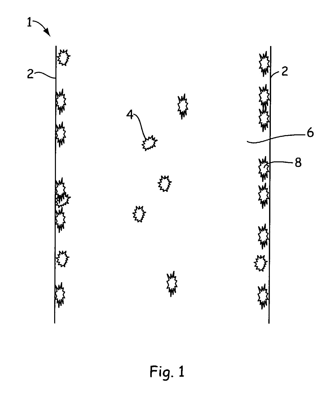
Methods for prevention of surface adsorption of biological materials to capillary walls in microchannels
a microchannel and biological material technology, applied in the direction of fluid pressure measurement, liquid/fluent solid measurement, peptides, etc., can solve the problems of peak tailing, unsatisfactory biasing of assay, and creating artifacts
- Summary
- Abstract
- Description
- Claims
- Application Information
AI Technical Summary
Benefits of technology
Problems solved by technology
Method used
Image

Examples
example 1
[0038]An assay screen is performed to identify inhibitors of an enzymatic reaction. An example of a microfluidic assay chip to be used is the nucleic acid (e.g., DNA LabChip® microfluidic chip device which is commercially available from Caliper Technologies Corp., for example, Colloidal silica particles were purchased as a 30% (by weight) from Aldrich Chemical Company (Milwaukee, Wis.) as Ludox® AM-30 colloidal silica particles (catalog no. 42,084). These particles have a very high surface area of approximately 220 square meters per gram of solid. This suspension was diluted 1:1 with pH 7.5 sodium HEPES buffer with 5 nM MgCl, and then mixed with equal volume of 1.22 micromolar solution of protein kinase-A-β enzyme (PKA-β) in the same buffer. The mixture containing enzyme and 7.5% colloidal silica was placed into one or more enzyme reservoir wells of the microfluidic assay chip. Into one or more other wells of the assay chip the same amount of enzyme was added without the colloidal s...
example 2
[0041]Ludox AM-30 colloidal particles (0.006 micron particle radius) were utilized in a microfluidic chip having channels similar to that employed in Example 1 above. In order to understand the shape of the channels (and thus the surface / volume ratio) it is useful to understand the method used to manufacture the microfluidic chips. The microfluidic chips utilized in this Example are made by isotropic etching (in HF) of a predetermined pattern of grooves into a quartz wafer substrate (about 1 mm thick) to a depth of about 12 microns by employing an etch mask width of 40 microns. The resulting groove has a widest dimension of about 64 microns. Enclosed channels are formed by fusing to the etched wafer surface a smooth, flat quartz wafer. The wafers are then diced into chips of desired size each incorporating one or more microchannels. Such microfluidic chips in general have at least one main channel and usually have one, or more, side channels that either add fluids to, or take fluids...
example 3
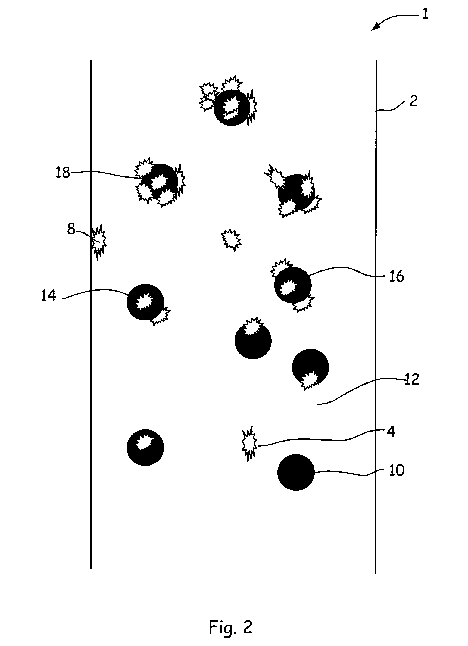
[0049]Colloidal silica particles as described above in Example 1 were again diluted 1:1 with pH 7.5 sodium HEPES buffer and then mixed with equal volume of 1.22 micromolar solutions of proteins kinase-A-β enzyme (PKA-β) in the same buffer. The mixture containing enzyme and 7.5% colloidal silica was placed into each of four enzyme wells of a sample microfluidic assay chip and the inhibitors again were assayed as described previously. The addition of the colloidal particles to the microfluidic microchannels having adsorbed enzyme removed the enzyme activity from the walls, leaving the walls free of such activity. Thus, colloidal particles can be used intermittently (or continuously) between successive inhibitor assays so as to remove enzyme residue and clean the walls to leave a clean surface for each assay. Intermittent injection of the colloidal silica particles can be accomplished by standard microfluidic techniques including multiport pressure control or electroosmotic flow induce...
PUM
| Property | Measurement | Unit |
|---|---|---|
| Percent by mass | aaaaa | aaaaa |
| Percent by mass | aaaaa | aaaaa |
| Length | aaaaa | aaaaa |
Abstract
Description
Claims
Application Information