Methods for crystallographic structure determination employing hydrogen exchange analysis

a technology of hydrogen exchange analysis and crystallography, which is applied in chemical methods analysis, material analysis using wave/particle radiation, instruments, etc., can solve the problems of insufficient time for study, inability to accurately characterize the three-dimensional structure of polypeptides, and inability to accurately characterize the critical structural features. , to achieve the effect of simplification of protein fragmentation methods and high resolution

Inactive Publication Date: 2007-10-09
RGT UNIV OF CALIFORNIA
View PDF3 Cites 3 Cited by
  • Summary
  • Abstract
  • Description
  • Claims
  • Application Information

AI Technical Summary

Benefits of technology

[0031]The present invention provides methods for crystallographic structure determination of a protein of interest through the use of hydrogen exchange analysis. Preferred methods of the present invention employ novel high resolution hydrogen exchange analysis. In some embodiments of the invention, methods of hydrogen exchange analysis comprise fragmentation of a labeled protein using methods described in U.S. Pat. Nos. 5,658,739; 6,331,400, and 6,291,189, the entire disclosures of which are incorporated herein by reference. In other embodiments of the invention, the hydrogen exchange analysis allows for high-throughput structural determinations due to simplifications of the protein fragmentation methods described in U.S. Pat. Nos. 5,658,739; 6,331,400, and 6,291,189.
[0037]In another preferred embodiment of the invention, hereinafter referred to as the “improved proteolysis” method, the process of determining the quantity of isotopic hydrogen and / or the rate of exchange comprises: (a) generating a population of sequence overlapping fragments of said labeled protein by treatment with at least one endopeptidase or combination of endopeptidases under conditions of slowed hydrogen exchange, and then (b) deconvoluting fragmentation data acquired from said population of sequence-overlapping endopeptidase-generated fragments. This improved method dramatically speeds and modulates the sites and patterns of proteolysis by endopeptidases so as to produce highly varied and highly efficient fragmentation of the labeled protein in a single step, thereby avoiding the use of carboxypeptidases completely.

Problems solved by technology
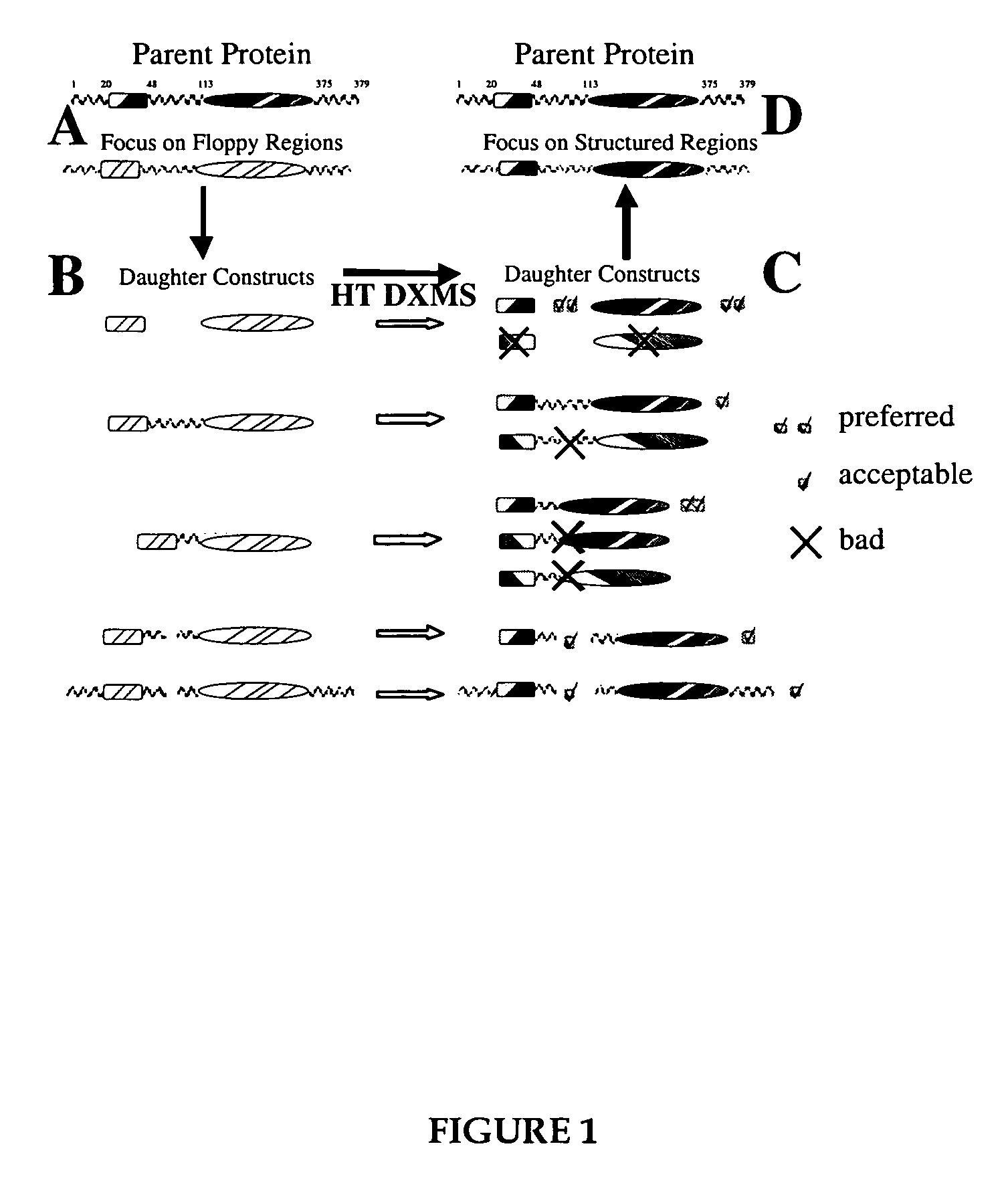
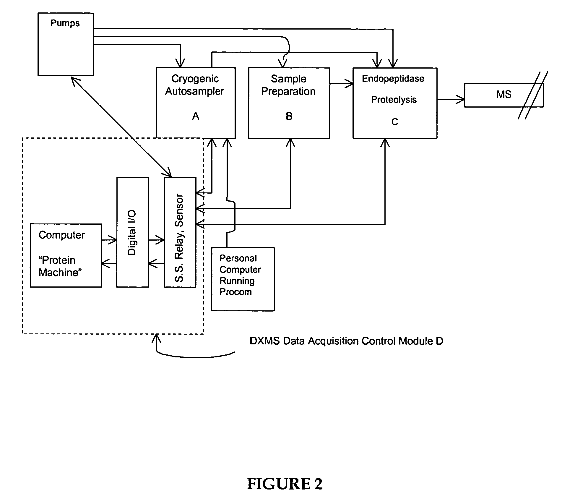
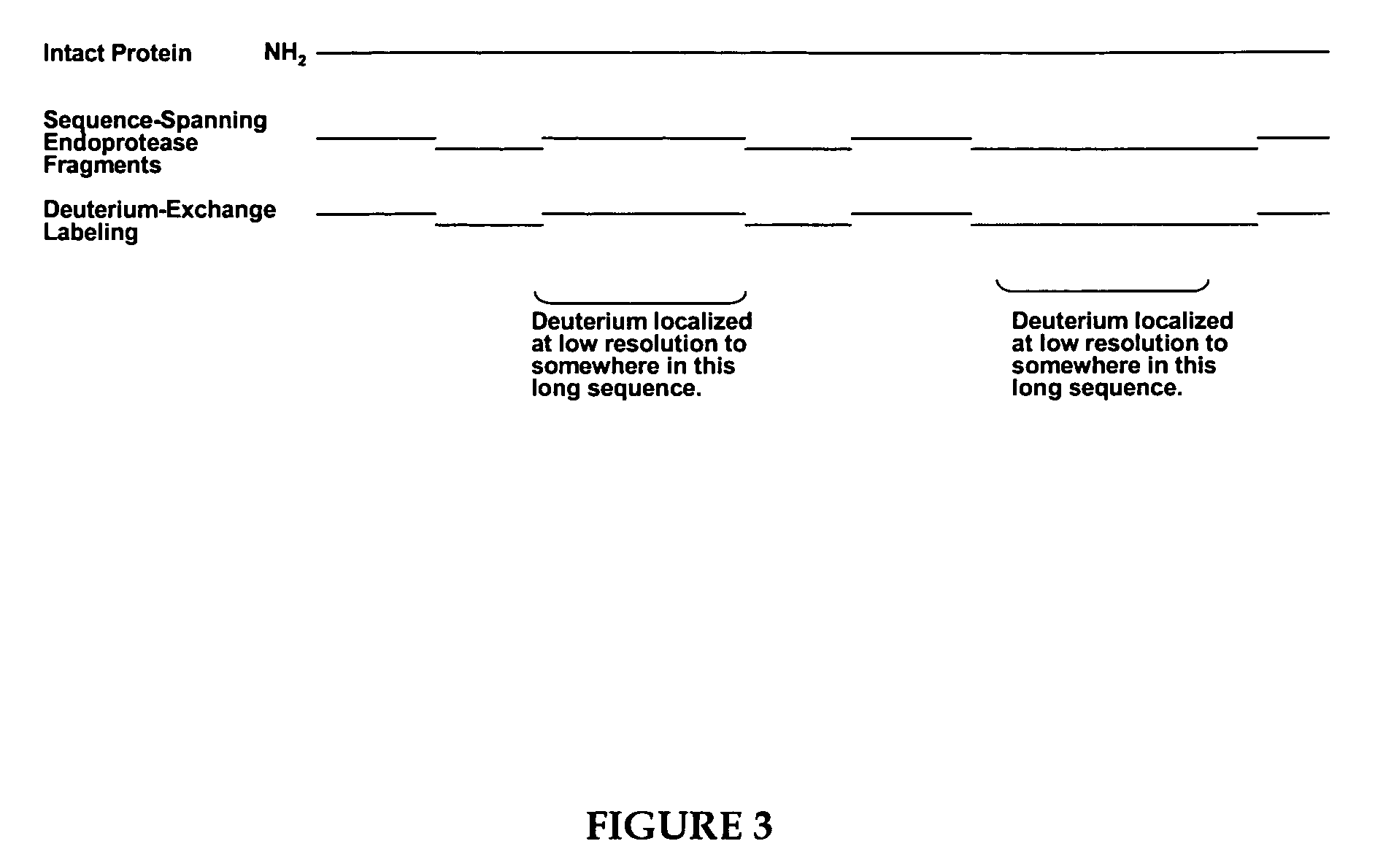
In general, the techniques that are the easiest to use and which give the quickest answers, result in an inexact and only approximate idea of the nature of the critical structural features.
Other techniques that are capable of finely characterizing polypeptide three-dimensional structure are considerably more difficult in practice.
While these techniques can ideally provide a precise characterization of relevant structural features, they have major limitations, including inordinate amounts of time required for study, inability to study large proteins, and, for X-ray analysis, the need for protein and / or protein-binding partner crystals.
The process of generating protein crystals suitable for structural analysis is commonly recognized as the most difficult and time-consuming step in the process of a crystallographic structure determination (see, e.g., Wiencek, Ann. Rev. Biomed. Eng.
Floppy, unstructured regions of proteins can play a dominant role in this problem; the energetics and kinetics of crystallization are often less favorable than for fully structured proteins, and additionally, these regions are often more susceptible to degradation during purification than are structured regions, thus promoting sample heterogeneity.
These studies do not allow a determination of the identity (location within the protein's primary amino acid sequence) of the exchanging amide hydrogens measured.
Again, determination of the identity of the particular peptide amides experiencing changes in their environment is not possible with these techniques.
However, the techniques described by Rosa and Richards were of marginal utility, primarily due to their failure to optimize certain critical experimental steps.
However, as the fragmentation of hemoglobin proceeds, each fragment's secondary and tertiary structure is lost and the unfolded peptide amide hydrogens become freely accessible to H2O in the buffer.
Moreover, in Englander's work, there is no appreciation that a suitably adapted exchange technique might be used to identify the peptide amides which reside in the contacting surface of a protein receptor and its binding partner.
Unfortunately, acid proteases are very nonspecific in their sites of cleavage, leading to considerable HPLC separation difficulties.
Even then, the fragments were “difficult to separate cleanly”.
Over the succeeding years since this observation was made, no advances have been disclosed which address these critical limitations of the medium resolution hydrogen exchange technique.
Efforts to improve the technology by employing other acid reactive proteases other than pepsin have not significantly improved the technique.
Furthermore, study of proteins by the NMR technique is not possible unless the protein is small (generally less than 30 kD), large amounts of the protein are available for the study, and computationally intensive resonance assignment work is completed.
The resolution of the deuterium-exchange mass spectrometry work disclosed in these publications therefore remained at the 10-14 amino acid level, with the primary limitation of their art being the ability to generate only a small number of peptides with the endopeptidase pepsin, as they employed it.
Despite the utility of such exchange data, the methods used to obtain it have remained labor intensive and time consuming, with substantial limitations in throughput, comprehensiveness and resolution.
Despite the availability of many enhancements that facilitate such efforts, high-throughput production of stable protein constructs that suitably crystallize continues to be a serious bottleneck.
While definition of successful constructs for protein production has long been a problem for conventional crystallography, the inadequacies of current approaches are particularly acute and costly for structural genomics efforts that presently show only a 10-20% success rate in target crystallization.
However, a switch to higher eukaryotes, such as mouse and human, will entail even lower success rates, due in part to more complex and higher molecular weight proteins.

Method used


Image

  • Methods for crystallographic structure determination employing hydrogen exchange analysis
  • Methods for crystallographic structure determination employing hydrogen exchange analysis
  • Methods for crystallographic structure determination employing hydrogen exchange analysis

Examples


example 1

DXMS Analysis used to Elucidate Phosphorylation-Driven Motions in the COOH-Terminal Src Kinase Csk

A. Background.

[0241]The Src family of nonreceptor protein tyrosine kinases (nrPTKs) bind to receptor protein tyrosine kinases (PTKs) where they phosphorylate down-stream protein targets associated with discrete signaling pathways (Superti-Furga and Courtneidge, Bioessays, 17:321-330, 1995; Neet and Hunter, Genes Cells 1:147-169, 1996; and Tatosyan and Mizenina, Biochemistry 64:49-58, 2000). While the Src enzymes comprise a large subfamily of nrPTKs, all are regulated through a single nrPTK, Csk (COOH terminal Src kinase). Csk down-regulates kinase activity by phosphorylating a single tyrosine residue in the C-terminus of the Src enzymes (Okada et al., J. Biol. Chem. 266:24249-24252, 1991; and Bergman et al., EMBO J. 11:2919-2924, 1992). Owing to this premier regulatory function, Csk has direct effects on many biological functions including T cell activation, neuronal development, cytosk...

example 2

DXMS Analysis used to Elucidate the Effects of cAMP and Catalytic Subunit Binding on cAPK Type IIβ Solvent Accessibility

A. Background.

[0254]A myriad of physiological processes are controlled by the stimulatory effects of cAMP on cAMP-dependent protein kinase (cAPK). The regulatory (R) subunits of cAPK serve as negative regulators of cAPK, as the inactive kinase exists as a tetramer composed of an R-subunit dimer bound to two catalytic (C) subunits. Binding of two cAMP molecules to each R-subunit causes dissociation of the holoenzyme complex and releases an active C-subunit. The R-subunits are known to exist in either one of two physiological states: in complex with the C-subunit or free and cAMP-saturated. A cAMP-free and C-subunit free state is believed to only exist transiently following translation due to the high affinity for cAMP and the intracellular cAMP concentrations.

[0255]Two general classes of R-subunits, type I and type II, are known to exist and differ by autophosphoryl...

example 3

DXMS Analysis Used to Refine Structure Determinations

[0282]DXMS analysis was attempted on the twenty-four Thermotoga maritima proteins listed below in Table 2, which exhibited either different degrees of resistance to crystallization or formed crystals that did not diffract X-rays sufficient for structure determination.

[0283]

TABLE 2T. maritima proteinsDid not crystallizeTIGR_description,MolecularTIGR_TMACnearest homolog.WeightLengthTM0212glycine cleavage system H protein13914.56124(gcvH) {Escherichia coli}cTM0855ribosome binding factor A15546.89131{Stigmatella aurantiaca}TM1171transcriptional regulator, crp family23394.46201{Pseudomonas stutzeri}TM1706transcription elongation factor,17848.31156greA / greB family {Bacillussubtilis}TM0160conserved hypothetical protein20551.31181{Aquifex aeolicus}TM1773conserved hypothetical protein63642.64538{Methanococcus jannaschii}Gave few crystalsMolecularTIGR_TMACTIGR_descriptionWeightLengthTM0913mazG protein {Haemophilus29804.83255influenzae}TM181...


PUM

PropertyMeasurementUnit
pHaaaaaaaaaa
cold temperatureaaaaaaaaaa
pHaaaaaaaaaa

Abstract

The present invention provides methods for crystallographic structure determination employing hydrogen exchange analysis. Hydrogen exchange analysis is used to identify unstructured regions of a protein, which are then deleted to obtain one or more modified proteins for crystallization and structure determination. Optionally, an initial hydrogen exchange stability map of a protein is compared to that of at least one modified form of the protein to identify unstructured regions, while retaining characteristic structure of the native protein. Hydrogen exchange analysis is performed by determining the quantity of isotope and / or rate of exchange of peptide amide hydrogen(s) with isotope on a labeled protein. Proteins with fewer unstructured regions, and thus improved hydrogen exchange structural maps, are optimal for high quality crystallization and structure determination.

Description

RELATED APPLICATIONS[0001]This application claims the benefit of U.S. Provisional Application No. 60 / 419,651, filed Oct. 18, 2002, the contents of which are incorporated herein in their entirety.FIELD OF THE INVENTION[0002]The present invention relates to methods for determining polypeptide structure using crystallography. In a particular aspect, the invention relates to methods for crystallographic structure determination that employ hydrogen exchange analysis.BACKGROUND OF THE INVENTION[0003]Considerable experimental work and time are required to precisely characterize the structure of a polypeptide of interest. In general, the techniques that are the easiest to use and which give the quickest answers, result in an inexact and only approximate idea of the nature of the critical structural features. Techniques in this category include the study of proteolytically generated fragments of the protein which retain binding function; recombinant DNA techniques, in which proteins are cons...

Claims


Application Information

Patent Timeline
Patent Type & Authority Patents(United States)
IPC IPC(8): G01N31/00G01N23/207G01N33/68G06F19/00
CPCG01N33/6803G01N23/207
Inventor WOODS, JR., VIRGIL L.
Owner RGT UNIV OF CALIFORNIA
Why Eureka
Try Eureka