Microfluidic systems and methods for transport and lysis of cells and analysis of cell lysate

a microfluidic system and cell technology, applied in the field of molecular biology, can solve the problems of insufficient lysis of erythrocytes, inability to quantify analytes in individual cells, slow lysis of individual cells, etc., and achieve the effect of considerable utility and high throughpu

Inactive Publication Date: 2008-09-02
UT BATTELLE LLC
View PDF20 Cites 28 Cited by
  • Summary
  • Abstract
  • Description
  • Claims
  • Application Information

AI Technical Summary

Benefits of technology

[0014]The microfluidic system and methods of this invention enable examination of the contents of single cells with high throughput. Consequently, this invention is expected to have considerable utility for facilitating research in the life sciences, and especially the pharmaceutical industry. For example, implementation of this invention in the pharmaceutical industry could expedite screening of cellular responses to large combinatorial libraries of potential novel drugs. In addition, the present invention can be used to advantage for the study of carcinogenesis or oncogenesis by assisting in the detection of rare cell mutations at an early stage, which is considered essential to the successful treatment of various forms of cancer. This invention may also be used to further elucidate metabolic pathways in cells.

Problems solved by technology

With the advances in Capillary Electrophoresis over the last two decades the quantitation of analytes in individual cells has become feasible albeit slow because of the intensive manual manipulations which have to be performed.
These reports indicate that applied electric fields resulting in cellular transmembrane potentials on the order of 1 Volt can result in lysis of erythrocytes.
However, these previously reported cell lysis techniques utilizing an electric field are typically carried out in a macroscale device, rather than a microchip device.
Consequently, such techniques are lacking in certain respects.
Specifically, the conditions employed in macroscale electric cell lysis devices do not consistently release relatively high molecular weight nucleic acid molecules, because such molecules do not readily pass through the pores created in the cell membrane by this lysis technique.

Method used


Image

  • Microfluidic systems and methods for transport and lysis of cells and analysis of cell lysate
  • Microfluidic systems and methods for transport and lysis of cells and analysis of cell lysate
  • Microfluidic systems and methods for transport and lysis of cells and analysis of cell lysate

Examples


example i

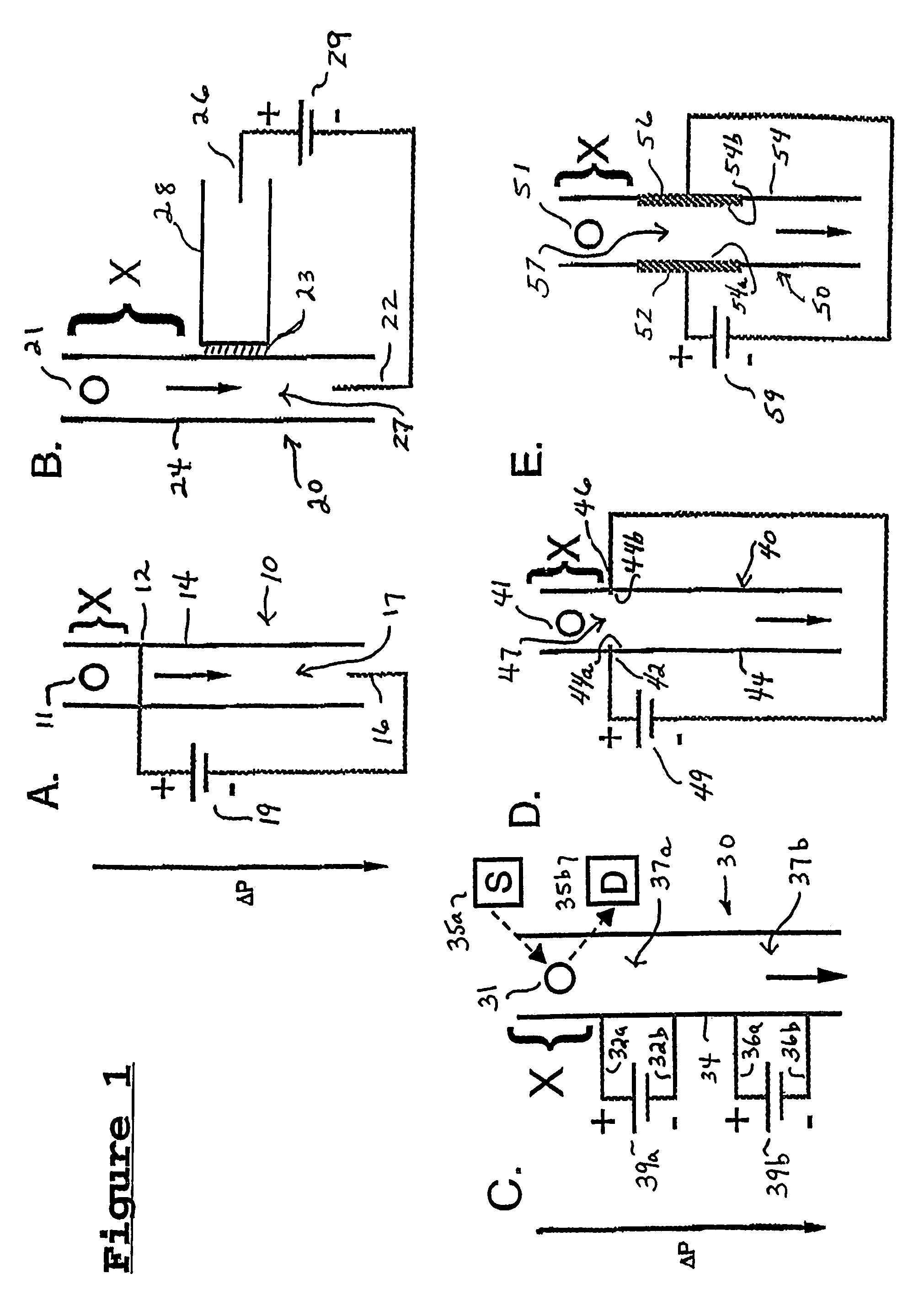
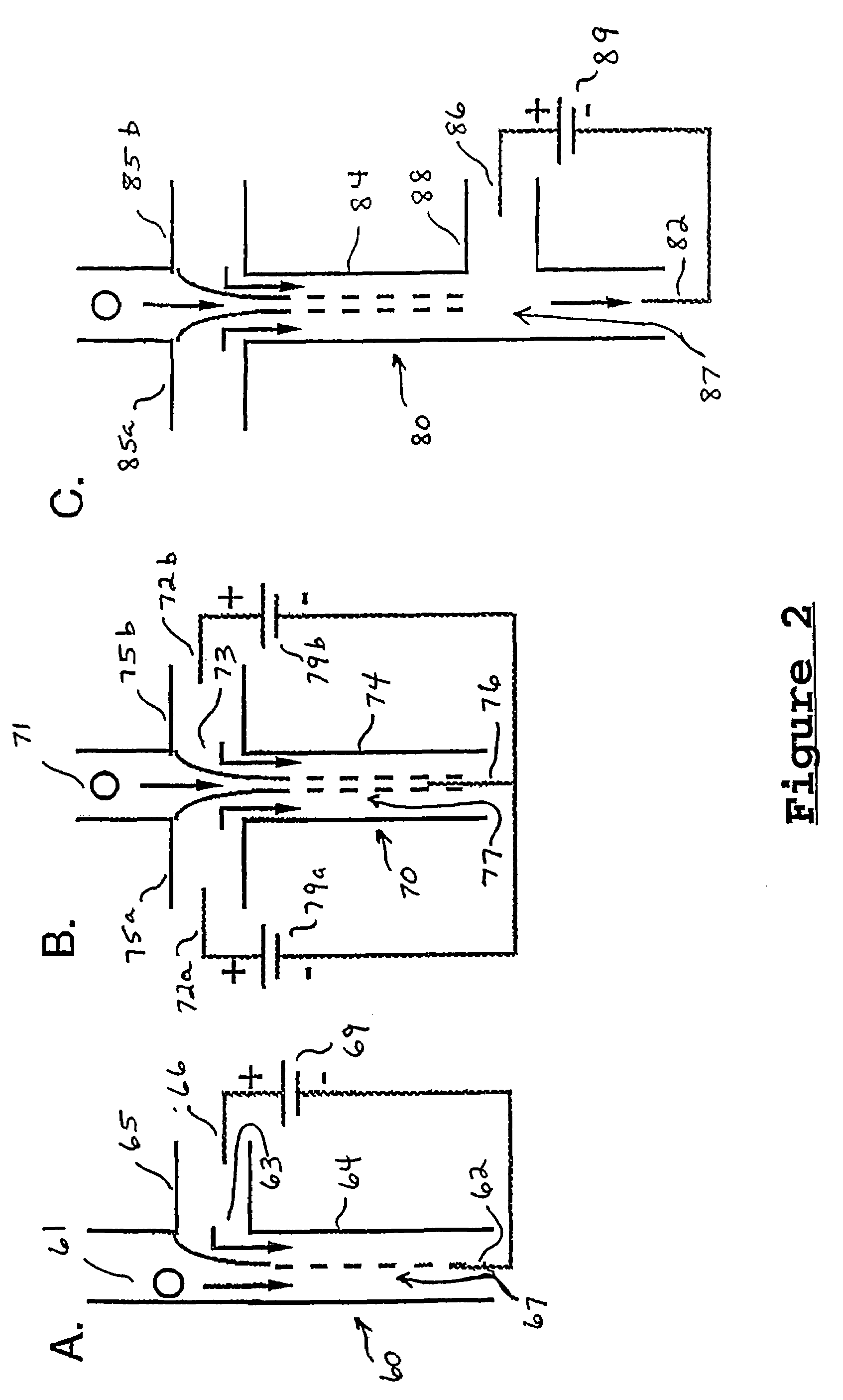
[0057]A microchip design of the type shown in FIG. 2B has been tested using the immortal Jurkat T lymphocyte cell line. The cells were labeled with calcein AM (Catalog #C-1430; Molecular Probes, Eugene, Oreg.) and contained in PBS. The cross intersection 73 was visualized using a video rate CCD camera (30 frames / sec) and the cells were hydraulically transported to the cross intersection by applying sub-ambient pressure to the lower segment of microchannel 74. The cells appear elongated in the images in FIG. 7 because the fluorescence signal from the calcein in the cell is integrated over 33 msec for each video frame. The streak length increases after the cell enters the lower segment of microchannel 74 because the flow rate in this channel segment is equal to the sum of the flow rates in the upper and side channel segments leading to intersection 73. A pulsed electric field (square wave) was applied between the side channel segments 75a, 75b and the lower segment of microchannel 74....

example ii

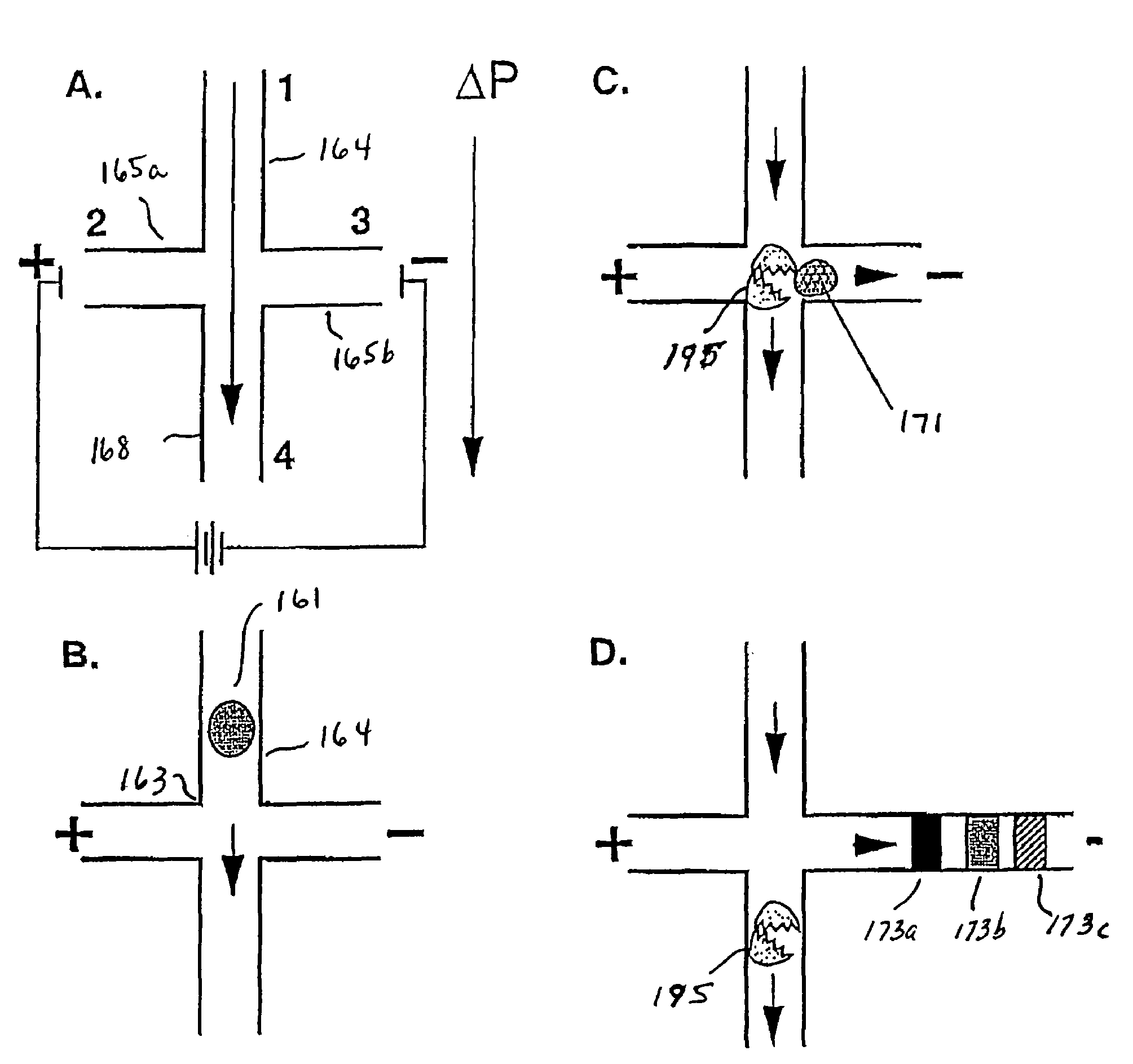
[0061]FIG. 8 shows the separation of several fluorescently labeled components released from a single cell utilizing a microchip design of the type depicted in FIG. 5, above. These components are Oregon green and some Oregon green degradation products. To generate this data, living Jurkat cells were loaded with the membrane permeable dye Oregon green diacetate. The cells 161 metabolized the dye transforming it into a membrane impermeable form that was trapped in the cell. The cell 161 was then transported through microchannel 164 to the cross intersection 163, where it was lysed by the electric field applied through channels 165a and 165b. A portion of the lysed contents in analyte plug 171 was then transported by the electric field into side channel 165b and electrophoretically separated into multiple analyte segments 173a, 173b, and 173c and detected by fluorescence.


PUM

PropertyMeasurementUnit
electric field strengthaaaaaaaaaa
axial lengthaaaaaaaaaa

Abstract

Microfluidic systems and methods are disclosed which are adapted to transport and lyse cellular components of a test sample for analysis. The disclosed microfluidic systems and methods, which employ an electric field to rupture the cell membrane, cause unusually rapid lysis, thereby minimizing continued cellular activity and resulting in greater accuracy of analysis of cell processes.

Description

RELATED U.S. APPLICATION DATA[0001]This application is a continuation of U.S. patent application Ser. No. 10 / 041,821, filed Oct. 19, 2001 now U.S. Pat No. 6,783,647, which is incorporated by reference herein in its entirety.[0002]This invention was made with Government support under contract DE-AC05-00OR22725 awarded by the United States Department of Energy to UT-Battelle, LLC. The Government has certain rights in this invention.FIELD OF THE INVENTION[0003]This invention relates to molecular biology, and in particular, to the use of an applied electric field in a microfluidic system for the manipulation of biological samples comprising cells and cell lysate(s) for subsequent analysis.BACKGROUND OF THE INVENTION[0004]Interest in microfabricated devices for chemical sensing and analysis has grown substantially over the past decade, primarily because these miniature devices have the potential to provide information rapidly and reliably at low cost. Microchips fabricated on planar subs...

Claims


Application Information

Patent Timeline
Patent Type & Authority Patents(United States)
IPC IPC(8): G01N27/453C12M1/33C12N13/00B01L3/00C12N1/06G01N27/447
CPCB01L3/502761G01N27/44743C12N1/066C12N13/00B01L2200/0647B01L2400/0415B01L2400/0487
Inventor CULBERTSON, CHRISTOPHER T.JACOBSON, STEPHEN C.MCCLAIN, MAXINE A.RAMSEY, J. MICHAEL
Owner UT BATTELLE LLC
Why Eureka
Try Eureka