

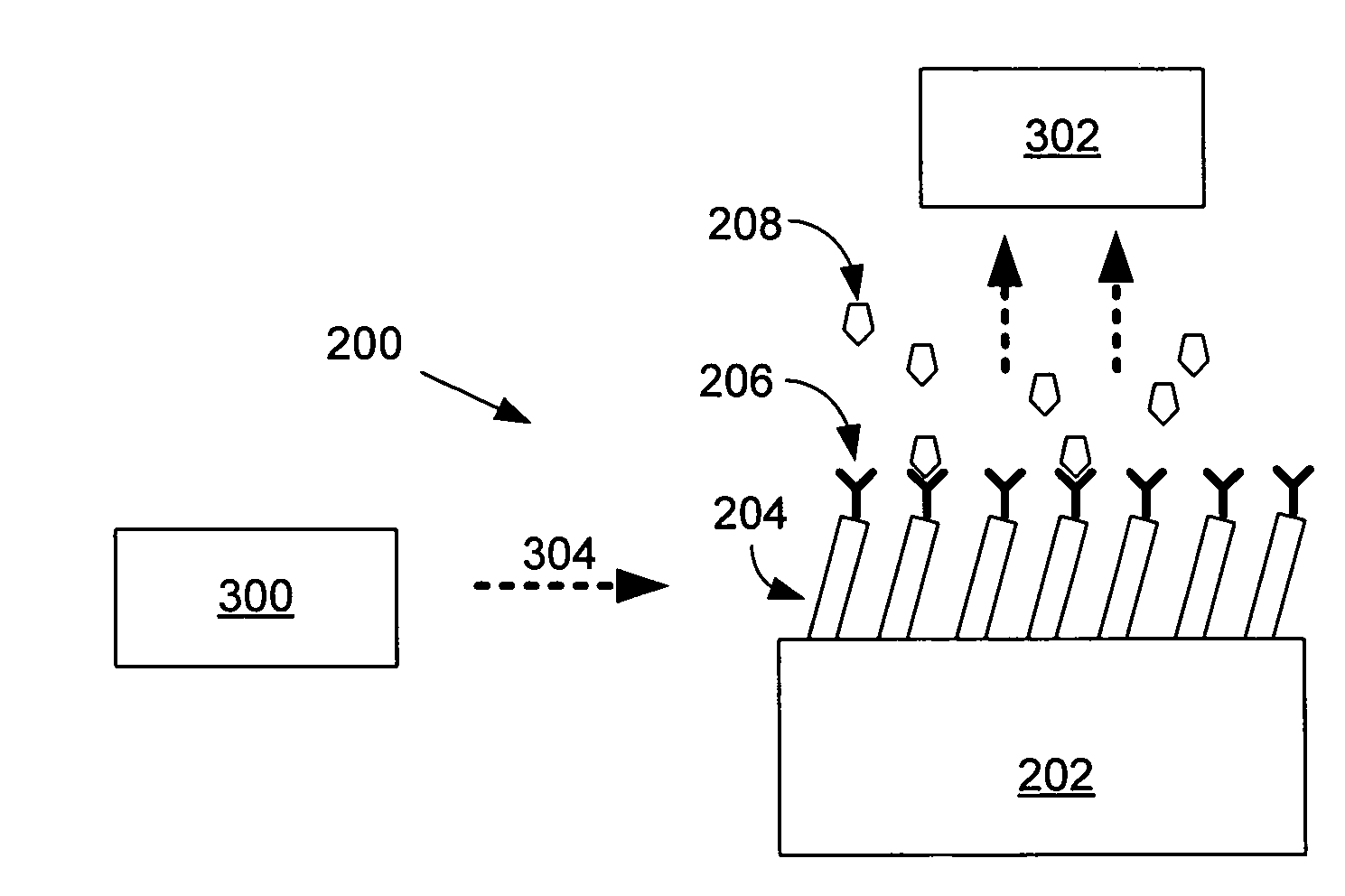
Surface enhanced raman spectroscopy (SERS) systems and methods of use thereof
a raman spectroscopic and enhanced technology, applied in the field of surface enhanced raman spectroscopy (sers) systems and methods, can solve the problems of difficult to obtain surface-enhanced raman data, unclear how this large enhancement effect might be exploited for applications in analytical chemistry, molecular biology, or medical diagnostics,
- Summary
- Abstract
- Description
- Claims
- Application Information
AI Technical Summary
Problems solved by technology
Method used
Image

Examples
example 1
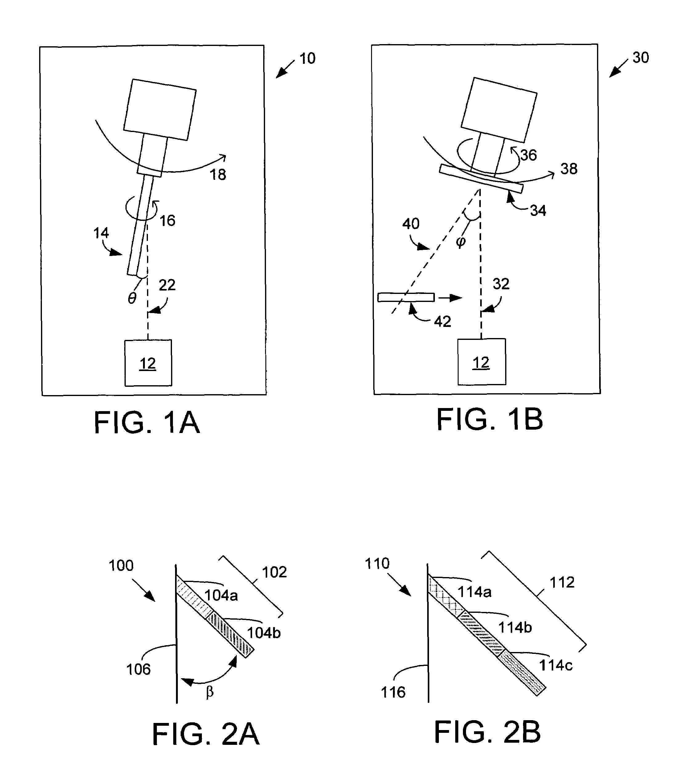
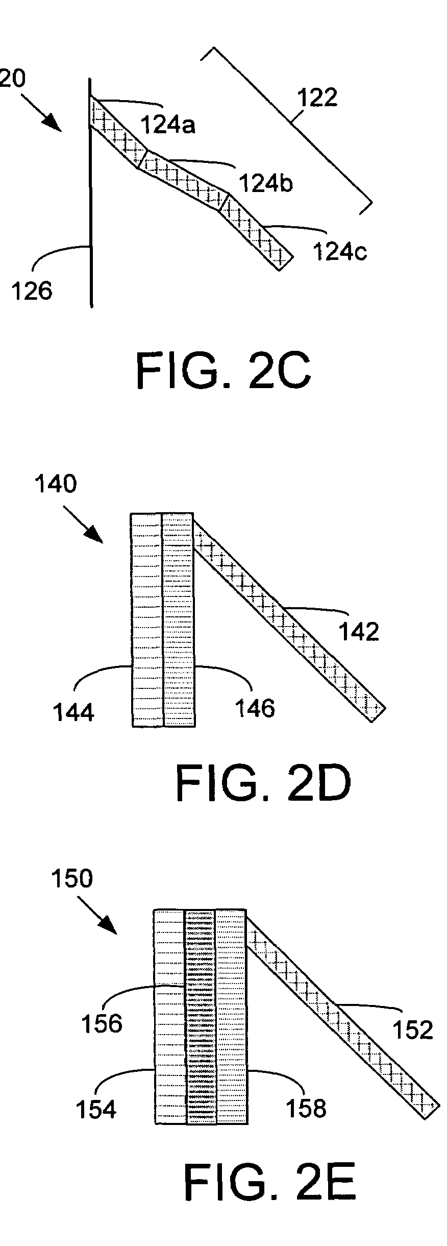
[0089]All of the samples were prepared using an electron beam / sputtering evaporation system (E-beam) that was custom built by Torr International. A schematic of the set-up is shown in FIG. 1A. A glass microscope slide with size 1×3″ and 1 mm thick (Gold Seal®) was used as a substrate 34. A custom shutter 42 was built that could be controlled externally by a feed through, and the shutter was used to selectively reveal increasing portions of the substrate 34 during the deposition process. This method can produce one single sample with 6 different active areas. As an example, one particular sample had a 50 nm thin film deposited at normal incidence and then it was rotated to an incident angle 4 of 86°. Then nanorods were deposited in steps of 200 nm; i.e. the shutter 42 was opened partially and 200 nm was deposited, then the shutter was opened slightly more exposing more of the substrate and another 200 nm was deposited while keeping the previously exposed area still ...
example 2
[0101]This example describes a method to prepare nanostructured SERS substrates that allow for rapid and sensitive detection of the molecular fingerprint of RNA and DNA viruses using Raman spectra, as well as providing structural and quantitative information about the viruses.
[0102]Development of diagnostic methods for rapid and sensitive identification of viruses is essential for defining the emergence of viral infection, determining the period that preventive measures should be applied, for evaluating drug and vaccine efficacy, for preventing epidemics, and determining agents of bioterrorism. Current diagnostic methods are either cumbersome, time-consuming, or have limited sensitivity. This example describes a nanofabrication technique to create novel SERS substrates that can be used to rapidly detect the Raman viral nucleic acid spectra of RNA and DNA viruses. Monoclonal antibodies reactive for individual RNA or DNA viruses were conjugated to these substrates, and it was demonstr...
example 3
Experimental Method
[0107]The concentrations of the prepared virus samples in Dulbecco's Modified Eagle Medium (DMEM) were 107 plaque forming units (pfus) / mL for HIV, 105 TCID[50] / mL for Rhinovirus and 106 TCID[50] / mL for the Adenovirus. After preparation, the virus samples were stored at −80° C. until the day of the experiment. The samples were thawed for about 5 minutes and an Eppendorf pipette was used to withdraw about 0.5 μL from the sample vial which was then allowed to spread onto the silver nanorod substrate corresponding to about 5000 plaque forming units (pfus) of HIV, about 350 pfus of Adenovirus and about 35 pfus of Rhinovirus. The virus droplet was allowed to dry and bind to the silver surface for ˜1 hour prior to the Raman experiment.
[0108]Surface Enhanced Raman spectra were acquired using a Kaiser Optical Systems confocal Raman microscope (Kaiser Optical Systems Incorporated, Ann Arbor, Mich.) equipped with a liquid nitrogen cooled Charge Coupled Device (CCD) camera (P...
PUM
| Property | Measurement | Unit |
|---|---|---|
| Angle | aaaaa | aaaaa |
| Angle | aaaaa | aaaaa |
| Angle | aaaaa | aaaaa |
Abstract
Description
Claims
Application Information