In vivo multiphoton diagnostic detection and imaging of a neurodegenerative disease

a neurodegenerative disease and multi-photon imaging technology, applied in the field of in vivo multi-photon diagnostic detection and imaging of neurodegenerative diseases, can solve the problems of untold anguish for millions, and amnesia accompanied by frustration and paranoia, and achieve the effect of promoting simultaneous multi-photon excitation of brain tissu

Inactive Publication Date: 2010-02-23
THE GENERAL HOSPITAL CORP +1
View PDF62 Cites 6 Cited by
  • Summary
  • Abstract
  • Description
  • Claims
  • Application Information

AI Technical Summary

Benefits of technology

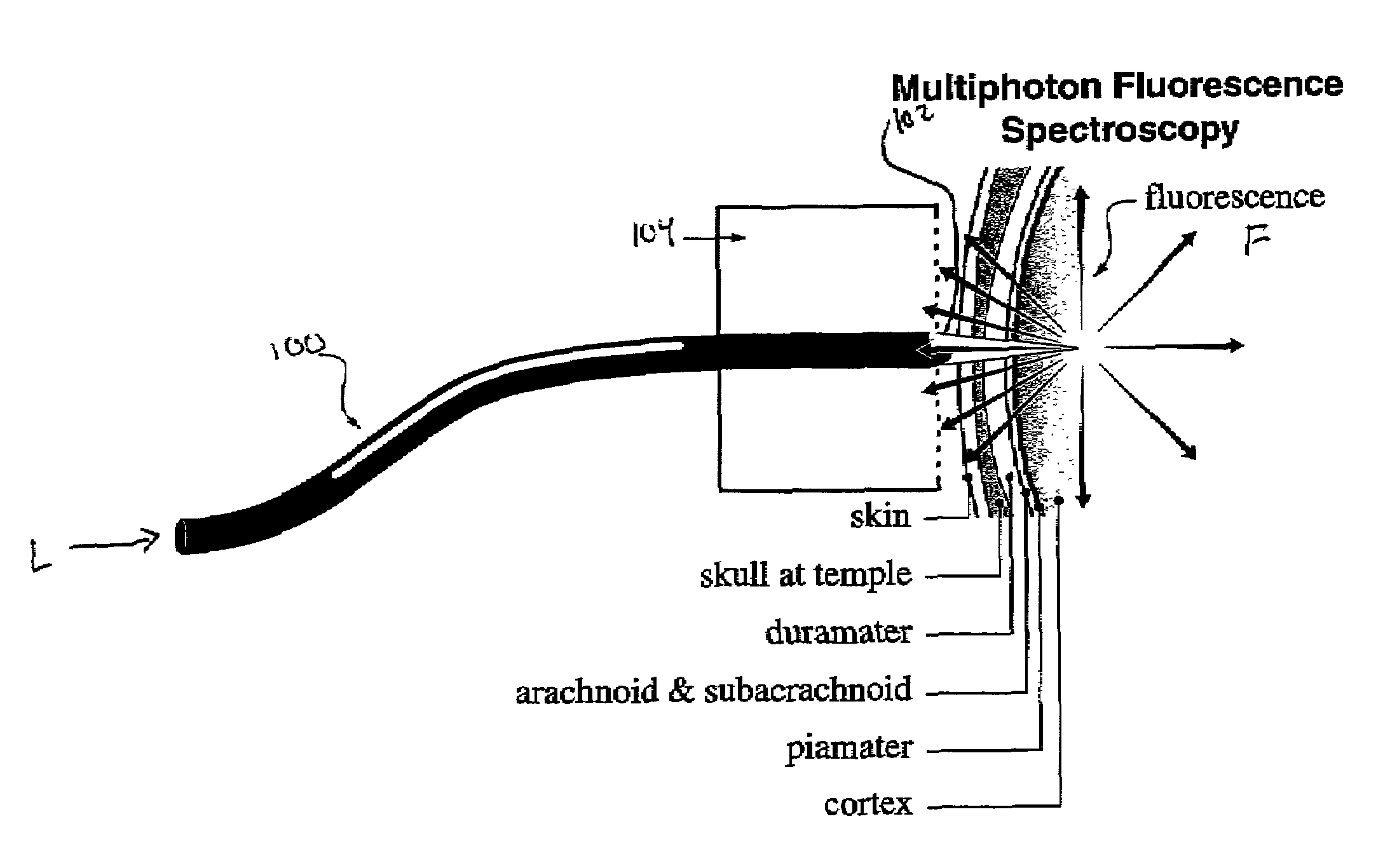
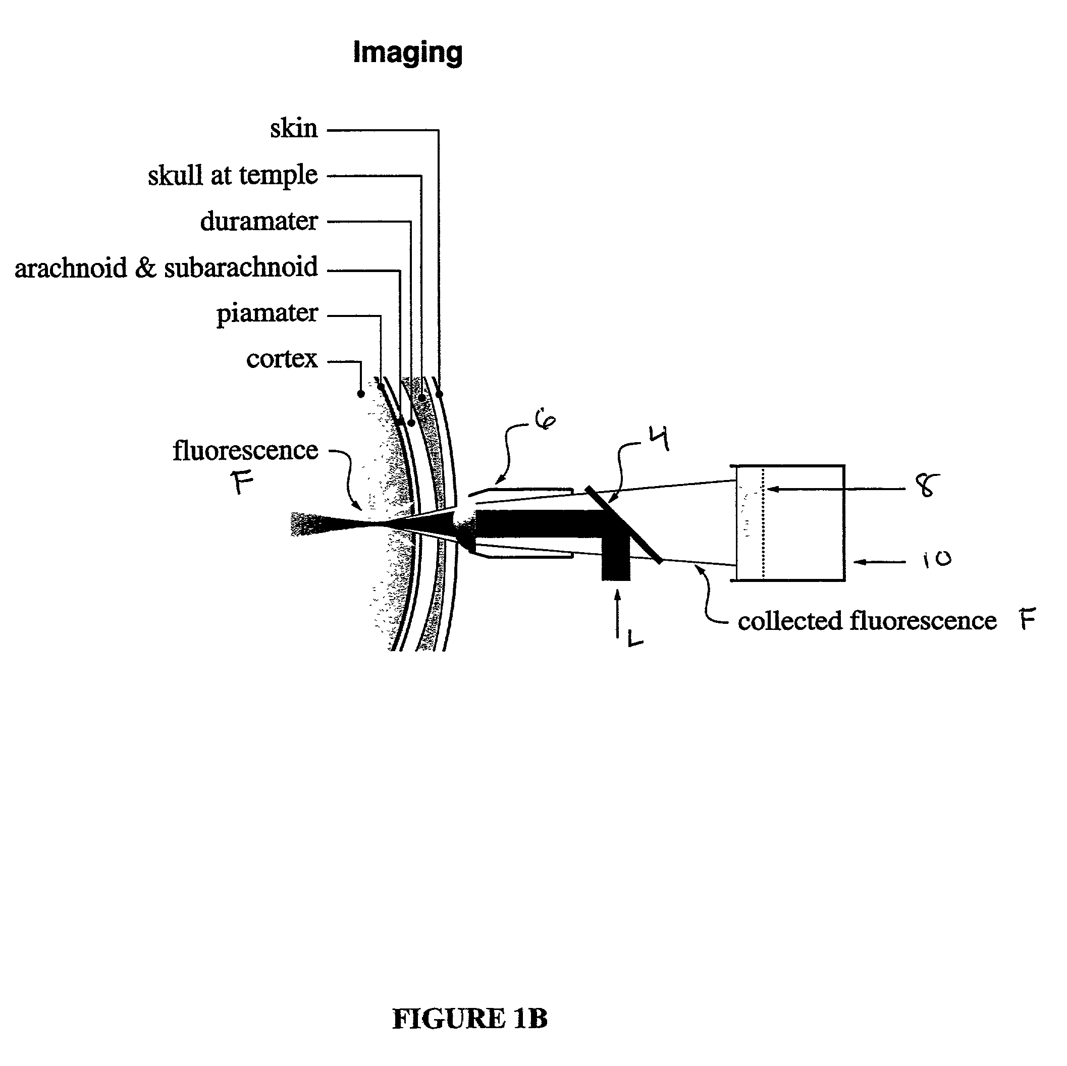
[0010]The present invention is directed to a method of detecting a neurodegenerative disease in a mammal by activating brain tissue of the mammal by application of radiation under conditions effective to promote a simultaneous multiphoton excitation of the brain tissue and to emit a fluorescence characteristic. The fluorescence characteristic is then compared to a standard fluorescence emitted by exciting healthy brain tissue of the mammal under the same conditions used to carry out the activating step. Brain tissue where the fluorescence characteristic differs from the standard fluorescence is identified as potentially having a neurodegenerative disease.
[0011]Another aspect of the present invention is directed to a method of producing an image of brain tissue from a mammal by activating brain tissue of a mammal with radiation applied under conditions effective to promote a simultaneous multiphoton excitation of the brain tissue and to produce fluorescence. The fluorescence is then collected to produce an image of the brain tissue.
[0012]The current state of the art is that diagnosis of Alzheimer's Disease occurs only after clinical manifestations of marked memory loss and other cognitive impairments occur. Neuropathological studies suggest that the disease process actually starts many years prior to these overt symptoms. An imaging technology that allows detection of the pathological process prior to clinical symptoms would allow treatments aimed at prevention of progression rather than simply treatment of symptoms. Ideally, a biomarker of this nature can be used for presymptomatic identification of patients, as a method of following the efficacy of therapy, and as a method to direct different therapies. Each of these applications would provide a substantial advance over the current state of affairs.

Problems solved by technology

Alzheimer's Disease is a devastating illness affecting nearly 4 million Americans, costing the country billions of dollars in direct care, lost income, and—most importantly—costing untold anguish for millions of American families.
Frequently this amnesia is accompanied by frustration and paranoia.
Ultimately patients lose all ability to care for themselves—to dress themselves, to bathe, even to control their bowel and bladder.
However, these changes are far too small (100 times too small) to be seen by even the most sophisticated clinical imaging devices such as computerized tomography or magnetic resonance imaging.

Method used


Image

  • In vivo multiphoton diagnostic detection and imaging of a neurodegenerative disease
  • In vivo multiphoton diagnostic detection and imaging of a neurodegenerative disease
  • In vivo multiphoton diagnostic detection and imaging of a neurodegenerative disease

Examples


example 1

In vivo Imaging of Amyloid Deposition

[0064]Nine male Tg2576 mice (mean age 18.6 months (Hsiao, et al., “Correlative Memory Deficits, Aβ Elevation, and Amyloid Plaques in Transgenic Mice,”Science, 274:99-102 (1996), which is hereby incorporated by reference in its entirety), were used for the in vivo imaging of plaques. These mice express human amyloid precursor precursor carrying the Swedish mutation under the hamster prion protein promoter. The skull was prepared 2-6 days prior to imaging. Mice were anesthetized with Avertin (Tribromoethanol, 250 mg / kg IP). A high-speed drill (Fine Science Tools, Foster City Calif.) was used to thin the skull in a circular region, approximately 1-1.2 mm in diameter (FIG. 2A), using a dissecting microscope for gross visualization of the site. Heat and vibration artifacts were minimized during drilling by frequent application of artificial cerebrospinal fluid (“ACSF”; 125 mM NaCl; 26 mM NaHCO3; 1.25 mM NaH2PO4; 2.5 mM KCl; 1 mM MgCl2; 1 mM CaCl2; 25 ...

example 2

Image Analysis

[0066]Montages were reconstructed into a single stack of images using Scion Image (Scion Corp, Frederick Md.). The area of individual plaque cross-sections was measured in each optical section by thresholding at two standard deviations above the mean of an adjacent background region. Plaques that did not satisfy criteria of imaging were eliminated from the measurement set. Plaques on the edge of the imaging area or on one of the montage lines were rejected due to the potential imprecision of moving the animal on the stage. Plaques whose intensity was not sufficiently above background for appropriate thresholding were also eliminated. This rejected many plaques, typically deep in the preparation, that appeared to be present, but were too faint to measure using the automatic threshold technique. Finally, plaques whose images contained any appreciable motion artifact were rejected. Maximal plaque diameter was then calculated from the cross-section of largest area for each...

example 3

Angiography

[0067]The tail of the animal was warmed on a heating pad to dilate the blood vessels, and approximately 0.05 ml fluorescein (25 mg / ml) in sterile PBS was injected into a tail vein of the mouse at least 20 minutes prior to imaging. The dye did not cross the blood-brain barrier and permitted concurrent visualization of blood vessels throughout the imaging volume in the brain.


PUM

PropertyMeasurementUnit
wavelengthaaaaaaaaaa
distanceaaaaaaaaaa
distanceaaaaaaaaaa

Abstract

The present invention is directed to a method of detecting a neurodegenerative disease in a mammal by activating brain tissue of the mammal by application of radiation under conditions effective to promote a simultaneous multiphoton excitation of the brain tissue and to emit a fluorescence characteristic. The fluorescence characteristic is then compared to a standard fluorescence emitted by exciting healthy brain tissue of the mammal under the same conditions used to carry out the activating step. Brain tissue where the fluorescence characteristic differs from the standard fluorescence is identified as potentially having a neurodegenerative disease. Another aspect of the present invention is directed to a method of producing an image of brain tissue from a mammal by activating brain tissue of a mammal with radiation applied under conditions effective to promote a simultaneous multiphoton excitation of the brain tissue and to produce fluorescence. The fluorescence is then collected to produce an image of the brain tissue.

Description

[0001]This application claims benefit of U.S. Provisional Patent Application Ser. No. 60 / 245,306, filed on Nov. 2, 2000.[0002]This invention was developed with government funding under National Institutes of Health Grant Nos. AG08487 and RR04224. The U.S. Government may have certain rights.FIELD OF THE INVENTION[0003]The present invention relates to the in vivo multiphoton diagnostic detection and imaging of a neurodegenerative disease.BACKGROUND OF THE INVENTION[0004]Alzheimer's Disease is a devastating illness affecting nearly 4 million Americans, costing the country billions of dollars in direct care, lost income, and—most importantly—costing untold anguish for millions of American families.[0005]The disease symptoms start insidiously, with relatively non-specific signs and symptoms. Subtle memory loss, occasional word-finding difficulties, irritability and aggressiveness can all be signs of a disease process from a clinical perspective, that has already affected the brain substa...

Claims


Application Information

Patent Timeline
Patent Type & Authority Patents(United States)
IPC IPC(8): A61B6/00A61B10/00A61B5/00A61B5/107A61K49/00G01N21/64
CPCA61B5/0059A61K49/0021A61K49/0041A61K49/0043A61K49/0056A61K49/0058G01N21/6456G01N21/6458G01N21/6486A61B5/1075A61B5/0042A61B5/4064A61B5/4088
Inventor HYMAN, BRADLEY T.CHRISTIE, RICHARDBACSKAI, BRIANWEBB, WATT W.ZIPFEL, WARREN R.
Owner THE GENERAL HOSPITAL CORP
Why Eureka
Try Eureka